A Field Guide to Amphibian Larvae and Eggs
of Minnesota, Wisconsin, and Iowa
U.S. Department of the Interior
U.S. Geological Survey
Information and Technology Report
Technical Report Series
The Biological Resources Division publishes scientific and technical articles and reports resulting from the research performed by our scientists and partners. These articles appear in professional journals around the world. Reports are published in two report series: Biological Science Reports and Information and Technology Reports.
Series Descriptions
Biological Science Reports ISSN 1081-292XThis series records the significant findings resulting from sponsored and co-sponsored research programs. They may include extensive data or theoretical analyses. Papers in this series are held to the same peer-review and high quality standards as their journal counterparts.
Information and Technology Reports ISSN 1081-2911
These reports are intended for publication of book-length monographs; synthesis documents; compilations of con-ference and workshop papers; important planning and ref-erence materials such as strategic plans, standard operating procedures, protocols, handbooks, and manuals; and data compilations such as tables and bibliographies. Papers in this series are held to the same peer-review and high quality standards as their journal counterparts
Suggested citation: Parmelee, J. R., M. G. Knutson, and J. E. Lyon. 2002. A field guide to amphibian larvae and eggs of Minnesota, Wisconsin, and Iowa. U.S. Geological Survey, Biological Resources Division, Information and Technology Report USGS/BRD/ITR-2002-0004, Washington, D.C. iv + 38 pp.
Additional copies of this report may be obtained from the National Technical Information Service, 5285 Port Royal Road, Springfield, VA 22161 (1-800-553-6847 or 703-487-4650). Also available to registered users from the Defense Technical Information Center, Attn: Help Desk, 8725 Kingman Road, Suite 0944, Fort Belvoir, VA 22060-6218 (1-800-225-3842 or 703-767-9050).
Front cover photo of Tiger Salamander eggs by Shawn Weick.
Back cover photo of Wood Frog by Allen Sheldon, Wood Frog eggs by Melinda Knutson.
Mention of trade names or commercial products does not constitute endorsement or recommendation for use by the U.S. Department of the Interior, U.S. Geological Survey.
A Field Guide to Amphibian Larvae and Eggs
of Minnesota, Wisconsin, and Iowa
By Jeffrey R. Parmelee, Melinda G. Knutson, and James E. Lyon Illustrations by Pearl Podgorniak
Information and Technology Report 2002-0004
June 2002
U.S. Department of the Interior
U.S. Geological Survey
Washington, D. C. 20240
About the authors
Jeff Parmelee is an assistant professor of biology at Simpson College in Indianola, Iowa, where he teaches herpetology, anatomy, and general biology. He obtained his Ph.D. from the University of Kansas in 1998 and his research interests include tropical herpetology and rattlesnake ecology (parmelee@simpson.edu). Melinda Knutson is a wildlife biologist at the USGS in La Crosse, Wisconsin, where she studies amphibians and birds and their habitats. She obtained her Ph.D. from Iowa State University in 1995 and her research interests include conservation biology and landscape ecology (melinda_ knutson@usgs.gov ; http://www.umesc.usgs.gov/). James Lyon is a biologist and naturalist with special interests in amphibians, reptiles, birds, and native plants. Pearl Podgorniak created the drawings of larvae and eggs for this publication as an undergraduate research assistant with Dr. Parmelee, supported by Simpson College.
Contents
Title ... 1
How to use the keys ... 3
Preserving amphibian eggs and larvae... 4
Raising larvae... 4
Field key to Amphibian Eggs ... 8
Field Key to Larval Salamanders... 10
Field Key to Larval Anurans... 12
Species descriptions ... 14
Blue-spotted Salamander (Ambystoma laterale)... 14
Spotted Salamander (Ambystoma maculatum)... 15
Small-mouthed Salamander (Ambystoma texanum)... 16
Tiger Salamander (Ambystoma tigrinum) ... 17
Four-toed Salamander (Hemidactylium scutatum)... 18
Mudpuppy (Necturus maculosus) ... 19
Eastern Newt (Notopthalmus viridescens) ... 20
Eastern Red-backed Salamander (Plethodon cinereus) ... 21
Northern Cricket Frog (Acris crepitans) ... 22
American, Canadian, Great Plains, Woodhouse’s, and Fowler’s Toads (Bufo americanus/B. hemiophrys/B. cognatus/B. woodhousii/B. fowleri) ... 23
Gray and Cope’s Gray Treefrog (Hyla versicolor/H. chrysoscelis) ... 25
Spring Peeper (Pseudacris crucifer) ... 26
Western Chorus and Boreal Chorus Frog (Pseudacris triseriata/P. maculata)... 27
Leopard, Pickerel, and Crawfish Frogs (Rana pipiens/R. sphenocephala/R. blairi/R. palustris/R. areolata) ... 28
American Bullfrog (Rana catesbeiana) ... 30
Green and Mink Frogs (Rana clamitans/R. septentrionalis)... 32
Wood Frog (Rana sylvatica)... 34
Plains Spadefoot (Spea bombifrons) ... 35
Acknowledgments... 36
Apparent worldwide declines in amphibian populations (Pechmann and Wake 1997) have stimulated interest in amphibians as bioindicators of the health of ecosystems. Because we have little information on the population status of many species, there is interest by public and private land management agencies in monitoring amphibian populations. Amphibian egg and larval surveys are established methods of surveying pond-breeding amphibians. Adults may be widely dispersed across the landscape, but eggs and larvae are confined to the breeding site during a specific season of the year. Also, observations of late-stage larvae or metamorphs are evidence of successful reproduction, which is an important indicator of the viability of the population. The goal of this guide is to help students, natural resources personnel, and
biologists identify eggs and larval stages of amphibians in the field without the aid of a microscope.
Anyone undertaking field identification of amphibians has a responsibility to avoid harming the amphibians or their habitats. Persons planning to sample amphibians should work in cooperation with state or federal wildlife professionals. Lack of knowledge about sensitive habitats or populations could result in the spread of diseases, damage to breeding habitats, or local reproductive failure of amphibian populations. State and federal laws protect amphibians from exploitation. Collection permits are required from the appropriate state or federal authorities before capturing, handling, or collecting amphibians. Permission for sampling should also be obtained from the landowner.
A Field Guide to Amphibian Larvae and Eggs
of Minnesota, Wisconsin, and Iowa
By Jeffrey R. Parmelee Simpson College Department of Biology 701 North C Street Indianola, Iowa 50125 and
Melinda G. Knutson and James E. Lyon
U.S. Geological Survey
Upper Midwest Environmental Sciences Center 2630 Fanta Reed Road
Research for this publication began with a study of farm ponds in southeastern Minnesota. We quickly recognized the need for a way to identify eggs and larvae of the amphibians encountered. We used our field knowledge of most of the species included here to produce this field guide. Additional information came from published keys for Wisconsin amphibians (Vogt 1981; Watermolen 1995; Watermolen and Gilbertson 1996) and descriptions from a variety of sources (e.g., Johnson 2000; Petranka 1998; Harding 1997; Russell and Bauer 1993; Stebbins 1985; Wright and Wright 1949).
There is no simple way to morphologically distinguish between many amphibian species during the egg and tadpole stage. This might be expected in closely related species such as the gray treefrogs Hyla chrysoscelis and Hyla versicolor, which vary in chromosome number and in subtle morphological traits. It is surprising that, to our knowledge, no one has reported a way to morphologically distinguish between larvae of several common frog species such as the Crawfish Frog and Southern Leopard Frog, which bear little resemblance to one another as adults. Even with the technical keys available to herpetologists, amphibian eggs and larvae are often difficult to identify to species. An excellent key to all species in the United States (Altig et al. 1998) is available on the internet (http: //www.pwrc.usgs.gov/tadpole/). The Altig key (along with earlier versions such as Altig [1970] for tadpoles and Altig and Ireland [1984] for salamander larvae) requires detailed knowledge of the anatomy of amphibian larvae. Altig et al. (1998) requires examination of tadpole mouthparts, which are only observable with dead specimens and the aid of a microscope. Tadpole biology is summarized by McDiarmid and Altig (1999). Additional research is needed to better describe and understand larval amphibians.
Three major factors make amphibian larvae, especially tadpoles, difficult to identify. First, many species are conservative in their anatomy and members of the true frog (Ranidae) and toad (Bufonidae) families are very similar in larval form. Second, morphology among tadpoles of a single species can vary geographically and with developmental stages. Third, larvae reveal differing anatomies depending on the physical and biotic environment where they are found. The proportions, coloration, and anatomy of larvae change as they grow from hatchlings to metamorphs. Larvae in early stages frequently do not show the characteristics necessary for their identification.
In one description, we summarize closely-related species that cannot be distinguished without the aid of a microscope. We believe that the weakness of specific identification is balanced by ease of use in the field. In many instances, identification to species is not necessary to meet land managers’ goals. However, professional herpetologists working for universities, museums, and state and federal agencies can assist with identifying larvae to species.
No key by itself can help identify every specimen; there will always be variant individuals. In identifying anurans, for example, knowledge of which species were calling at the pond may help sort out identification questions. It also is helpful to note the geographic distribution of species; you can reasonably rule out Canadian Toads if the site is in southern Iowa. Ruling out a species by geography is not easy or reliable, however, if your survey site is near the edge of a species’ range. You could also return several times to the breeding site to observe late-stage metamorphs. Amphibians often are easier to identify once they develop mature traits. If a specimen cannot be identified
using this key, preserve the specimen and attempt identification using a more detailed key such as Altig et al. (1998). If still not confident in the identification, contact the senior author of this key or another herpetologist in your area for assistance. Another approach, if adequate time and facilities are available, is to raise a few eggs or larvae to metamorphosis and identify the late-stage metamorphs or adults from their mature characteristics. We anticipate that rapid molecular DNA techniques suitable for field identification will be available within the next few decades. Until then, keys will be needed.
How to use the keys
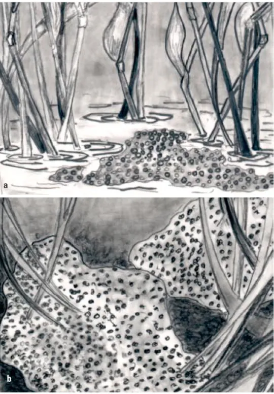
Most amphibians lay their eggs in clusters or strings. Four general types of amphibian egg masses are illustrated (Figure 1); globular clusters are large and easier to see than eggs in long strings, whereas eggs laid singly or in small clusters are difficult to find, especially in dense vegetation.
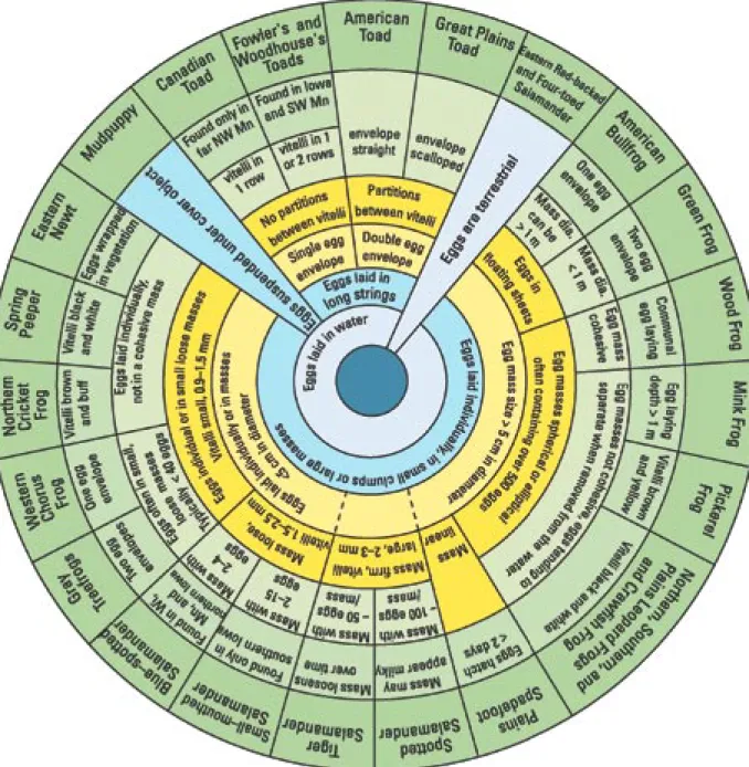
To use the circular keys (Figures 2–4), start in the center and choose among the available options, ending in the outer circle with the identification of a species or group of species. In some instances, dashed lines are used if a species exhibits multiple traits. These traits are described to the left and right of the section enclosed by the dashed line. After making an identification, use the species description to confirm the identification. If the identification seems in error (e.g., the collection location is geographically distant from the range of that species, the specimen looks nothing like the illustration, or the description doesn’t match the specimen), return to the key and work backwards to find another species that more closely fits your specimen.
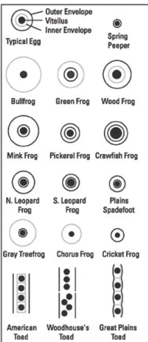
The jellies of most amphibian eggs are
very small when first laid, but quickly swell with water. All amphibian eggs in this key are pigmented and have one or several gelatinous envelopes surrounding them (Figure 2). The gelatinous capsules are difficult to see, but it helps to use a magnifying lens and adjust the lighting. The egg key groups most ranids together, and mole salamanders also have similar eggs.
Salamanders (Figure 3) have four limbs and feathery external gills, whereas tadpoles (Figure 4) have four limbs only when they are close to metamorphosis and their gills are internal. The location of the eyes on the tadpole is an important character separating the treefrogs (hylids) from the true frogs (ranids). Eyes dorsal means the eyes do not interrupt the lateral margin of the head when viewed from above (superior view; Figure 4a). Another early characteristic in the key is whether the vent is medial or dextral (Figure 4b). This is difficult to see on a squirming live tadpole; however, hold it with the bottom (ventral) side up, straighten the tail fin, and look carefully at the location of the opening of the vent. A magnifying lens will help.
The scale bar by each drawing in the species description indicates 1 cm. Drawings of typical individuals are presented, but all specimens will not look exactly like the drawings because of natural variation. The size ranges (TL = total length from tip of snout to tip of tail, SVL = snout to vent length) may help rule out some larvae. The range maps were produced from information at Minnesota and Wisconsin DNR web sites (http://www.dnr.state.mn.us; http://www.dnr.state.wi.us) as well as from Christoffel et al. (2001) and Casper (1996) for Wisconsin, Oldfield and Moriarty (1994) and J. Moriarity (personal communication) for Minnesota, and Christiansen and Bailey (1991) and J. L. Christiansen (personal communication)
for Iowa. The range maps are color-coded as follows: dark green indicates the current range of the species and red indicates the former range of the species (range contraction). Common and scientific names follow Crother (2000).
Preserving amphibian eggs and larvae
Samples of amphibian eggs and larvae can easily be preserved to make a voucher or reference collection or to send to a specialist for positive identification. Most states require collection permits issued by the state Department of Natural Resources or other similar agency. Remember to observe all wildlife laws and only collect where it is legal and where the collection of a few individuals will not affect the population.
Larvae should be anesthetized according to the procedure recommended by Green (2001). There is no perfect preservative and techniques for preserving specimens are still debated (McDiarmid and Altig 1999). We recommend preserving amphibian eggs and larvae by placing them in a small vial filled with a 10% formalin solution. Alcohol is more pleasant to work with and safer than formaldehyde, but tends to dehydrate specimens. Whatever preservative you use, read the relevant Material Safety Data Sheets (MSDS) to learn how to safely handle and store that chemical. Larvae can be placed individually, or as a lot of 5-20 individuals, in screw top vials. Do not place too many individuals in one container. Immediate labeling is a must; use pencil or indelible ink on all submerged tags. Field tags should be linked to corresponding field notes; labels with detailed information must be kept with the specimens. Do not rely on memory as a record of locality, date, and habitat information. The minimum information includes date, locality (kilometers
from a crossroad or other landmark, or GPS coordinates), habitat description, and name of the collector. We recommend maintaining a numbered log that links to tags on the vials. Other important information includes notes on live coloration (specimens quickly lose color in preservative). Specimens should be deposited in a museum or university collection where they can be appropriately cataloged, maintained, and available for researchers worldwide.
Raising Larvae
If a specific identification cannot be accomplished in the field, raising amphibian eggs or larvae to metamorphosis is one alternative. To appropriately provide for the needs of captive larvae, consult Mattison (1993). We provide a general description of larval care here to familiarize the reader with the procedures involved.
Raising amphibian larvae in an aquarium is not difficult, but avoid overcrowding (raise only one or two animals per ~4 liters [~1 gallon] of water). Water quality is the most important factor. Treat the water as you would for aquarium fish or collect natural pond water or rain water. Tap water contains chlorine, which will kill larvae as well as many fish. Water can be effectively de-chlorinated by letting it stand uncovered for several days. Change the water every few days or as needed. An aquarium or large, shallow container is preferred; the larger the surface area the more oxygen will be available. An airstone is usually not necessary. Do not clean the tank with soap or other cleansers because residues may kill the animals.
Salamander larvae are carnivorous and must be fed an animal diet. Smaller salamanders can be fed “zooplankton” (small invertebrates) netted out of streams or ponds. Larger salamander
larvae can be fed small worms (earthworms or tubifex), tadpoles, feeder fish, and even pieces of liver if you wriggle it in front of their mouths. If you feed them liver, remember to not leave any in the container, as it will quickly foul the water. Frog and toad tadpoles can be raised on a diet of fish flakes or rabbit pellets. Boiled lettuce or spinach, naturally collected algae (be careful not to introduce predaceous invertebrates such as dragonfly larvae or diving beetles), and small amounts of cooked egg yolk can also be fed to tadpoles. Offer these foods daily, but be sure to replace the aquarium water frequently.
The length of time required for metamorphosis varies from several years for American Bullfrogs to a few weeks in the Plains Spadefoot. Some Tiger Salamanders never metamorphose and remain aquatic. Most treefrogs and toads take about two months to develop from eggs to metamorphs. Salamander metamorphosis is less dramatic than frog metamorphosis. Salamanders absorb their gills and any tail fins when they metamorphose. Because amphibians are ectothermic, the temperature of the environment affects their development time. Raise larvae at room temperature or slightly warmer. Light will help maintain the algae that tadpoles feed on; direct sunlight may cause overheating.
If your tadpoles are healthy and developing as expected, you will notice the appearance of hind limbs, then a reshaping of the body, and finally, front limb emergence. At this stage, the tadpole will begin to absorb its tail and many changes will occur both internally and externally as it develops into a juvenile frog. Feeding will cease at some point; the metamorphosing frog is quite vulnerable at this stage. It can no longer swim well, but is not yet able to survive on land. Provide a solid surface area for the metamorphosing animals to climb onto and cover the container to prevent escape. Tilting
the container is an easy way to offer both land and water. You may now be able to identify the frog from the developing adult characteristics. If you need to continue to raise your larvae for identification or other purposes, you should feed the froglet or newly-terrestrial salamander tiny insects such as pinhead crickets, wingless fruit flies, or bloodworms, which can be purchased in pet stores.
To prevent the spread of disease to native populations, any frogs or salamanders you raise should not be released back into the environment. Lab-raised amphibians can be anesthetized and euthanized with benzocaine or tricaine methanesulfonate (MS 222, Green 2001). If you anticipate difficulty complying with this guidance, you should not undertake raising larvae in captivity.
a
b
Figure 1. Egg masses of amphibians; (a) a thin raft of eggs (e.g., Green Frog), (b) a globular cluster of eggs (e.g., Leopard Frog), (c) eggs laid singly or in small clusters (e.g., Chorus Frog), (d) eggs laid
c
d
Figure 2a. Frog and toad eggs. Dark lines represent distinct egg envelope borders, gray lines represent indistinct borders.
Figure 3a. Tail fin and costal grooves.
Figure 3b. Mole salamander digits (front feet shown).
Figure 4a. Eye location (dorsal view).
Figure 4b. Vent location (ventral view).
Species Descriptions
Blue–spotted Salamander
Ambystoma laterale
Status: Wisconsin – Common Minnesota – Common Iowa – Endangered
Size at hatching, 8 – 10 mm total length; at metamorphosis, ~ 34 mm snout–vent length
1 cm
The Blue-spotted Salamander is one of four mole salamanders in our region. Other mole salamanders include the Spotted, Small-mouthed, and Tiger Salamanders. As adults, mole salamanders live under cover objects such as rotting logs or in burrows in the forest floor (Parmelee 1993). In early spring (March and April), adults migrate to temporary ponds to breed. Larvae develop during spring and summer and usually metamorphose in the fall.
The Blue-spotted Salamander is a woodland species of northern North America. The eggs are laid in small clumps (7–40 eggs) attached to vegetation or debris at the bottom of ponds. The larvae are similar to Spotted Salamanders but are more darkly colored with the fins mottled with black, and dark blotches on the dorsum. Blue-spotted Salamanders metamorphose at about the same size as Spotted Salamanders but are darker, sometimes with flecks of blue.
Spotted Salamander
Ambystoma maculatum
Status: Wisconsin – Locally abundant
Minnesota – Status to be determined
Size at hatching, 12 – 17 mm; at metamorphosis, 49 – 60 mm total length
1 cm
The Spotted Salamander is present only in the northeastern part of our range (where it is sympatric with up to two other mole salamanders). Females deposit eggs in a firm oval mass (60–100 mm in diameter) attached to vegetation near the surface of the water. The eggs (1–250 per mass) are black, but the egg mass may be clear or milky, with a greenish hue because of symbiotic algae. The larvae have a light chin and throat with a dull dorsum and tail
fin lacking blotches. Spotted Salamanders gain their spots shortly after metamorphosis.
Small-mouthed Salamander
Ambystoma texanum
Status: Iowa – Declining
Size at hatching, 7 -14 mm; at metamorphosis, 48 – 61 mm total length
1 cm
The Small-mouthed Salamander is found in the southern third of Iowa. The adult of this species resembles its common name in having a relatively smaller head and mouth compared with other mole salamanders. The eggs are laid singly or in small loose clusters on vegetation or debris in ponds (rarely in streams). Larger masses may be sausage-shaped. The larvae are similar to other mole salamanders but have pigmentation on their chin. They are a uniform brown after metamorphosis.
Tiger Salamander
Ambystoma tigrinum
Status: Wisconsin – Common Minnesota – Common Iowa – Common
Size at hatching, 13 – 17 mm; at metamorphosis, 7.5 – 12.5 cm, sometimes reaching 24 cm total length
1 cm
The Tiger Salamander has the widest distribution of any salamander in the United States (Conant and Collins 1991). It tolerates human disturbance and survives in agricultural regions. Adults migrate to breeding ponds very early in the spring (March), as soon as ponds are free of ice at northern latitudes. Eggs are laid singly or in masses of 18–110 eggs. Tiger Salamanders use any fish-free body of water from ponds to cattle tanks. Fresh egg masses are firm (6–7 cm diameter)
and older egg masses are flimsy. In older egg masses, you can identify the developing embryos as salamanders, not frogs, because they have external gills and an elongated shape. The eggs of Tiger Salamanders are large, 2–3 mm in diameter, with three gelatinous envelopes. The digits of larval Tiger Salamanders are quite different than the other three species of mole salamanders. They are flattened and pointed from base to tip (Figure 3b). Late-stage Tiger Salamander larvae reach > 100 mm total length versus < 61 mm for the other mole salamanders.
Four–toed Salamander
Hemidactylium scutatum
Status: Wisconsin – Special Concern Minnesota – Endangered
Size at hatching: 11 – 15 mm. Size at metamorphosis: 17 – 25 mm total length
1 cm
As the name suggests, this salamander has only four toes on the hind feet instead of five as in other salamanders (except the Mudpuppy). This species is in the large family of lungless salamanders, many of which lay their eggs on land and do not have a free-swimming larval stage. Four-toed Salamanders usually lay their eggs in moss near the edges of bogs. The female will stay with the eggs, guarding them. After the eggs hatch, the larvae drop into the water and spend the next several months as free-swimming larvae before
metamorphosing into terrestrial adults. The larvae also have four toes, but are much smaller than Mudpuppy larvae and have a dorsal fin that extends onto their body (Figure 3c).
Mudpuppy
Necturus maculosus
Status: Wisconsin – Locally abundant, but possibly in decline Minnesota – Uncommon
Iowa – Uncommon
Size at hatching, 21 – 25 mm; adult features appear at 13 – 15 cm total length
1 cm
The Mudpuppy is a paedomorphic species; it retains larval characteristics in its adult form. Mudpuppies are only found in large lakes and rivers. The eggs are easily identified, laid in nests constructed by females on the underside of cover objects such as rocks, boards, and other sunken debris. The eggs are suspended individually in an area 15–30 cm in diameter; each egg is approximately 5–6.5 mm in diameter. The larvae are striped, with large external gills,
four toes on the hind feet, and a dorsal fin extending only onto the tail, not onto the body (Figure 3a). Mudpuppies remain in the larval stage for several years and never acquire a terrestrial form.
Eastern Newt
Notopthalmus viridescens
Status: Wisconsin – Common Minnesota – Common Iowa – Endangered
Size at hatching, 7 – 9 mm; at metamorphosis, 21 – 38 mm total length
1 cm
The Eastern Newt is widely distributed over the eastern United States and is found in ponds in its aquatic form. The female lays the eggs singly, attaching them to sticks, leaves, and stems in the pond. The eggs have a tough, rubbery jelly, are often oval, and have three envelopes with a sticky outer envelope that attaches to vegetation. The larvae are unique among our salamanders in that they lack costal grooves (Figure 3a) and have a dark stripe through the eye. They also have yellow spots along the dorsum.
Eastern Red-backed Salamander
Plethodon cinereus
Status: Wisconsin – Locally common Minnesota – Locally common
|
Hatchling size, 2 cm total length; adults, 6-10 cm total length(Drawing of a female attending her eggs) _____ _____1 cm |
This species is our only amphibian that foregoes the aquatic larval stage, laying terrestrial eggs that develop directly into juvenile salamanders. The eggs can be distinguished from the Four-Toed Salamander because there will be an attending female Eastern Redback Salamander with her eggs. Three to 17 eggs are laid in rotten logs, or in damp soil under rocks or logs. The eggs are laid individually but in a cluster in subterranean cavities, usually naturally occurring cracks and crevices. The female
remains coiled around the egg cluster until they hatch. The young hatch in about two months and are approximately 2 cm long.
Northern Cricket Frog
Acris crepitans
Status: Wisconsin – Endangered Minnesota – Endangered Iowa – Declining
2.7 – 4.4 cm total length
1 cm
Northern Cricket Frogs lay eggs singly or in loose clusters of 7–40 eggs near the surface of streams and other bodies of water. Cricket Frogs have distinctive tadpoles when their tail tips are black. No other tadpole will have such an obvious black tail in contrast to the rest of the tail and body. This trait may or may not be present depending on conditions. For example, tadpoles that developed in ponds with predaceous dragonfly larvae had black tails (directs attacks away from the body), whereas those developing in lakes or streams with fish
had clear tails (less visible; Caldwell 1982). Tadpoles of this species can also be identified by their eyes, which are located between dorsal and lateral, and the bands of pigment that lie along the dorsal edge of the tail musculature.
Northern Cricket Frogs are the least arboreal of the tree frog family in our area. The adults are found along muddy banks of streams, ponds, and lakes. There is considerable concern over the disappearance of this species in the northern parts of its range (indicated in red). A range contraction has occurred in the last few decades and the cause is under investigation (Lannoo 1998; Hay 1998). There is some evidence that this species is more susceptible to UV radiation than the leopard frogs, toads, and other tree frogs (Van Gorp 2002).
American, Canadian, Great Plains, Woodhouse's, and
Fowler's Toads
American Toad Bufo americanus Canadian Toad Bufo hemiophrys Great Plains Toad Bufo cognatus Woodhouse's Toad Bufo woodhousii Fowler's Toad Bufo fowleri
Status: B. americanus – Common to locally abundant in Wisconsin, Minnesota, and Iowa.
B. hemiophrys – Locally abundant in Minnesota B. cognatus – Locally abundant in Minnesota and Iowa B. woodhousii – Locally abundant in Iowa
B. fowleri – Locally abundant in Iowa
1.8 – 2.4 cm total length (American toad tadpole)
1 cm
Toad eggs and tadpoles can be found in almost any aquatic situation, from muddy farm ponds to clear swampy areas, and are distinctively different from those of other frogs. Toads lay their eggs in long strings that may be benthic or entwined in (but not attached to) vegetation. You should be able to distinguish the eggs of any species of toad based on site location and characteristics of the envelope surrounding the eggs. Canadian Toads are found in northwestern Minnesota and have a single tubular envelope enclosing the eggs. The American Toad has a double envelope surrounding the eggs, with eggs separated by partitions. Woodhouse’s and Fowler’s Toads, are found in western and southeastern Iowa and have a single tubular membrane not separated by partitions. Great Plains Toads are found in western Iowa and Minnesota and have a scalloped egg string.
The larvae of different toad species are very similar. Toad tadpoles are small and black or dark brown, often with some bright metallic spots.
Toad Range Maps
B.americanus B. hemiophrys B. cognatus B. woodhousii (western Iowa) B. fowleri (eastern Iowa)They have a clear fin that is rounded at the tip. The eyes are dorsal and the vent is medial. Metamorphosing toads quickly acquire their unique warty skin and squatty shape.
Gray and Cope's Gray Treefrogs
Gray Treefrog Hyla versicolor
Cope's Gray Treefrog Hyla chrysoscelis
Status: Wisconsin – Common
Minnesota – Common Iowa – Common
3.2 – 3.8 cm total length 1 cm
Gray Treefrogs and Cope’s Gray Treefrogs cannot be distinguished based on morphology; herpetologists use calling rate or chromosomes for identification (Oberfoell and Christiansen 2001). Differences in eggs and tadpoles are unknown. The adults spend most of the year in trees and vegetation and descend to small ponds to breed in May–June. They lay clumps of 30–40 light brown eggs attached to floating vegetation near the surface. The outer jelly layer is weak and indistinct. Tree frog tadpoles are striking, with
high (1.5 times depth of tail musculature) tail fins that often have a red-orange tinge (this color quickly disappears in preservative). Their tails usually end in a distinct flagellum. Eyes are lateral, and older larvae have very heavily pigmented tail fins with the pigment covering both the fins and musculature. Metamorphs have smooth skin and large toe pads.
Spring Peeper
Pseudacris crucifer
Status: Wisconsin – Common Minnesota – Common
Iowa – Common adjacent to Mississippi River
~ 3.4 cm total length
1 cm
Spring Peeper adults are associated with forested or brushy areas. Peepers lay eggs in shallow water singly or in small clusters (2-3 eggs) attached to dead leaves, sticks, and grasses. Spring Peeper tadpoles are highly variable. They have lateral eyes and the
pigmentation of the tail can be heavy or almost non-existent. When there is pigmentation on the tail it is concentrated along the outer edge and there is a clear area near the musculature.
Western Chorus and Boreal Chorus Frogs
Western Chorus Frog Pseudacris triseriata Boreal Chorus Frog Pseudacris maculata Status: Wisconsin – Common
Minnesota – Common Iowa – Common
2.6 – 3.7 cm total length
1 cm
The Western Chorus Frog and the Boreal Chorus Frog are difficult to distinguish and have overlapping ranges (Platz 1989). The exact range of the Boreal Chorus Frog has not been determined. Differences in eggs and larvae are unknown and we treat them together in this account. The Chorus Frog is often one of the first frogs to call and lay eggs in the spring. They breed in ponds and smaller bodies of water and lay eggs in loose, irregular clumps of 5–20 eggs (occasionally
over 100 eggs) attached to vegetation or debris in shallow water. The tadpoles can be confused with other tree frogs such as Spring Peepers, which also have lateral eyes and a high tail fin. Chorus Frog tadpoles are brownish, black, or gray above and bronze or silvery below, and usually have clear fins with a bicolored tail musculature (dark on the dorsal half, light on the ventral half).
Leopard, Pickerel, and Crawfish Frogs
Northern Leopard Frog Rana pipiens
Southern Leopard Frog Rana sphenocephala
Plains Leopard Frog Rana blairi
Pickerel Frog Rana palustris
Crawfish Frog Rana areolata
Status: R. pipiens – Common in Wisconsin, Minnesota and Iowa R. sphenocephala – Uncommon and declining in Iowa R. blairi – Locally abundant but declining in Iowa R. palustris – Special Concern in Wisconsin, uncommon in Iowa and Minnesota R. areolata – Extirpated in Iowa (?)
4.5 – 8.5 cm total length (Northern Leopard Frog tadpole)
1 cm
We group the eggs and larvae of the ranid species above together in this account. Geography and observation of adults may be necessary to help with identification. Southern Leopard Frogs are incidental in extreme southeastern Iowa and the Crawfish Frog has not been seen in extreme southern Iowa in over 60 years (Christiansen 1998). The eggs of these species are laid in globular masses attached to vegetation or on the bottom in shallow water. The Northern Leopard Frog and Pickerel Frog can be distinguished from each other because the Northern Leopard Frog has eggs that are black on one side and white on the other, whereas the Pickerel Frog has eggs that are brown on one side and yellowish on the other. The tadpoles are greenish or brownish with dorsal eyes and mottling on the tail. The intestinal coil is at least partly visible through the skin on the belly. Pickerel Frog tadpoles have been described as green or olive green with large dark blotches on their tail musculature and fins. Leopard Frogs are green or yellowish green without the large dark blotches.
Leopard, Pickerel, and Crawfish Frog Range Maps
R. pipiens R.blairi
R. palustris R. areolata
American Bullfrog
Rana catesbeiana
Status: Wisconsin – Common
Minnesota – Range expanding through introductions Iowa – Increasingly common
~16.2 cm total length
1 cm
American Bullfrogs are the largest frogs in our area, produce the greatest number of eggs, and have very large larvae that usually overwinter at the bottom of ponds and mature after a couple of years in the tadpole stage. They are now found statewide in Iowa because of introductions with stocked fish. Introductions of American Bullfrogs in the western United States have caused the decline of native species (Hayes and Jennings 1986) because adult bullfrogs will eat other frogs, as well as other large prey. American
Bullfrogs are at home along permanent bodies of water and are able to sustain populations in the presence of fish predators. American Bullfrogs and Green Frogs have either skin toxins or behavioral defenses against larval predation (Kats et al. 1988; Werner and McPeek 1994).
American Bullfrogs lay their eggs in large (may exceed 1 m diameter), flat (about one egg deep) floating rafts in June or July. The eggs are small (0.12–0.17 mm in diameter) with up to 20,000 eggs in a mass. The only other frog with a large floating mass is the Green Frog, but this species has smaller eggs (0.10–0.15 mm diameter) and fewer of them (< 5,000 eggs) in the mass. American Bullfrogs have only
one weak jelly layer; Green Frog eggs have two jelly layers per egg. American Bullfrog tadpoles get quite large (16 cm or larger) and are greenish brown with dorsal eyes. Intestines are visible in small tadpoles, but the belly becomes opaque in larger larvae. American Bullfrogs have distinct black spots on the dorsum of the body and on the tail, these spots are concentrated on the dorsal half of the tail. The spots are round and distinct, whereas the Green Frog has fuzzier, less distinct spotting.
Green and Mink Frogs
Green Frog
Rana clamitans
Mink Frog Rana septentrionalis
Status: R. clamitans – Common in Wisconsin, Minnesota and Iowa R. septentrionalis – Locally abundant Wisconsin and Minnesota
1 cm
Green Frogs are found near springs, streams, and ponds and Mink Frogs are often found among lily pads in ponds, swamps, and streams. Adult Green Frogs superficially resemble American Bullfrogs as adults but are smaller and have dorsolateral folds along the back. Mink Frog adults can be difficult to distinguish from Green Frogs, but mature Green Frogs have bands, “tiger stripes,” on their hind legs instead of the indefinite patterns of spots and blotches found in Mink Frogs. Also, Mink Frogs emit an odor similar to rotten onions when their skin is rubbed roughly. Mink Frogs are found only in northern Minnesota and Wisconsin within our range. Mink Frog and Green Frog tadpoles are similar,
although Mink Frogs have been reported to have pink-buff colored spots on their tail.
Mink Frogs lay eggs in loose globular masses (500–3,000 eggs in a mass), often in deep water (> 1 m depth). Mink Frogs need cold, well-oxygenated water for egg development because the egg jelly is thick, and they are not laid on the surface of the water as are Green Frogs and American Bullfrogs. Green Frogs lay eggs in a large floating film with up to 5,000 eggs. Green Frog and Mink Frog tadpoles have a muscular tail with low fins. The belly is completely opaque and the greenish bodies have numerous dark blotches consisting of fuzzy dots, dashes, and splotches, but not distinct round dots.
Green and Mink Frog Range Maps
R.clamitans
Wood Frog
Rana sylvatica
Status: Wisconsin – Common Minnesota – Common
4.2 – 4.8 cm total length
1 cm
Adult Wood Frogs have the same general shape as other members of their genus—but are brown instead of green—with a dark brown eye mask. They breed early in spring and their eggs are often the first ranid eggs laid in the year. The eggs (500–3,000) are laid in loose, round globular masses (5-12 cm diameter) attached to submerged vegetation, often near the surface. Many females lay their eggs in the same vicinity. Egg masses are rather flimsy (although less flimsy than
Northern Leopard Frog egg masses) and lose their shape when lifted from the water. A large amount of clear jelly in the egg mass causes the eggs to be widely separated. Larval Wood Frogs have clear tail fins that end in a sharp point. The fins are higher than other ranid tadpoles. The musculature of the body and tail is colored uniformly brownish with a greenish sheen. The belly is usually cream-colored with a cream line along the edge of the mouth.
Plains Spadefoot
Spea bombifrons
Status: Iowa – Locally abundant
4.0 – 4.8 cm total length
Plains Spadefoots are present in extreme western Iowa. Adults spend most of the year underground and breed explosively (all within a few days or weeks) in small pools after heavy spring rains (even depressions in agricultural fields) and occasionally in more permanent ponds. The eggs and tadpoles develop quickly; eggs hatch in about 20 hours at 30oC (Justus et al. 1977), and a population
in Oklahoma required only 13–14 days from egg to metamorphosis (King 1960). The eggs
are laid in a cylindrical or elliptical mass of 10–250 eggs that is attached to submerged vegetation. Plains Spadefoot tadpoles have a medial vent, a characteristic they share only with toads in our region. The body is dark brown or bronze in color, and is large and bulbous (broadest just behind the eyes) with mostly clear tail fins. Pigmented “veins” are usually found on the clear fins. The spiracle is found lower on the body than our other frogs (Figure 4c). Tadpoles may also be rapidly developing “carnivorous morphs,” with larger heads and mouths than the typical omnivorous forms (Pfennig 1992). A black “spade” will be observable on the hind feet well before metamorphosis.
Acknowledgments
Funding was provided through the Minnesota Environment and Natural Resources Trust Fund as recommended by the Legislative Commission on Minnesota Resources (LCMR), the USGS Upper Midwest Environmental Sciences Center, the USGS Amphibian Research and Monitoring Initiative (ARMI), and Simpson College. The drawings, with one exception, are by Pearl Podgorniak from specimens in the collections of the University of Kansas, Bell Museum of Natural History, or the personal collection of Jeff Parmelee. The illustration of the Eastern Red-backed Salamander is by Sian Marcone from a photo by Erica Crespi.Bill Duellman and John Simmons at the University of Kansas, and Andrew Simons at the Bell Museum lent us specimens in their care. We thank R. W. Van Devender for his excellent photographs of a larval Mudpuppy, Four-toed Salamander, Eastern Newt, and Small-mouthed Salamander, which served as models for the drawings. We thank Jim Christiansen and John Moriarty for helping us with the distribution maps of amphibians. We borrowed the idea of the circular key from Oldfield and Moriarty (1994) and the general format from Christoffel et al. (2001). We also thank Jerry Cox, Shawn Weick, Josh Kapfer, William Richardson, John Moriarty, Carl Korschgen, Sam Bourassa, Ben Campbell, Joel Jahimiack, and Andy Kimball for logistical and field assistance. We also thank the private landowners who participated in the farm pond research study that led to the development of this field guide.
References
Altig, R. 1970. A key to the tadpoles of the continental United States and Canada. Herpetologica 26(2):180–207.
Altig, R. and P. H. Ireland. 1984. A key to salamander larvae and larviform adults of the United States and Canada. Herpetologica 40(2):212–218.
Altig, R., R. W. McDiarmid, K. A. Nichols, and P. C. Ustach. 1998. Tadpoles of the United States and Canada: A Tutorial and Key. Contemporary Herpetology Information Series 1998 (2). http:// www.pwrc.usgs.gov/tadpole/
Caldwell, J. P. 1982. Disruptive selection: A tail color polymorphism in Acris tadpoles in response to differential predation. Canadian Journal of Zoology 60:2818– 2827.
Casper, Gary S. 1996. Geographic distributions of the amphibians and reptiles of Wisconsin. Milwaukee Public Museum, Inc. Milwaukwee, Wisconsin. 87 pp.
Christiansen, J. L. 1998. Perspectives on Iowa’s declining amphibians and reptiles. Journal of the Iowa Academy of Science 105(3):109–114.
Christiansen, J. L. and R. M. Bailey. 1991. The Salamanders and Frogs of Iowa. Iowa Department of Natural Resources, Des Moines. 24 pp.
Christoffel, R., R. Hay, and M. Wolfgram. 2001. Amphibians of Wisconsin. Bureau of Endangered Resources, Wisconsin Department of Natural Resources. Madison, Wisconsin.
Conant, R. and J. T. Collins. 1991. A field guide to reptiles and amphibians of Eastern and Central North America. 3rd
Ed., Houghton Mifflin Co., Boston. xviii + 450 pp.
Crother, Brian I. (Chair). 2000. Scientific and Standard English Names of Amphibians and Reptiles of North America North of Mexico, With Comments Regarding Confidence In Our Understanding. Society for the Study of Amphibians and Reptiles Herpetological Circular No. 29. iii + 82 pp.
Green, D. E. 2001. USGS Amphibian Research and Monitoring Initiative Standard Operating Procedures Pertaining to Amphibians. National Wildlife Health Center, Madison, Wisconsin USA. 37 pp. http://www.nwhc.usgs.gov/research/ amph_dc/amph_sop.html.
Harding, James H. 1997. Amphibians and reptiles of the Great Lakes Region. University of Michigan Press. Ann Arbor, Michigan. 378 pp.
Hay, B. 1998. Blanchard’s cricket frogs in Wisconsin: a status report. Pages 79–90 in Status and conservation of Midwestern amphibians (M. J. Lannoo, Ed.).
University of Iowa Press, Iowa City, Iowa USA.
Hayes, M. P. and M. R. Jennings. 1986. Decline of ranid frog species in western North America: Are bullfrogs (Rana catesbeiana) responsible?Journal of Herpetology 20 (4):490–509.
Johnson, T. R. 2000. The amphibians and reptiles of Missouri, 2nd Ed. Missouri
Dept. of Conservation, Jefferson City, Missouri.
Justus, J. T., M. Sandomir, T. Urquhart, and B. O. Ewan. 1977. Developmental rates of two species of toads from the desert southwest. Copeia 1977(3):592–594. Kats, L. B., J. W. Petranka, and A. Sih. 1988.
Antipredator defenses and the persistence of amphibian larvae with fishes. Ecology 69(6):1865–1870.
King, O. M. 1960. Observations on Oklahoma toads. Southwestern Naturalist 5(2): 102–103.
Lannoo, M. J. 1998. Amphibian conservation and wetland management in the upper midwest: a catch 22 for the cricket frog. Pages 331–339 in Status and Conservation of Midwestern Amphibians (M. J. Lannoo, Ed.). University of Iowa Press, Iowa City. Mattison, C. 1993. Keeping and breeding
amphibians. Sterling Publ. Co., New York. 224 pp.
McDiarmid, R. W. and R. Altig (Eds.) 1999. Tadpoles: the biology of anuran larvae. Univ. Chicago Press, Chicago, IL. 444 pp. Oberfoell, E. C. and J. L. Christiansen.
2001. Identification and distribution of the treefrogs Hyla versicolor and Hyla chrysoscelis in Iowa. Journal of the Iowa Academy of Science 108(3):79–83. Oldfield, B. and J. J. Moriarty. 1994.
Amphibians and reptiles native to
Minnesota. University of Minnesota Press, Minneapolis. xv + 237 pp.
Parmelee, J. R. 1993. Microhabitat segregation and spatial relationships among four species of mole salamanders (Genus Ambystoma). Occasional Papers of the Museum of Natural History, University of Kansas 160:1–33.
Pechmann, J. H. K. and D. B. Wake. 1997. Declines and disappearances of amphibian populations. In: Principles of Conservation Biology (G. K. Meffe, C. R. Carroll, and contributors), pp. 135–137. Sinauer Associates, Inc., Sunderland, MA. Petranka, J. W. 1998. Salamanders of the
Institution Press, Washington D.C. xvi + 587 pp.
Pfennig, D. W. 1992. Polyphenism in spadefoot toad tadpoles as a locally adjusted evolutionarily stable strategy. Evolution 46(5):1408–1420.
Platz, J. E. 1989. Speciation within the chorus frog Pseudacris triseriata: morphometric and mating call analyses of the boreal and western subspecies.Copeia. 1989(3): 704–712.
Russell, A. P. and A. M. Bauer. 1993. The amphibians and reptiles of Alberta. A field guide and primer of boreal herpetology. Univ. Calgary Press. Calgary, Alberta Canada. x + 264 pp.
Stebbins, R. C. 1985. A field guide to Western amphibians and reptiles. Houghton Mifflin Co., Boston. xiv + 336 pp.
Watermolen, D. J. 1995. A key to the eggs of Wisconsin’s amphibians. Wisconsin Department of Natural Resources Research Report (165).
Watermolen, D. J. and H. Gilbertson. 1996. Keys for the identification of Wisconsin’s
larval amphibians. Wisconsin Endangered Resources Report (109), Wisconsin DNR. Werner, E. E. and M. A. McPeek. 1994. Direct
and indirect effects of predators on two anuran species along an environmental gradient. Ecology 75:1386–1392. Wright, A. H. and A. A. Wright. 1949.
Handbook of frogs and toads of the United States and Canada.Comstock, Ithaca, NY, xiv + 640 pp.
Van Gorp, C. D. 2002. Changes in the
distribution of Acris crepitans blanchardi, with studies of nesting microhabitat and the possible impact of ultraviolet radiation. Master’s Thesis, Drake University, Des Moines, Iowa. Vogt, R. C. 1981. Natural history of
amphibians and reptiles of Wisconsin. Milwaukee Public Museum, Milwaukee, Wisconsin. 205 pp.
Upper Midwest Environmental Sciences Center
CENTER DIRECTOR
Leslie E. Holland-Bartels
ACTING CHIEF, TERRESTRIAL SCIENCES BRANCH
Thomas W. Custer
REPORT EDITOR