Article
CD93 Marks a Non-Quiescent Human Leukemia
Stem Cell Population and Is Required for
Development of MLL-Rearranged Acute Myeloid
Leukemia
Graphical Abstract
Highlights
d
Cell surface lectin CD93 is a functional marker of LSCs in
MLL-rearranged AML
d
CD93
+LSCs are cycling, non-quiescent leukemia-initiating
cells
d
LSC expression of CD93 is essential for MLL-mediated
leukemogenesis
d
CD93 regulates LSC self-renewal by silencing CDKN2B in
MLL leukemia
Authors
Masayuki Iwasaki, Michaela Liedtke,
Andrew J. Gentles, Michael L. Cleary
Correspondence
mcleary@stanford.edu
In Brief
Iwasaki et al. demonstrate that leukemia
stem cells (LSCs) in a distinctive genetic
subtype of leukemia are non-quiescent.
Although human LSCs are typically
enriched in the highly quiescent
CD34
+CD38 phenotypic compartment,
co-expression of the lectin CD93 further
demarcates LSCs as a discrete
subpopulation of actively cycling,
non-quiescent AML cells.
Accession Numbers
GSE64773
GSE64776
Iwasaki et al., 2015, Cell Stem Cell17, 412–421 October 1, 2015ª2015 Elsevier Inc.
Cell Stem Cell
Article
CD93 Marks a Non-Quiescent Human Leukemia Stem
Cell Population and Is Required for Development
of MLL-Rearranged Acute Myeloid Leukemia
Masayuki Iwasaki,1Michaela Liedtke,2Andrew J. Gentles,3and Michael L. Cleary1,*1Department of Pathology
2Department of Medicine
3Department of Radiology
Stanford University School of Medicine, Stanford, CA 94305, USA
*Correspondence:mcleary@stanford.edu
http://dx.doi.org/10.1016/j.stem.2015.08.008
SUMMARY
Leukemia stem cells (LSCs) are thought to share
several properties with hematopoietic stem cells
(HSCs), including cell-cycle quiescence and a
capac-ity for self-renewal. These features are hypothesized
to underlie leukemic initiation, progression, and
relapse, and they also complicate efforts to eradicate
leukemia through therapeutic targeting of LSCs
without adverse effects on HSCs. Here, we show
that acute myeloid leukemias (AMLs) with genomic
rearrangements of the MLL gene contain a
non-quies-cent LSC population. Although human CD34
+CD38
LSCs are generally highly quiescent, the C-type lectin
CD93 is expressed on a subset of actively cycling,
non-quiescent AML cells enriched for LSC activity.
CD93 expression is functionally required for
engraft-ment of primary human AML LSCs and
leukemogen-esis, and it regulates LSC self-renewal predominantly
by silencing CDKN2B, a major tumor suppressor
in AML. Thus, CD93 expression identifies a
predomi-nantly cycling, non-quiescent leukemia-initiating
cell population in MLL-rearranged AML, providing
opportunities for selective targeting and eradication
of LSCs.
INTRODUCTION
Acute myeloid leukemia (AML) is organized as a cellular hierarchy, relying on self-renewing leukemia stem cells (LSCs) at the apex to generate non-self-renewing blasts and differentiated progeny cells. LSCs typically comprise a small minority of cells within the hierarchy, but their relative abundance may be clinically signifi-cant since recent studies suggest that detection of an LSC or ‘‘stemness’’ gene expression signature in AML is associated
with a poor prognosis (Gentles et al., 2010; Eppert et al., 2011).
From a clinical perspective, it has been demonstrated that LSCs are resistant to standard chemotherapy and are retained and
give rise to relapse (Jordan et al., 2006; Ishikawa et al., 2007;
Konopleva and Jordan, 2011). Thus, LSCs are the ultimate
thera-peutic target to eradicate AML without killing normal hematopoi-etic stem cells (HSCs), but this strategy depends on identifying and exploiting biomarkers or pathways that are differentially ex-pressed by LSCs and may functionally sustain leukemogenesis.
Both HSCs and LSCs share the properties of self-renewal,
quiescence, and expression of a CD34+CD38
immunopheno-type (Bhatia et al., 1997; Bonnet and Dick, 1997; Lapidot et al.,
1994). However, in some cases LSCs may be phenotypically
distinguished from HSCs by preferential expression of cell surface proteins such as CD123, CD44, CLL-1, CD96, CD47,
CD25, CD32, or TIM-3 (Jordan et al., 2000; Jin et al., 2006; van
Rhenen et al., 2007; Hosen et al., 2007; Jaiswal et al., 2009; Majeti et al., 2009; Saito et al., 2010; Kikushige et al., 2010; Jan et al., 2011). Expression of TIM-3 is particularly discrimina-tory, capable of distinguishing highly mutated LSCs from less
mutated pre-leukemic HSCs (Jan et al., 2011), suggesting a
po-tential function underlying the progressive evolution of mutated HSCs to bona fide LSCs. Other differentially expressed antigens function in immune recognition or its evasion, thus providing a mechanistic rationale as candidate targets for immunotherapies (Jin et al., 2006, 2009; Majeti et al., 2009; Kikushige et al., 2010). However, not all AML subtypes consistently express the fore-going antigens, raising the issue of whether alternative surface markers may be selectively expressed by LSCs in specific genetic subtypes of AML. Furthermore, surface markers that distinguish LSCs versus HSCs have not generally been mecha-nistically linked with essential LSC functions, particularly those that underlie self-renewal or cell-cycle regulation. Although LSCs are enriched in a highly quiescent phenotypic compart-ment of the AML hierarchy, they must nevertheless actively cycle to self-renew, sustain disease, and facilitate clinical relapse. It is likely that LSC phenotypes and cycling characteristics vary with different genetic subtypes of AML, which are remarkably diverse in terms of developmental and pathological characteristics.
Here we report that the cell surface lectin CD93 is a functional marker of LSCs in a specific genetic subtype of AML with
genomic rearrangements of theMLL(Mixed Lineage Leukemia)
gene. CD93 selectively marks and essentially maintains LSCs and identifies them as predominantly cycling, non-quiescent leukemia-initiating cells. CD93 promotes LSC proliferation in part through silencing of CDKN2B, a major cell-cycle inhibitor and tumor suppressor whose expression is silenced in a majority of AMLs with poor prognosis.
RESULTS
Identification of Surface Markers Differentially Expressed by LSCs
A leukemia cell fraction highly enriched for LSCs was generated in a mouse model of AML induced by co-expression of MLL
target genesHoxA9andMeis1. Limiting dilution transplantation
analyses performed on various prospectively isolated leukemia cell subpopulations revealed that cells capable of transplanting AML into syngeneic recipient mice (the operational definition of
LSCs) were highly enriched in the leukemia cell fraction,
display-ing an immunophenotype (Lin Sca1 c-Kit+CD16/32+CD34+)
comparable to that of normal granulocyte macrophage
progen-itors (GMPs) (Figures S1A and S1B), referred to as L-GMPs.
Gene expression profiling identified 2,291 genes that were differentially expressed (at least 1.5-fold) in L-GMPs versus GMPs (1,027 upregulated versus 1,264 downregulated). Ten of the significantly upregulated genes encoded candidate cell
sur-face molecules (Figure S1C). These included CD93, a C-type
lectin implicated in cellular adhesion, whose surface expression
A B C D MLLr AML CD34+CD38- gated CD34+CD38- gated Cord Blood CD34+CD38- gated Non MLLr AML CD93 SSC CD34 CD38 CD93 SSC CD34 CD38 CD93 SSC CD34 CD38 P < 0.0001 P < 0.0003
Figure 1. CD93 Expression on Human CD34+CD38–AML and Cord Blood Cells (A and B) Flow cytometry plots show CD93 expression on CD34+
CD38 AML cells of MLLr patient #2 (A) and non-MLLr patient #19 (B). (C) Flow cytometry plot shows CD93 expression on Lin CD34+
CD38 cord blood cells. (D) Dot plot summarizes the relative CD93 expression on CD34+
CD38 cells from the indicated sources. See alsoFigures S1andS2andTable S1.
CD34+CD38- CD93- CD34+CD38-0 100 200 300 400 500 600 0 100 200 300 400 500 600 CD93+ CD93-
CD34+CD38-Figure 2. CD34+CD38–CD93+ Human AML Cells Are Highly Enriched in LSC Activity (A) Bar graph shows CFC frequencies for FACS-sorted CD34+
CD38 CD93+
or CD34+
CD38 CD93 cells from primary MLLr AMLs (n = 5) plated in methylcellulose for 12 days. Data are the mean number of colonies (±SEM) per 5,000 plated AML cells. *p = 0.0006.
(B) Representative colony morphologies are shown for the experiment in (A).
(C) LSC frequencies were determined by limiting-dilution transplantation of FACS-sorted CD34+
CD38 CD93+
or CD34+
CD38 CD93 AML
sub-populations.
appeared strongly correlated withMeis1levels in co-expression
experiments with eitherHoxA9orMLLoncogenes (Figure S2).
Thus, we focused our subsequent studies on CD93 as a potential
cell surface antigen to functionally delineate LSCs inHox
-asso-ciated leukemia containingMLLrearrangements.
CD93 Is a Functional Marker of LSCs in MLL-Rearranged AML
In most human AMLs, LSCs are enriched in the CD34+CD38
cell fraction (Bonnet and Dick, 1997), which also contains normal
HSCs and multipotent progenitors. Therefore, we evaluated the
CD93 expression profile on CD34+CD38 cells comprising
various human AMLs (Table S1) and normal cord blood by
flow cytometry (representative results are shown in Figures
1A–1C). CD93 was expressed on a significant, albeit variable,
percentage of cells in the CD34+CD38 fraction of
MLL-rear-ranged (MLLr) leukemias (Figure 1D). In contrast, the
compara-ble subpopulations within non-MLLr leukemias or cord blood
cells (Lin CD34+CD38 fraction) generally lacked significant
expression of CD93 (Figure 1D) although the bulk AML
popula-tion in some non-MLLr leukemias contained cells with high CD93 expression. Thus, CD93 is selectively expressed on a sub-set of cells within the stem/progenitor cell-enriched subpopula-tion of MLLr AML.
Since CD93 expression phenotypically subdivides the
CD34+CD38 population of human MLL leukemia, we assessed
whether its presence may correlate with functional properties of LSCs. Colony-forming assays performed on prospectively iso-lated MLLr AML cells revealed that clonogenic activity was at
least 10-fold higher in the CD93+ fraction compared with the
CD93 fraction of CD34+CD38 cells (Figure 2A and
Fig-ure S3A). The compact morphology of many colonies generated
by CD34+CD38 CD93+cells versus uniformly diffuse colonies
induced by CD34+CD38 CD93 cells (Figure 2B) suggested
that the former may be composed of a high proportion of
prolif-erating cells. CD93+also enriched for CFC (colony-forming cell)
activity within the CD34+CD38+fraction albeit to a lesser extent
(Figure S3A). Very high CD93 levels (CD93++) were present on
terminally differentiated non-clonogenic myeloid cells in the
un-fractionated AML population (Figure S3B). To determine
whether LSCs were more enriched in the CD93+cell fraction,
limiting dilution xenotransplantation experiments were per-formed in NSG recipient mice. Consistent with the CFC assays,
prospectively isolated CD34+CD38 CD93+cells induced
leuke-mia much more efficiently than CD34+CD38 CD93 cells (Table
S2). Estimation of LSC frequencies based on Poisson statistical
analysis indicated that LSCs were at least 100-fold more
preva-lent in the CD34+CD38 CD93+ subpopulation (Figure 2C).
CD93 also enriched for LSCs outside of the CD34+CD38
subpopulation (CD34+CD38+) of AML (Table S3). The xenograft
leukemia cells were of human origin (hCD45+), displayed
myeloid immunophenotypes, and consisted of CD34/CD38/ CD93 subpopulations similar to the primary human AMLs (Figure S4). Leukemias serially engrafted secondary transplant recipients (data not shown), indicating that the originally injected
CD34+CD38 CD93+ cells were composed of self-renewing
LSCs. Taken together, these results indicate that CD93 expres-sion further enriches for a subpopulation of MLLr AML cells with functional properties of LSCs.
CD93+LSCs in MLL-Rearranged AML Are Non-Quiescent
Global gene expression profiling analysis was performed to identify the genes and pathways that correlated with CD93 expression in MLLr LSCs. Comparison of gene expression
profiles of the LSC-enriched (CD34+CD38 CD93+) versus
LSC-depleted (CD34+CD38 CD93 ) populations from within
individual leukemias revealed 374 differentially expressed (at least 1.3-fold) probe sets (258 upregulated versus 116 downregulated). Classification based on gene ontology (GO) showed that the ten most significant annotation groups for
upregulated genes in CD34+CD38 CD93+ cells consisted
of cell-cycle genes (Figure 3A). Furthermore, gene set
enrichment analysis (GSEA) revealed significant enrichment Figure 3. The Gene Expression Profile of the CD93+LSC
Subpopula-tion Strongly Correlates with Cell-Cycle-Related Genes
(A) GO analysis of genes upregulated in the LSC-enriched CD34+
CD38 CD93+
AML subpopulation based on their biological process annota-tions.
(B) GSEA plots show upregulation of cell-cycle-process-related genes in the LSC-enriched CD34+CD38 CD93+AML subpopulation.
for cell-cycle-related gene sets, including Rb- and
E2F-regulated genes (Figure 3B). Thus, the CD93+ LSC-enriched
subpopulation of MLLr AML differentially expresses activated cell-cycle genes.
The gene expression profiling results prompted studies to determine whether human MLLr LSCs are quiescent as sug-gested for most cancer stem cells or cycling, non-quiescent cells. Cell-cycle status was quantified by flow cytometry based on Hoechst and Pyronin Y staining in human MLLr AML cells comprising phenotypic subpopulations that differentially ex-press CD34 and CD38. Consistent with previous studies, the
CD34+CD38 subpopulation, which has been reported
as LSC-enriched in AML, was the most quiescent of the
phenotypic subpopulations (Figure 4A). However, further
sub-division based on CD93 expression showed that despite the
apparent quiescence of the overall CD34+CD38 fraction,
cycling cells were highly enriched in the CD34+CD38 CD93+
subpopulation, consistent with the microarray results (Figures
4B and 4C). Prospective subfractionation of CD34+
CD38 CD93+ cells based on their cell-cycle status showed
that G1 fraction (cycling) cells formed about 30-fold more
col-onies than G0 quiescent cells (Figure 4D). This contrasted with
normal cord blood cells, which contained more CFCs in the G0 quiescent fraction compared to the G1 cycling fraction of Lin
A B C D E F CD34+CD38- gated CD34+CD38-CD93+ gated CD34+CD38-CD93- gated CD34-CD38+ gated CD34+CD38+ gated CD34+CD38- gated CD34-CD38- gated CD34 CD38 Hoechst Hoechst Hoechst Hoechst Pyronin Y Pyronin Y Pyronin Y Pyronin Y 12.8 75.8 10.6 0.8 G0 G1 S/G2/M 68.4 15.6 15.1 0.9 61.7 35.8 2.1 0.36 66.8 31.6 0.55 0.98 MLLr AML Hoechst Hoechst Pyronin Y Pyronin Y CD93 SSC 54.1 45.1 0 0.31 G0 G1 S/G2/M 69.5 6.74 23.0 0.68 MLLr AML
Relative colony number
0 10 20 30 40 G0 G1 CD34+CD38-CD93+ 0 50 100 150 200 250 300 0 20 40 60 80 100 Survival (%)
Time post-transplant (days)
G1 CD34+CD38-CD93+ G0 CD34+CD38-CD93+ Cord blood 0 0.2 0.4 0.6 0.8 1.0 1.2
Relative colony number
G0 G1 CD34+CD38-0 20 40 60 80 100 Percentage of cells MLLr AML CD93- CD93+ CD34+CD38-G0 G1 S/G2/M
Figure 4. MLL LSCs Are Enriched for G1 Cycling Cells, Not G0 Quiescent Cells (A and B) Representative flow cytometry plots show Hoechst/Pyronin Y staining profiles for the indicated AML cell subpopulations.
(C) Bar graph shows the cell-cycle profiles for the indicated MLLr AML cell subpopulations as determined by flow cytometry. Data are mean values from three human MLLr AMLs with statisti-cally significant differences in G0 (p = 0.0133) and G1 (p = 0.0111) subpopulations.
(D) Bar graph denotes the relative colony numbers generated by G0 versus G1 phase cells isolated by FACS from human MLLr CD34+CD38 CD93+cells. Error bars indicate SDs of five independent ex-periments. *p = 0.0184.
(E) Bar graph denotes the relative colony numbers generated by G0 versus G1 phase cells isolated by FACS from CD34+CD38 cord blood cells. Error bars indicate SDs of three independent experi-ments. *p = 0.0141.
(F) Survival curves are shown for cohorts of mice transplanted with G0 or G1 phase cells (1,000 cells) isolated from the CD34+CD38 CD93+ subpopula-tion of MLLr AML cells by FACS with Hoechst/Py-ronin Y staining (n = 9 per cohort representing three human MLLr AML injected into three mice each). Acute leukemia was confirmed by flow cytometry (engraftment) and necropsy.
See alsoFigure S5andTables S4–S6.
CD34+CD38 cells (Figure 4E).
Xeno-transplantation of prospectively isolated
G0 versus G1 phase CD34+CD38
CD93+ cells from MLLr AML showed
that G1 cells induced AML with higher incidence and shorter latency than G0
quiescent cells (Figure 4F andTable S4). In contrast, engrafting
LSCs of non-MLLr AMLs were only present in the G0
subfrac-tion of CD34+CD38 cells (Table S5). Flow cytometry analysis
of the xenograft AMLs demonstrated similar CD34/CD38
pro-files and composition of cycling CD93+ G1 cells (Figure S5)
to those in primary patient AMLs (Figures 4A and 4B). The
G1 phase CD34+CD38 CD93+ cells from xenograft MLLr
AML serially engrafted secondary recipients, indicating that the G1 cells have strong stem cell function and self-renew to
produce themselves whereas G0 cells do not (Table S6). Taken
together, these studies indicate that LSCs in human MLLr AML are highly enriched for actively cycling cells, in contrast to the quiescence of non-MLLr LSCs and cancer stem cells in general.
CD93 Is Essential for MLL Leukemogenesis
To assess whether CD93 may be more than a passive marker for cycling LSCs and thus required for maintenance of MLL leuke-mogenesis, its expression was knocked down in human MLLr leukemia cells using shRNA techniques. Efficient (>70%–80%) knockdown of CD93 expression dramatically reduced the
clono-genic growth of human MLLr leukemia cells (Figures 5A and 5B)
and promoted terminal leukemia cell differentiation compared
or normal cord blood cells, which were unaffected (Figure 5B). Xenotransplantation experiments were performed to assess the potential in vivo dependence of primary human AML cells on CD93. Following lentiviral transduction of shRNAs and transplantation of unselected cells into NSG recipient mice, engraftment and disease progression were monitored by flow cytometry to quantify the relative proportions of human AML
cells (hCD45+) that were knocked down for CD93 (mCherry+) in
bone marrow (BM) at 7–9 weeks post-transplant. CD93 knock-down markedly inhibited in vivo leukemia cell growth of MLLr
AML with minimal effects on non-MLLr AML (Figure 5D). These
results demonstrate that the cell surface lectin CD93, which marks cycling LSCs, contributes an essential function that sus-tains human MLL leukemogenesis.
CD93 Is Required for Initiation and Establishment of Hox-Associated Leukemia
The role of CD93 in MLL leukemogenesis was further investi-gated using a murine model that recapitulates the features of human MLLr leukemia. Similar to human MLLr AML, LSCs
were enriched in the CD93+ subpopulation (Figure S6) and
CD93 knockdown substantially decreased clonogenicity and
growth of MLL leukemia cells in vitro (Figures 6A–6C). The
requirement for CD93 appeared to be selective for myeloid cells transformed by oncoproteins that dysregulate the Hox pathway
including MLL and NUP98-HOXA9 (Figure 6D). CD93
knock-down colonies were smaller, more diffuse, and composed of morphologically more differentiated cells, supporting a role for
CD93 in promoting maturation arrest and self-renewal (
Fig-ure 6E). CD93 knockdown AML cells were unable to induce
leukemia in secondary transplant assays (Figure 6F) consistent
with their more differentiated phenotype (lower c-Kit and higher
Mac1 and Gr-1 expression) (Figure 6G). Together, these data
demonstrate that CD93 is an essential regulator that selectively maintains MLL LSC oncogenic potential through enhanced self-renewal and maturation arrest.
CD93 Regulates LSC Self-Renewal by Suppression of Cdkn2bin MLL Leukemia
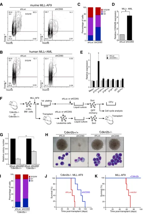
To assess the mechanistic role of CD93 in regulating LSC self-renewal, AML cells were analyzed by flow cytometry for cell-cycle status following CD93 knockdown. Both human and murine knockdown AML cells were more quiescent (increased G0 cells) compared with control cells, suggesting that CD93 plays a crucial role in modulating cell cycle in MLL
leukemia cells (Figures 7A–7C). Notably, qRT-PCR analysis of
candidate cell-cycle regulators demonstrated substantial
upre-gulation of the cell-cycle inhibitor Cdkn2b (p15Ink4b) in CD93
knockdown versus control AML cells (Figures 7D and 7E).
These results suggested a potential model wherein CD93 may functionally suppress Cdkn2b expression and prevent cell-cycle quiescence to promote LSC self-renewal. To test this model, we attempted to bypass the cell-cycle suppression
caused by CD93 knockdown using cells fromCdkn2b / mice
(Figure 7F). MLL-transformed cells deficient for Cdkn2b were significantly more resistant to the loss of clonogenicity induced
by CD93 knockdown (Figure 7G) and were able to circumvent
loss of maturation arrest as evidenced by colony morphology
and cellular cytology (Figure 7H). Furthermore,Cdkn2b
defi-ciency significantly attenuated the shift from G1 to G0 induced
by CD93 knockdown (Figure 7I compared to Figure 7C) and
rescued the leukemogenic potential of CD93 knockdown AML
cells (Figure 7J compared toFigure 6F). Consistent with these
results, overexpression of Cdkn2b in MLL leukemia cells
induced morphologic differentiation and markedly suppressed oncogenic activity in colony-forming assays and
transplanta-tion assays (Figure 7K and Figure S7). Thus, CD93 sustains
LSC oncogenic potential of MLLr AML primarily through
sup-pression ofCdkn2b.
DISCUSSION
We identified the cell-surface lectin CD93 as an LSC-specific sur-face molecule in MLL-rearranged AML. The various genetic
A B C D 0 0.2 0.4 0.6 0.8 1 1.2 Input BM shLuc shCD93 0 0.2 0.4 0.6 0.8 1 1.2 Input BM Normalized %mCherry
MLLr AML Non MLLr AML
P < 0.0001 C D shLuc shCD93 Relative CD93 expression 0 0.2 0.4 0.6 0.8 1.0 shLuc shCD93
Relative colony number
0 0.2 0.4 0.6 0.8 1.0 shLuc shCD93 shLuc shCD93
MLLr AML Cord Blood
B
C D
Relative colony number
0 0.2 0.4 0.6 0.8 1.0 hL hCD93 hL shCD93
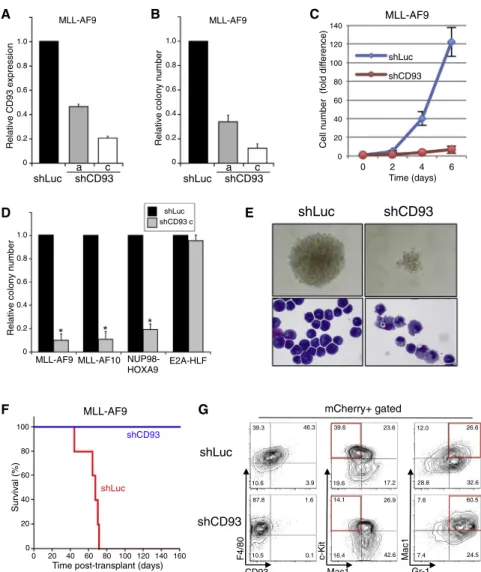
Figure 5. CD93 Is Required for MLL Leukemogenesis
(A) Bar graph denotes transcript levels of CD93 as assayed by qRT-PCR of primary human MLLr leukemia cells transduced with lentiviral vectors en-coding the indicated shRNAs.
(B) Bar graph shows the relative CFC activity of human MLLr AML cells and cord blood cells transduced with the indicated shRNAs. Error bars indicate SDs of five (human MLLr AML) and three (cord blood) independent experi-ments. *p < 0.0001.
(C) May-Grunwald-Giemsa-stained cytopreps show the morphologic features of human MLLr AML cells expressing the indicated shRNAs after methylcel-lulose culture (day 12).
(D) Graphs show the relative numbers of human leukemia cells transduced with the indicated shRNAs present in the BM of NSG recipient mice after seven (MLLr) or nine (non MLLr) weeks post-transplantation, respectively. Data shown are representative of two cases each of MLLr and non-MLLr AMLs. Error bars indicate SDs of three independent mice.
subtypes of AML display remarkable diversity in their develop-mental characteristics and pathological phenotypes, and a sub-type-specific LSC marker has not been previously described. Furthermore, the LSCs marked by CD93 expression are predom-inantly cycling and comprise a minor component of an otherwise
quiescent CD34+CD38 compartment of human AML. The
cycling character of CD93+ LSCs was evident in their gene
expression profiles and confirmed by xenotransplantation of FACS-sorted cycling versus quiescent cells from the same pa-tient AML samples. The fact that LSCs in human MLLr AML are actively cycling contrasts with previous studies demonstrating
that cancer stem cells are predominantly quiescent (Jordan
et al., 2006) and our own analyses of non-MLLr AMLs (Table
S5), but it is consistent with previous studies in a mouse model
of MLL leukemia (Somervaille et al., 2009). Nevertheless, we
cannot exclude the possibility of a rare quiescent LSC population in MLLr AML. Importantly, CD93 is more than a passive marker co-expressed on LSCs, but rather is required for LSC function, constituting an essential role for a lectin in LSC biology. By contrast, the C-type lectin-like molecule CLL-1, which is ex-pressed on the whole blast compartment of many AMLs, has
not been shown to be essential or selectively enrich LSCs (van
shLuc shCD93 0 20 40 60 80 100 120 140 0 2 4 6 shLuc shCD93 MLL-AF9
Cell number (fold dif
ference) Time (days) A B C D E F G MLL-AF9 0 0.2 0.4 0.6 0.8 1.0 Relative CD93 expression shLuc shCD93 a c MLL-AF9 0 0.2 0.4 0.6 0.8 1.0
Relative colony number
shLuc shCD93 a c
MLL-AF9 MLL-AF10 NUP98- E2A-HLF HOXA9
shLuc shCD93 c
Relative colony number
0 0.2 0.4 0.6 0.8 1.0 0 20 40 60 80 100 120 140 160
Time post-transplant (days)
Survival (%) 0 20 40 60 80 100 MLL-AF9 shCD93 shLuc shLuc mCherry+ gated F4/80 c-Kit Mac1 shCD93 Gr-1 Mac1 CD93 39.3 46.3 10.6 3.9 39.6 23.6 19.6 17.2 12.0 26.6 28.8 32.6 87.8 1.6 10.5 0.1 14.1 26.9 16.4 42.6 7.6 60.5 7.4 24.5
Figure 6. CD93 Is Required for Murine Hox-Associated Leukemia Development (A) Bar graph shows relative CD93 transcript levels determined by qRT-PCR for primary murine MLL-AF9 leukemia cells transduced with the indicated shRNAs.
(B) Bar graph shows the relative CFC activity of murine MLL-AF9 AML cells transduced with the indicated shRNAs.
(C) Graph shows cell numbers (expressed as fold-change relative to day 0) for culture of murine MLL-AF9 leukemia cells expressing control or CD93 (construct c) shRNAs. Error bars indicate SDs of three independent experiments.
(D) Bar graph denotes the relative CFC activities of murine AML cells induced by the oncogenes listed below and expressing the indicated shRNAs. Error bars indicate SDs of at least three independent experiments. *p < 0.0001.
(E) Representative images are shown for typical colonies and cellular morphology (May Grunwald Giemsa stain) for murine MLL-AF9 AML cells ex-pressing CD93 or control shRNAs after methyl-cellulose culture (day 5).
(F) Survival curves are shown for mice transplanted with murine MLL-AF9 AML cells (5,000 cells) transduced with shRNAs targeting CD93 (n = 5) or luciferase (n = 5).
(G) Flow cytometry profiles show representative expression levels for the indicated surface markers on murine MLL-AF9 AML cells expressing CD93 or control shRNAs after methylcellulose culture (day 5). Red boxes highlight significant changes in myeloid antigens.
See alsoFigure S6.
Rhenen et al., 2007). We hypothesize that when CD93 expression falls below a certain threshold CDKN2B is activated, cells exit the cycle, and they initiate differentiation out of the LSC compartment.
The association of CD93 with LSCs in MLLr AML, but not other genetic subtypes, likely reflects the fact that high CD93 expression correlates with elevated levels of MEIS1, an essential downstream effector in the transcriptional cascade subordinate to MLL onco-proteins, although CD93 does not appear to be a direct target gene of MEIS1. Consistent with our results, CD93 was reported as one of 25 human candidate genes that discriminated LSCs
from HSCs (Saito et al., 2010). Although early studies reported
CD93 expression on normal HSCs (Danet et al., 2002),
CD93-deficient mice are viable and display no major developmental
abnormalities (Norsworthy et al., 2004), suggesting that CD93 is
non-essential for HSC function, consistent with our observations that human cord blood stem/progenitor cells do not express and are not dependent on CD93, in contrast to MLLr LSCs.
Xenotransplantation assays showed that G1 phase CD34+
CD38 CD93+cells retained strong LSC function in serial
trans-plant assays, whereas G0 phase CD34+CD38 CD93+ cells
from MLLr AML inefficiently engrafted primary recipients. Since G0 reconstituted mice showed similar CD34/CD38 profiles and
the G0 fraction was likely contaminated with G1 AML cells despite the extensive FACS procedure employed to prospec-tively isolate rare CD34/CD38/CD93 subpopulations based on their cell-cycle status. This is consistent with the long latency and reduced penetrance for AMLs induced by the enriched G0
subpopulation (Table S4).
CD93 is a C-type lectin transmembrane receptor that has been implicated in the modulation of phagocytosis, inflammation,
and cell adhesion (Nepomuceno et al., 1997; Norsworthy
et al., 2004; McGreal et al., 2002). It is expressed on cells involved in inflammation, including myeloid cells, endothelial
cells, and platelets (Fonseca et al., 2001; Petrenko et al.,
1999; Nepomuceno and Tenner, 1998). However, its molecular function has remained elusive. Although CD93 was originally identified as a receptor for complement molecule C1q (C1qRp), subsequent studies reported that CD93 alone did
not bind to C1q (Nepomuceno et al., 1997; McGreal et al.,
2002). The CD93 requirement for AML cell maintenance in
shLuc shCD93 shLuc shCD93 Cdkn2b+/+ Cdkn2b-/-A C B E F G H I J human MLLr AML Hoechst Pyronin Y Hoechst Pyronin Y G1 G0 S/G2/M shLuc shCD93 60.8 13.1 25.6 0.53 5.52 41.7 0.29 52.4 murine MLL-AF9 shLuc shCD93 Hoechst Pyronin Y Hoechst Pyronin Y G1 G0 S/G2/M 50.8 32.3 16.2 0.62 39.7 14.1 43.5 2.61 Relative expression 0 1 2 3 4 5 shCD93 c shLuc 1.0 0.8 0.6 0.4 0.2 0
Relative colony number
Cdkn2b+/+ Cdkn2b-/-shLuc shCD93 Cdkn2b-/-shLuc shCD93 shCD93 shLuc 20 40 60 80 100 0 120
Time post-transplant (days)
Survival (%) 0 20 40 60 80 100 Cdkn2b-/- MLL-AF9 G0 G1 S/G2/M 0 20 40 60 80 100 Percentage of cells shLuc shCD93 0 20 40 60 80 100 Percentage of cells G0 G1 S/G2/M shLuc shCD93 Cdkn2b-/-D MLLr AML shLuc shCD93 0 1 2 3 4 5 Relative CDKN2B expression * 1st plating Liquid culture Sort GFP mCherry MLL-AF9 c-Kit+ BM cells puro CFU
Cell cycle analysis
Leukemia cells shLuc or shCD93 Liquid culture Sort Cdkn2b-/- or Cdkn2b+/+ Transplant Transplant shLuc or shCD93 K 0 20 40 60 80 100 0 20 40 60 80 100 Survival (%)
Time post-transplant (days)
MLL-AF9 +Cdkn2b
+vector
Figure 7. CD93 Regulates the Cell Cycle through Suppression of Cdkn2b in MLL Leukemia
(A and B) Representative flow cytometry profiles are shown for Hoechst/Pyronin Y staining of murine MLL-AF9 AML (A) and human MLLr AML (B) cells expressing control or CD93 shRNAs 6 days following transduction.
(C) Bar graph shows cell-cycle stages of murine MLL-AF9 leukemia cells with (shCD93) or without (shLuc) CD93 knockdown after 6 days culture. Data are mean values from three independent experiments with statistically significant differ-ences in G0 (p = 0.0231) and G1 (p = 0.0294) subpopulations.
(D) Bar graph shows relative levels ofCDKN2B
transcript determined by qRT-PCR of human MLLr AML cells expressing shLuc or shCD93. Results are the means of three leukemias and are ex-pressed relative to shLuc control. *p = 0.0141. (E) Bar graph shows relative levels of the indicated transcripts determined by qRT-PCR of murine MLL-AF9 leukemia cells expressing shLuc or shCD93. Results are the means of four leukemias in each category and are expressed relative to shLuc control. *p = 0.041.
(F) BM progenitor/stem cells harvested from wild-type or Cdkn2b /
mice were transduced with the MLL-AF9 oncogene. Immortalized cells from the third to fifth round of replating were then transduced a second time with either shCD93 or shLuc-expressing lentivirus. FACS-sorted mCherry+
/GFP+
cells were plated in methylcellulose for 5 days to determine CFC frequencies.
(G) Bar graph showing the relative CFC activity of murine MLL-AF9 immortalized cells derived from
Cdkn2b+/+
orCdkn2b /
BM cells compared with (shCD93) or without (shLuc) CD93 knockdown. Error bars indicate SDs of five independent ex-periments. *p < 0.001.
(H) Representative images are shown for typical colonies and morphologies (May Grunwald Gi-emsa stain) of murine MLL-AF9 immortalized cells derived fromCdkn2b+/+
orCdkn2b / BM cells with (shCD93) or without (shLuc) CD93 knockdown after methylcellulose culture (day 5). (I) Bar graph shows cell-cycle stages ofCdkn2b / MLL-AF9-immortalized cells with (shCD93) or without (shLuc) CD93 knockdown after 6 days in culture. Data are representative of three indepen-dent experiments.
(J) Survival curves are shown forCdkn2b /
mice transplanted with murine MLL-AF9 AML cells (5,000 cells) transduced with shRNAs targeting CD93 or luciferase.
(K) Survival curves are shown for mice transplanted with murine MLL-AF9 AML cells transduced withCdkn2b(n = 5) or control vector (n = 5). See alsoFigure S7.
our culture assays suggests that its unknown ligand is present in vitro. One possibility is that AML cells may produce the ligand to sustain an autocrine loop, or alternatively CD93 may mediate essential homotypic cellular interactions to stim-ulate LSC self-renewal divisions. Future studies are warranted to address these possibilities.
Our studies link CD93 with cell-cycle regulation, and this occurs in part through suppression of the cell-cycle inhibitor p15Ink4b. Loss ofCDKN2B(p15Ink4b) expression through muta-tion or epigenetic silencing plays a prominent role in leukemia
pathogenesis. Hyper-methylation of theCDKN2Bpromoter
oc-curs frequently in AML (Drexler, 1998; Krug et al., 2002; Herman
et al., 1996; Chim et al., 2001) and is correlated with poor
prog-nosis (Wong et al., 2000; Shimamoto et al., 2005; Aggerholm
et al., 2006; Teofili et al., 2003). Furthermore,Cdkn2b-deficient mice display increased susceptibility to retrovirus-induced AML and develop non-reactive monocytosis and hematologic
fea-tures resembling chronic myelomonocytic leukemia (Wolff
et al., 2003; Bies et al., 2010). Thus, CDKN2B is a tumor suppres-sor for myeloid leukemia; however, little is known about the signals driving its repression in leukemogenesis. Our studies indi-cate that lectin CD93, through currently undefined signaling pathways, provides a novel upstream mechanism to suppress
CDKN2Bexpression and regulate LSC oncogenic potential in
MLLr AML.CDKN2Bis one of several downstream mediators
of transforming growth factorb(TGF-b) signaling, which
main-tains a pool of quiescent HSCs (Dao et al., 2002), but the specific
role ofCDKN2Bis not clear and MLLr LSCs are not malignant
counterparts of HSCs, but rather, more downstream progenitors.
Previous studies have shown thatCDKN2Blevels are
undetect-able in CD34+cells and upregulated during normal and leukemic
myeloid differentiation (Teofili et al., 2000). This suggests that
suppression of LSC cell division in MLLr leukemia cells by cell-cycle inhibitors triggers the default outcome in the myeloid line-age, which is differentiation. This may account for why there appear to be few if any quiescent LSCs in MLLr leukemia. Recently it was reported that cell-cycle regulator CDK6, but not its functional homolog CDK4, is a crucial effector in MLLr AML (Placke et al., 2014). Since CDKN2B forms a complex with CDK6 to prevent its activation, our studies support a cell-cycle regulatory pathway in which CD93 suppresses CDKN2B, thereby freeing its target kinase CDK6 to drive cell-cycle progression and unrestrained LSC self-renewal.
In summary, we identified lectin CD93 as an LSC-specific
cell-surface molecule in the genetic subtype of AML defined byMLL
rearrangements. LSCs marked by CD93 are actively cycling in contrast to previous studies of quiescent cancer stem cells. LSCs functionally require CD93, which regulates their differenti-ation, self-renewal activity, and in vivo progression by modu-lating the cell cycle through CDKN2B. These results suggest that CD93 may be a useful candidate therapeutic target for anti-LSC therapy and prognostic marker for quantitation of
minimal residual disease (MRD) forMLL-rearranged AML.
EXPERIMENTAL PROCEDURES Human Samples
Human AML samples (Table S1) and cord blood cells were obtained from patients at the Stanford Medical Center with informed consent and
institu-tional review board approval. Mononuclear cells were purified by Ficoll-density gradient centrifugation enrichment.
Mice
Inbred C57BL/6 mice were used for syngeneic transduction/transplantation experiments. NSG mice (NOD.Cg-Prkdcscid
IL2rgtm1Wjl
/SzJ) were obtained from the Jackson Laboratory and bred in a pathogen-free environment.
Cdkn2b /
mice were maintained on a C57BL/6 background. All experiments on mice in this study were performed with the approval of and in accordance with Stanford University’s Administrative Panel on Laboratory Animal Care. Cell Culture
Immortalized mouse myeloid cells or leukemia cells were cultured in RPMI 1640 medium supplemented with 20% fetal calf serum (FCS) and 20% WEHI conditioned medium or cytokines (10 ng/ml IL-6, 20 ng/ml SCF). 293T and Phoenix cells were cultured in DMEM supplemented with 10% FCS. In Vitro Colony Forming Assays
Mouse cells were plated in methylcellulose medium (M3231, Stem Cell Tech-nologies) with cytokines as described previously (Lavau et al., 1997) and counted after 5 days. Human cells were plated in H4535 (Stem Cell Technol-ogies) and counted after 11–14 days of culture.
Flow Cytometry
Antibodies for flow cytometry were as follows. Anti-mouse antibodies used were CD93-PE-Cy7 or Biotin (AA4.1), Mac1/CD11b-PE or FITC (M1/70), Gr1-PE (RB6-8C5), B220-PE (RA3-6B2), F4/80-PE (BM8), NK1.1-PE (PK136), TER119-PE (TER-119), IL-7Ra-PE (SB14), CD3-PE (145-2C12), CD4-PE (GK1.5), CD8-PE (53-6.7), CD117-APC (2B8), Sca1-PE-Cy5 (D7), CD16/32-PE-Cy7 (2.4G3), and CD34-FITC (RAM34) (all from eBioscience). The lineage cocktail included the following mAbs: Mac1, Gr-1, B220, TER119, IL-7Ra, NK1.1, CD3, CD4, and CD8. Anti-human antibodies were CD34-APC (8G12) (BD Biosciences), CD38-PE-Cy7 (HIT2) (eBioscience), CD93-PE or Biotin (R3) (eBioscience), CD45-APC or FITC (2D1 or H130) (eBioscience and BD Bioscience), CD3-PE-Cy5 (UCHT1) (BD Biosciences) and CD64-PE (10.1) (BioLegend). Analyses and cell sorting experiments were performed using a FACS Vantage or FACS Aria II (BD Biosciences) equip-ped with Diva software (BD), and data were analyzed using FlowJo (Tree Star). DNA Constructs and Virus Production
Retroviral constructs encoding HoxA9, Meis1, MLL-AF9, MLL-AF10, NUP98-HOXA9, and E2A-HLF were reported previously (Wong et al., 2007; Somer-vaille and Cleary, 2006; Iwasaki et al., 2005; Ayton and Cleary, 2003). The pSicoR lentiviral vector (Ventura et al., 2004) with either mCherry or GFP marker was used for knockdown studies. shRNAs were designed using pSicoOligomaker v1.5 (developed by A. Ventura, Jacks Laboratory) and se-quences are listed in theSupplemental Experimental Procedures. Retrovirus and lentivirus production was performed as described previously (Wong et al., 2007; Somervaille et al., 2009).
In Vivo Leukemogenesis Assays
FACS-sorted mouse AML cells were transplanted intravenously into suble-thally irradiated (4.5Gy) C57BL/6 mice. Development of acute leukemia was confirmed by blood smear, peripheral blood leukocyte counts, FACS analysis, histology, or some combination thereof. For xenotransplant experiments, FACS-sorted human AML cells were transplanted into sublethally irradiated (2.5Gy) NSG mice, which were monitored for development of AML. In some ex-periments, BM was analyzed for engraftment (hCD45+
) at 2–4 months. See the Supplemental Experimental Proceduresfor more details of xenotransplant experiments.
Microarray and Bioinformatics Analysis
Total RNA was extracted from prospectively isolated cells using Trizol reagent (Invitrogen) followed by RNeasy MinElute Cleanup Kit (QIAGEN). Microarray experiments were performed in the Stanford PAN Facility with mouse Genome 430 2.0 arrays (Affymetrix) and human Gene 1.0 ST arrays (Affymetrix). Micro-array data were normalized with Expression Console software (Affymetrix) us-ing RMA algorithms, then further normalized with dChip (Li and Wong, 2001).
GSEAs were performed using GSEA software (http://www.broad.mit.edu/ gsea) with a t test metric for gene ranking and 1,000 data permutations. GO analysis was performed using MGI Gene Ontology Term Finder (http://proto. informatics.jax.org/prototypes/GOTools/web-docs/MGI_Term_Finder.html). Cell-Cycle Analysis
Cells were incubated with cell surface markers to enable our detection of phenotypic subpopulations and then incubated with Hoechst 33342 (Sigma-Aldrich) and Pyronin Y (Sigma-(Sigma-Aldrich) as previously described (Passegue´ et al., 2005).
qRT-PCR
RNA from sorted cell populations was purified using Trizol (Life Technologies). cDNA was synthesized by SuperScript III First-Strand Synthesis System for RT-PCR (Life Technologies) according to the manufacturer’s instructions. Real-time PCR was performed with TaqMan probes (Life Technologies) using b-Actin or b-ACTINas internal control. TaqMan probes for the following mouse and human genes were purchased from Life Technologies:
Cd93 (Mm00440239_g1), Meis1 (Mm00487664_m1), Cdkn1a (p21) (Mm01303209_m1), Cdkn1b (p27) (Mm00438167_g1), Cdkn2a (p19Arf ) (Mm01257348_m1),Cdkn2b(p15Ink4b ) (Mm00483241_m1),Cdkn2c(p18Ink4c ) (Mm00483243_m1), b-Actin (Mm00607939_s1); CD93 (Hs00362607_m1), andb-ACTIN(HS01060665_g1). Primers for mousep16Ink4a
were reported previously (Zhang et al., 2003) and purchased from Life Technologies. Statistical Analysis
Statistics were calculated using Prism 6 software (GraphPad software, Inc). Data were presented as the mean ± SD. The significance of the differences be-tween groups was determined via Student’s t test.
ACCESSION NUMBERS
The accession numbers for the expression data reported in this paper are GEO: GSE64773 and GEO: GSE64776.
SUPPLEMENTAL INFORMATION
Supplemental Information for this article includes seven figures and six tables and can be found with this article online athttp://dx.doi.org/10.1016/j.stem. 2015.08.008.
AUTHOR CONTRIBUTIONS
M.I. planned the study; conducted, performed, and interpreted experiments; and co-wrote the manuscript. M.L. provided patient AML samples. A.J.G. as-sisted with bioinformatics data interpretation. M.L.C. directed the studies and co-wrote the manuscript.
ACKNOWLEDGMENTS
We thank Cita Nicolas and Maria Ambus for technical support, Norm Cyr for graphical assistance, and Ravindra Majeti and Lawrence Okumoto for AML patient samples. We thank Nicholas Leeper and Juraj Bies for providing Cdkn2b knockout mice. We acknowledge support from the Children’s Health Initiative of the Packard Foundation, the California Institute for Regenerative Medicine, and Public Health Service (PHS) grant CA116606.
Received: January 15, 2015 Revised: May 28, 2015 Accepted: August 10, 2015 Published: September 17, 2015 REFERENCES
Aggerholm, A., Holm, M.S., Guldberg, P., Olesen, L.H., and Hokland, P. (2006). Promoter hypermethylation of p15INK4B, HIC1, CDH1, and ER is frequent in myelodysplastic syndrome and predicts poor prognosis in early-stage pa-tients. Eur. J. Haematol.76, 23–32.
Ayton, P.M., and Cleary, M.L. (2003). Transformation of myeloid progenitors by MLL oncoproteins is dependent on Hoxa7 and Hoxa9. Genes Dev.17, 2298– 2307.
Bhatia, M., Wang, J.C., Kapp, U., Bonnet, D., and Dick, J.E. (1997). Purification of primitive human hematopoietic cells capable of repopulating immune-defi-cient mice. Proc. Natl. Acad. Sci. USA94, 5320–5325.
Bies, J., Sramko, M., Fares, J., Rosu-Myles, M., Zhang, S., Koller, R., and Wolff, L. (2010). Myeloid-specific inactivation of p15Ink4b results in monocyto-sis and predisposition to myeloid leukemia. Blood116, 979–987.
Bonnet, D., and Dick, J.E. (1997). Human acute myeloid leukemia is organized as a hierarchy that originates from a primitive hematopoietic cell. Nat. Med.3, 730–737.
Chim, C.S., Liang, R., Tam, C.Y.Y., and Kwong, Y.L. (2001). Methylation of p15 and p16 genes in acute promyelocytic leukemia: potential diagnostic and prognostic significance. J. Clin. Oncol.19, 2033–2040.
Danet, G.H., Luongo, J.L., Butler, G., Lu, M.M., Tenner, A.J., Simon, M.C., and Bonnet, D.A. (2002). C1qRp defines a new human stem cell population with hematopoietic and hepatic potential. Proc. Natl. Acad. Sci. USA99, 10441– 10445.
Dao, M.A., Hwa, J., and Nolta, J.A. (2002). Molecular mechanism of trans-forming growth factor beta-mediated cell-cycle modulation in primary human CD34(+) progenitors. Blood99, 499–506.
Drexler, H.G. (1998). Review of alterations of the cyclin-dependent kinase inhibitor INK4 family genes p15, p16, p18 and p19 in human leukemia-lym-phoma cells. Leukemia12, 845–859.
Eppert, K., Takenaka, K., Lechman, E.R., Waldron, L., Nilsson, B., van Galen, P., Metzeler, K.H., Poeppl, A., Ling, V., Beyene, J., et al. (2011). Stem cell gene expression programs influence clinical outcome in human leukemia. Nat. Med. 17, 1086–1093.
Fonseca, M.I., Carpenter, P.M., Park, M., Palmarini, G., Nelson, E.L., and Tenner, A.J. (2001). C1qR(P), a myeloid cell receptor in blood, is predominantly expressed on endothelial cells in human tissue. J. Leukoc. Biol.70, 793–800. Gentles, A.J., Plevritis, S.K., Majeti, R., and Alizadeh, A.A. (2010). Association of a leukemic stem cell gene expression signature with clinical outcomes in acute myeloid leukemia. JAMA304, 2706–2715.
Herman, J.G., Jen, J., Merlo, A., and Baylin, S.B. (1996). Hypermethylation-associated inactivation indicates a tumor suppressor role for p15INK4B. Cancer Res.56, 722–727.
Hosen, N., Park, C.Y., Tatsumi, N., Oji, Y., Sugiyama, H., Gramatzki, M., Krensky, A.M., and Weissman, I.L. (2007). CD96 is a leukemic stem cell-spe-cific marker in human acute myeloid leukemia. Proc. Natl. Acad. Sci. USA104, 11008–11013.
Ishikawa, F., Yoshida, S., Saito, Y., Hijikata, A., Kitamura, H., Tanaka, S., Nakamura, R., Tanaka, T., Tomiyama, H., Saito, N., et al. (2007). Chemotherapy-resistant human AML stem cells home to and engraft within the bone-marrow endosteal region. Nat. Biotechnol.25, 1315–1321. Iwasaki, M., Kuwata, T., Yamazaki, Y., Jenkins, N.A., Copeland, N.G., Osato, M., Ito, Y., Kroon, E., Sauvageau, G., and Nakamura, T. (2005). Identification of cooperative genes for NUP98-HOXA9 in myeloid leukemogenesis using a mouse model. Blood105, 784–793.
Jaiswal, S., Jamieson, C.H., Pang, W.W., Park, C.Y., Chao, M.P., Majeti, R., Traver, D., van Rooijen, N., and Weissman, I.L. (2009). CD47 is upregulated on circulating hematopoietic stem cells and leukemia cells to avoid phagocy-tosis. Cell138, 271–285.
Jan, M., Chao, M.P., Cha, A.C., Alizadeh, A.A., Gentles, A.J., Weissman, I.L., and Majeti, R. (2011). Prospective separation of normal and leukemic stem cells based on differential expression of TIM3, a human acute myeloid leuke-mia stem cell marker. Proc. Natl. Acad. Sci. USA108, 5009–5014. Jin, L., Hope, K.J., Zhai, Q., Smadja-Joffe, F., and Dick, J.E. (2006). Targeting of CD44 eradicates human acute myeloid leukemic stem cells. Nat. Med.12, 1167–1174.
Jin, L., Lee, E.M., Ramshaw, H.S., Busfield, S.J., Peoppl, A.G., Wilkinson, L., Guthridge, M.A., Thomas, D., Barry, E.F., Boyd, A., et al. (2009). Monoclonal
antibody-mediated targeting of CD123, IL-3 receptor alpha chain, eliminates human acute myeloid leukemic stem cells. Cell Stem Cell5, 31–42. Jordan, C.T., Upchurch, D., Szilvassy, S.J., Guzman, M.L., Howard, D.S., Pettigrew, A.L., Meyerrose, T., Rossi, R., Grimes, B., Rizzieri, D.A., et al. (2000). The interleukin-3 receptor alpha chain is a unique marker for human acute myelogenous leukemia stem cells. Leukemia14, 1777–1784. Jordan, C.T., Guzman, M.L., and Noble, M. (2006). Cancer stem cells. N. Engl. J. Med.355, 1253–1261.
Kikushige, Y., Shima, T., Takayanagi, S., Urata, S., Miyamoto, T., Iwasaki, H., Takenaka, K., Teshima, T., Tanaka, T., Inagaki, Y., and Akashi, K. (2010). TIM-3 is a promising target to selectively kill acute myeloid leukemia stem cells. Cell Stem Cell7, 708–717.
Konopleva, M.Y., and Jordan, C.T. (2011). Leukemia stem cells and microen-vironment: biology and therapeutic targeting. J. Clin. Oncol.29, 591–599. Krug, U., Ganser, A., and Koeffler, H.P. (2002). Tumor suppressor genes in normal and malignant hematopoiesis. Oncogene21, 3475–3495.
Lapidot, T., Sirard, C., Vormoor, J., Murdoch, B., Hoang, T., Caceres-Cortes, J., Minden, M., Paterson, B., Caligiuri, M.A., and Dick, J.E. (1994). A cell initi-ating human acute myeloid leukaemia after transplantation into SCID mice. Nature367, 645–648.
Lavau, C., Szilvassy, S.J., Slany, R., and Cleary, M.L. (1997). Immortalization and leukemic transformation of a myelomonocytic precursor by retrovirally transduced HRX-ENL. EMBO J.16, 4226–4237.
Li, C., and Wong, W.H. (2001). Model-based analysis of oligonucleotide arrays: expression index computation and outlier detection. Proc. Natl. Acad. Sci. USA98, 31–36.
Majeti, R., Chao, M.P., Alizadeh, A.A., Pang, W.W., Jaiswal, S., Gibbs, K.D., Jr., van Rooijen, N., and Weissman, I.L. (2009). CD47 is an adverse prognostic factor and therapeutic antibody target on human acute myeloid leukemia stem cells. Cell138, 286–299.
McGreal, E.P., Ikewaki, N., Akatsu, H., Morgan, B.P., and Gasque, P. (2002). Human C1qRp is identical with CD93 and the mNI-11 antigen but does not bind C1q. J. Immunol.168, 5222–5232.
Nepomuceno, R.R., HenschenEdman, A.H., Burgess, W.H., and Tenner, A.J. (1997). cDNA cloning and primary structure analysis of C1qR(p), the human C1q/MBL/SPA receptor that mediates enhanced phagocytosis in vitro. Immunity6, 119–129.
Nepomuceno, R.R., and Tenner, A.J. (1998). C1qRP, the C1q receptor that enhances phagocytosis, is detected specifically in human cells of myeloid line-age, endothelial cells, and platelets. J. Immunol.160, 1929–1935.
Norsworthy, P.J., Fossati-Jimack, L., Cortes-Hernandez, J., Taylor, P.R., Bygrave, A.E., Thompson, R.D., Nourshargh, S., Walport, M.J., and Botto, M. (2004). Murine CD93 (C1qRp) contributes to the removal of apoptotic cells in vivo but is not required for C1q-mediated enhancement of phagocytosis. J. Immunol.172, 3406–3414.
Passegue´, E., Wagers, A.J., Giuriato, S., Anderson, W.C., and Weissman, I.L. (2005). Global analysis of proliferation and cell cycle gene expression in the regulation of hematopoietic stem and progenitor cell fates. J. Exp. Med. 202, 1599–1611.
Petrenko, O., Beavis, A., Klaine, M., Kittappa, R., Godin, I., and Lemischka, I.R. (1999). The molecular characterization of the fetal stem cell marker AA4. Immunity10, 691–700.
Placke, T., Faber, K., Nonami, A., Putwain, S.L., Salih, H.R., Heidel, F.H., Kra¨mer, A., Root, D.E., Barbie, D.A., Krivtsov, A.V., et al. (2014). Requirement for CDK6 in MLL-rearranged acute myeloid leukemia. Blood 124, 13–23.
Saito, Y., Kitamura, H., Hijikata, A., Tomizawa-Murasawa, M., Tanaka, S., Takagi, S., Uchida, N., Suzuki, N., Sone, A., Najima, Y., et al. (2010). Identification of therapeutic targets for quiescent, chemotherapy-resistant hu-man leukemia stem cells. Sci. Transl. Med.2, 17ra9.
Shimamoto, T., Ohyashiki, J.H., and Ohyashiki, K. (2005). Methylation of p15(INK4b) and E-cadherin genes is independently correlated with poor prog-nosis in acute myeloid leukemia. Leuk. Res.29, 653–659.
Somervaille, T.C.P., and Cleary, M.L. (2006). Identification and characteriza-tion of leukemia stem cells in murine MLL-AF9 acute myeloid leukemia. Cancer Cell10, 257–268.
Somervaille, T.C.P., Matheny, C.J., Spencer, G.J., Iwasaki, M., Rinn, J.L., Witten, D.M., Chang, H.Y., Shurtleff, S.A., Downing, J.R., and Cleary, M.L. (2009). Hierarchical maintenance of MLL myeloid leukemia stem cells employs a transcriptional program shared with embryonic rather than adult stem cells. Cell Stem Cell4, 129–140.
Teofili, L., Morosetti, R., Martini, M., Urbano, R., Putzulu, R., Rutella, S., Pierelli, L., Leone, G., and Larocca, L.M. (2000). Expression of cyclin-dependent ki-nase inhibitor p15(INK4B) during normal and leukemic myeloid differentiation. Exp. Hematol.28, 519–526.
Teofili, L., Martini, M., Luongo, M., Diverio, D., Capelli, G., Breccia, M., Lo Coco, F., Leone, G., and Larocca, L.M. (2003). Hypermethylation of GpG islands in the promoter region of p15(INK4b) in acute promyelocytic leukemia represses p15(INK4b) expression and correlates with poor prognosis. Leukemia17, 919–924.
van Rhenen, A., van Dongen, G.A.M.S., Kelder, A., Rombouts, E.J., Feller, N., Moshaver, B., Stigter-van Walsum, M., Zweegman, S., Ossenkoppele, G.J., and Jan Schuurhuis, G. (2007). The novel AML stem cell associated antigen CLL-1 aids in discrimination between normal and leukemic stem cells. Blood 110, 2659–2666.
Ventura, A., Meissner, A., Dillon, C.P., McManus, M., Sharp, P.A., Van Parijs, L., Jaenisch, R., and Jacks, T. (2004). Cre-lox-regulated conditional RNA inter-ference from transgenes. Proc. Natl. Acad. Sci. USA101, 10380–10385. Wolff, L., Garin, M.T., Koller, R., Bies, J., Liao, W., Malumbres, M., Tessarollo, L., Powell, D., and Perella, C. (2003). Hypermethylation of the Ink4b locus in murine myeloid leukemia and increased susceptibility to leukemia in p15(Ink4b)-deficient mice. Oncogene22, 9265–9274.
Wong, I.H.N., Ng, M.H.L., Huang, D.P., and Lee, J.C.K. (2000). Aberrant p15 promoter methylation in adult and childhood acute leukemias of nearly all morphologic subtypes: potential prognostic implications. Blood95, 1942– 1949.
Wong, P., Iwasaki, M., Somervaille, T.C.P., So, C.W.E., and Cleary, M.L. (2007). Meis1 is an essential and rate-limiting regulator of MLL leukemia stem cell potential. Genes Dev.21, 2762–2774.
Zhang, S., Qian, X., Redman, C., Bliskovski, V., Ramsay, E.S., Lowy, D.R., and Mock, B.A. (2003). p16 INK4a gene promoter variation and differential binding of a repressor, the ras-responsive zinc-finger transcription factor, RREB. Oncogene22, 2285–2295.