Paul J. Christo and Danesh Mazloomdoost
Department of Anesthesiology & Critical Care Medicine, Division of Pain Medicine, The Johns Hopkins University School of Medicine, Baltimore, Maryland, USA
Pain ranges in prevalence from 14–100% among cancer patients and occurs in 50–70% of those in active treatment. Cancer pain may result from direct invasion of tumor into nerves, bones, soft tissue, ligaments, and fascia, and may induce visceral pain through distension and obstruction. Cancer pain is multifaceted. Clinicians may describe cancer pain as acute, chronic, nociceptive (somatic), visceral, or neuropathic. Despite imple-mentation of the WHO guidelines, reports of undertreatment of cancer pain persist in various clinical settings and in spite of decades of work to reduce unnecessary discom-fort. Substantial obstacles to adequate pain relief with opioids include specific concerns of patients themselves, their family members, physicians, nurses, and the healthcare system. The WHO analgesic ladder serves as the mainstay of treatment for the relief of cancer pain in concert with tumoricidal, surgical, interventional, radiotherapeutic, psychological, and rehabilitative modalities. This multidimensional approach offers the greatest potential for maximizing analgesia and minimizing adverse effects. Primary therapies are directed at the source of the cancer pain and may enhance a patient’s function, longevity, and comfort. Adjuvant therapies include nonopioids that confer analgesic effects in certain medical conditions but primarily treat conditions that do not involve pain. Nonopioid medications (over-the-counter agents) are useful in the man-agement of mild to moderate pain, and their continuation through step 3 of the WHO ladder is an option after weighing a drug’s risks and benefits in individual patients. Symptomatic treatment of severe cancer pain should begin with an opioid, regardless of the mechanism of the pain. They are very effective analgesics, titrate easily, and offer a favorable risk/benefit ratio. Cancer pain remains inadequately controlled despite the diagnostic and therapeutic means of ensuring that patients feel comfortable during their illness. Therefore, all practitioners need to make control of cancer pain a professional duty, even if they can only use the most basic and least expensive analgesic medications, such as morphine, codeine, and acetaminophen, to reduce human suffering.
Key words: cancer; pain; analgesia; opioids; addiction; barriers to pain relief; cancer pain; adjuvant therapies; nonopioid therapies; co-analgesics; malignancy; therapies; WHO 3-step analgesic ladder
Scope of the Problem
Pain ranges in prevalence from 14–100%
among cancer patients1 and occurs in 50–
70% of those in active treatment.2 The
liter-ature reports pain figures as high as 60–90%
Address for correspondence: Paul J. Christo, M.D., M.B.A., Assistant Professor, Director, Multidisciplinary Pain Fellowship Director, Pain Treat-ment Center, DepartTreat-ment of Anesthesiology & Critical Care Medicine, Division of Pain Medicine, The Johns Hopkins University School of Medicine, 550 N. Broadway, Suite 301, Baltimore, MD 21205. Voice: +410-955-1818; fax:+410-502-6730. pchristo@jhmi.edu
for patients with advanced stages of cancer.2–4
Two-thirds of this pain in advanced disease is due to tumor infiltration, and almost one-fourth is a consequence of cancer treatments. No cures exist for many patients with advanced systemic cancers; yet, pain therapies do exist that can ease the suffering related to an in-dividual’s course of illness. This knowledge is critically important to communicate given that patients with advanced cancer commonly ex-perience and fear pain. The World Health Or-ganization (WHO) recognized the global need
Ann. N.Y. Acad. Sci. 1138: 278–298 (2008).C 2008 New York Academy of Sciences.
Figure 1. World Health Organization 3-Step Analgesic Ladder with examples of anal-gesics. Adapted from Management of Cancer Pain: Clinical Practice Guideline Number 9. Rockville, MD: U.S. Department of Health and Human Services; 1994, AHCPR Pub No. 94-0592.
to establish guidelines for basic pain control in cancer patients and thereby developed an el-emental “3-step analgesic ladder” in 1986 for
use among practitioners.5The WHO made an
important step in disseminating critical con-cepts of pain management through education and opioid availability. Increasingly, patients, healthcare providers, and healthcare accred-itation bodies are demanding greater atten-tion to the burden of pain and, in particular, cancer-related pain. However, despite appli-cation of the WHO “3-step analgesic lad-der” (Fig. 1), advancing pain research, and expansive interventional modalities, as many as 50% of cancer patients with pain may re-main undertreated.6In response to the signifi-cant problem of unrelieved pain in cancer and other disease states, the Joint Commission on Accreditation of Health Organizations, an in-dependent, nonprofit organization that evalu-ates and accredits healthcare organizations in the United States, created comprehensive stan-dards for pain management in 1999.
Health-care institutions must fulfill these standards in order to meet the requirements for reaccredita-tion. Perhaps this initiative will move clinicians closer to the overarching goal that, “no can-cer patient should live or die with unrelieved pain.”7
The following review will discuss important principles of managing pain in malignant dis-ease. Concepts relating to the sources of pain in cancer, types of pain in cancer, barriers to ef-fective pain control, measuring pain in cancer patients, and pharmacotherapeutic approaches to pain control will be discussed.
Sources of Cancer Pain
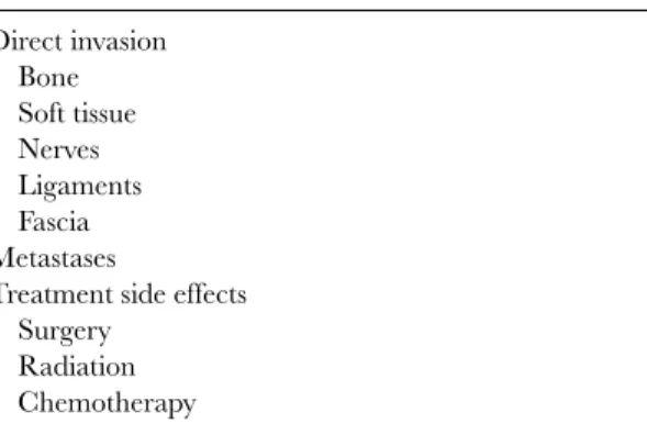
Cancer pain may result from direct inva-sion of tumor into nerves, bones, soft tissue, ligaments, and fascia, and may induce vis-ceral pain through distension and obstruction (Table 1). While over two-thirds of cancer pain usually results from the tumor burden, a quarter of pain experienced by cancer
TABLE 1. Sources of Cancer Pain Direct invasion Bone Soft tissue Nerves Ligaments Fascia Metastases
Treatment side effects Surgery
Radiation Chemotherapy
patients can be attributed to the cancer-related treatments.3 For instance, surgery, radiation,
and chemotherapeutics may all elicit acute pain that diminishes in time, while other therapies
may cause chronic pain conditions.8
Radia-tion treatment frequently causes acute mus-cle stiffness and aching, but carries the risk of chronic pain secondary to nerve injury, chronic inflammation, osteoradionecrosis, or myofas-cial injury. Surgery-associated pain may result from direct nerve injury, inflammation, postam-putation phantom pain conditions, and even the development of Complex Regional Pain Syndrome. Many chemotherapeutic agents are known to cause pain. Several classes, such as the alkaloids, platinum-based compounds, and the antimitotics, are known to contribute to periph-eral neuropathies.
Types of Pain
Cancer pain is multifaceted, as illustrated in Table 2. Clinicians may describe cancer pain as acute, chronic, nociceptive (somatic), vis-ceral, or neuropathic. Alternatively, some have proposed just three prime categories of can-cer pain: nociceptive, neuropathic, and
psy-chogenic.9 Furthermore, multiple taxonomies
of pain exist including a research-oriented and treatment-oriented classification of pain that groups together patients with similar pain mechanisms.10Clearly, no individual classifica-tion is optimal in truly capturing the multidi-mensional phenomenon of cancer pain.
Clin-TABLE 2. Types of Cancer Pain
Examples of neuropathic pain Tumor compression of plexi Tumor invasion into nerves Tumor invasion into spinal cord Chemotherapy-induced neuritis Radiation-induced nerve injury Examples of somatic/nociceptive pain
Tumor invasion into bone Pathologic fracture Postsurgical pain Examples of visceral pain
Tumor invasion into organs Obstruction (e.g., biliary, intestinal) Organ rupture (e.g., bowel, bladder)
ically, patients experience pain with varying degrees of intensity, frequency, anatomic loca-tion, duraloca-tion, and body system involvement. Further, they may describe features of both nociceptive and neuropathic pain rather than distinctive elements of a single process. It is instructive, nevertheless to understand com-mon terminology often applied to cancer pain. For instance, nociceptive pain arises from ac-tivation of nociceptors (free nerve terminals of primary afferent fibers that respond to painful stimuli) that are located in all tissues except the central nervous system. Neuropathic pain results from a primary lesion or dysfunction in the central or peripheral nervous system.11The following terms help distinguish varying physi-ological types of cancer pain.12
Nociceptive pain—associated with tis-sue injury from surgery, trauma, inflam-mation, or tumor. The pain is caused by stimulation of pain receptors in cutaneous and deeper musculoskeletal structures. It is often proportional to the degree of no-ciceptor activation. Both somatic and vis-ceral pain conditions may be character-ized as nociceptive.
Somatic pain—arising from direct in-jury to bones, tissue, or tendons. Some consider somatic and nociceptive pain to be synonymous. Somatic pain is described as aching or dull and sometimes stabbing.
It tends to be very focal. This cate-gory often includes metastatic bone pain, postsurgical incisional pain, and muscu-loskeletal inflammation and spasm.
Visceral pain—arising from organ damage or tumor infiltration, compres-sion, or distortion of organs within the pelvis, abdomen, or thorax. It is de-scribed as a pressure-like sensation, inter-nal squeezing, or crampiness. It tends to be vague and diffuse and may be associ-ated with distension/stretching of organs, nausea, vomiting, and sweating. The pain may be referred to superficial locations that are distant from the affected organ.
Neuropathic pain—may be directly related to the malignant disease, such as tumor infiltration of peripheral nerves, plexi, roots, or spinal cord. It may arise from efforts to treat the disease, such as surgery, chemotherapy, or other drug-induced neuropathy or neuritis, and even from radiation-induced injury to periph-eral nerves and the spinal cord. This type of pain is invariably associated with sen-sory changes caused by injury to the cen-tral or peripheral nervous system and may be incompletely responsive to opioid ther-apy. Patients typically describe this pain as burning, shooting, pins/needles, elec-trical, or numbness, and it tends to radiate over dermatomal distributions.
Barriers to Treating Pain
Despite implementation of the WHO guide-lines and in spite of decades of work to re-duce unnecessary discomfort, reports of un-dertreatment of cancer pain persist in various clinical settings.13 Substantial obstacles to ad-equate pain relief with opioids include specific concerns of patients themselves, their family members, physicians, nurses, and the health-care system.14–17 Many of these barriers focus
on psychosocial factors related to the fear of opi-oid addiction and physical dependence,
con-cerns about adverse effects of medications, and patient fears of disappointing their physician by reporting pain.18–20 Healthcare providers,
patients, and their families report distinct but sometimes overlapping concerns.
Healthcare Providers as Barriers to Pain Relief
Physicians are often reluctant to prescribe opioids and nurses may express concern about administering opioids to patients. Physicians tend to feel that managing pain with opi-oids and other controlled substances leads to documentation woes, entails frequent prescrip-tion refills, requires onerous telephone calls, and exposes themselves to intense regulatory scrutiny.21Moreover, many healthcare
profes-sionals still lack appropriate knowledge of anal-gesic (primarily opioid) pharmacology with re-spect to dosing, timing, alternative routes of administration (such as rectal, subcutaneous, epidural, intrathecal), and converting from in-travenous to oral therapies. Coupled with an over-exuberant fear of respiratory compromise and a pervasive fear of addiction, physicians and other healthcare providers leave many pa-tients inadequately treated.
While understandable, fears of opioid ad-verse effects and complications related to respi-ratory depression need not paralyze practition-ers from prescribing opioids. Opioids do affect both the rate and depth of respiration. Data from studies on mice indicate that both the analgesic and respiratory depressive features of morphine are linked to the mu opioid recep-tor22,23 in a dose-dependent fashion (that is,
increasing the dose produces greater analgesia and greater depression of respiration). A recent acute-pain study in healthy human volunteers shows similar effects and notes that respiratory depression is possible irrespective of concomi-tant severe pain.24In contrast, many clinicians find that respiratory depression rarely precedes analgesia when administering opioids to re-lieve chronic pain. That is, physicians treating chronic pain patients with opioids report that
patients typically feel comfortable before expe-riencing respiratory compromise. This clinical phenomenon provides some comfort in esca-lating doses of opioids in patients who continue to experience pain. Further, cancer pain may in fact more accurately mimic chronic rather than acute pain models, thereby attenuating the risk of respiratory depression in this popu-lation. Nonetheless, serious adverse effects can be mitigated by attention to dosing, frequency of dosing, duration of pain (periodic or con-stant), co-administration of psychoactive sub-stances (such as benzodiazepines, barbiturates, alcohol, other opioids), and proper supervision of patients on chronic opioid therapy. Practi-tioners can safely tailor opioid therapy in can-cer patients by considering the delicate balance between depression of respiration (from factors such as opioid therapy, sleep deprivation, and sedation from co-administered sedatives) and stimulation of respiration (from pain, arousal, stress, anxiety, inflammation, and other causes). A myriad of healthcare providers use the risk of opioid addiction as justification for minimiz-ing or withholdminimiz-ing appropriate opioid therapy. Three medical societies (The American So-ciety of Addiction Medicine, The American Pain Society, and The American Academy of Pain Medicine) define addiction as “a primary, chronic, neurobiological disease, with genetic, psychosocial, and environmental factors influ-encing the development and manifestations. It is characterized by behaviors that include one of more of the following: impaired control over drug use, compulsive use, continued use despite harm, and craving.”25In the context of treating patients in chronic pain with opioids, addiction may be viewed as a combination of observa-tions that suggest maladaptive behaviors rather than pharmacologic phenomena, such as tol-erance, physical dependence, and dose escala-tion. These latter conditions are expected to occur during the course of pain treatment. Ac-cordingly, addiction may be more specifically defined in patients on chronic opioid therapy as a series of behavioral observations that sug-gest: adverse consequences due to the use of
TABLE 3. Warning Signs of Addiction among Patients Treated with Opioids
Apathy to adverse consequences Loss of control
Preoccupation with opioids despite psychological dependence
Aberrant drug-related behaviors Manipulation of healthcare provider Seeking drugs from other providers Use of unsanctioned drugs
drugs, loss of control over drug use, preoccupa-tion with obtaining opioids despite the presence of adequate analgesia, evidence of psychologi-cal dependence, and demonstration of aberrant drug-related behaviors, such as obtaining addi-tional drug by manipulating the treating physi-cian or medical system, procuring drugs from other medical or nonmedical sources, and use of unsanctioned drugs during opioid therapy (see Table 3).26,27
Regrettably, overestimation of addiction in cancer patients treated with opioids has led to widespread undertreatment of pain in this pop-ulation.28 Unfortunately, some countries have
even enacted laws and regulations that impede the availability of opioids for medical purposes because of this excessive fear of addiction.29 It is difficult to interpret the results of many studies designed to estimate prevalence of ad-diction in cancer patients on long-term opi-oid therapy because few studies exist and many fail to clearly define the terms used to evalu-ate addiction. However, the evidence thus far suggests that addiction or related problematic opioid use ranges in prevalence from 0–7.7% in cancer patients.30–33One of these studies found
an addiction rate as low as 0.2%.33These rates suggest that cancer-pain patients should receive sufficient opioid treatment to relieve their dis-comfort without an undue fear of addiction. In chronic, nonmalignant-pain patients, the risk of addiction requires continuous monitoring dur-ing the course of treatment with opioids. As the longevity of cancer patients grows due to improvements in chemotherapy and other an-tineoplastic agents, their pain conditions will
become longer-lasting and in fact may mimic those of the chronic, nonmalignant-pain popu-lation receiving opioid therapy. Therefore, ra-tional use of opioids in cancer patients who receive opioids for chronic treatment demands continual monitoring for addictive behaviors, notwithstanding the low rates of addiction in this group.
Other barriers to effective pain control in cancer patients include insufficient education of adjuvant analgesics, such as tricyclic antide-pressants (TCAs) and anticonvulsants. Both of these classes of agents may be may be useful at many stages of disease and especially in eas-ing the symptoms associated with neuropathic pain. Finally, educational efforts that expose the entire healthcare team (including physicians, nurses, social workers, and pharmacists) to the array of targeted interventional therapies for cancer pain would help to deconstruct another barrier to pain relief in patients suffering from cancer.
Patients and Families as Barriers to Pain Relief
Patients and their families may also present obstacles to proper pain control (Table 4). For instance, patients worry that alerting physicians to their pain may divert attention away from their cancer treatment, and that “good” pa-tients do not complain of pain. Furthermore, some patients and their families share a mis-taken belief that neither medications nor inter-ventions can alleviate their discomfort.
More specific evidence of these beliefs comes from work by Ward and colleagues in their sur-vey of 270 patients with cancer. This investiga-tion focused on reasons that patients may be to reluctant to report pain or use pain-relieving
medications.20 Almost 80% of patients cited
fear of addiction with pain medications as a prime concern, and up to 85% reported be-lieving that side effects of pain medications can not be controlled. Approximately 60% of pa-tients stated that a choice needed to be made between treating the pain and treating the
dis-TABLE 4. Patient Barriers to Pain Control
Fear of addiction
Fear of developing tolerance Fear of masking disease progression Fear of physician fatigue or annoyance
ease. An equally high percentage felt that pain medication should be reserved for severe pain; otherwise, it might be ineffective when needed. Finally, nearly half of the patients feared an-noyance from the physician if they complained of pain. Clearly, healthcare providers must ad-dress the need to dispel these myths. Educating both patients and families on the proper use of pain medications and communicating the truth about their risks should be the duty of all clinical practitioners. Addiction concerns can be addressed with patients and their families in a way that reflects the known risk and de-scribes methods of minimizing that risk through assessment, evaluation, and monitoring. Like-wise, clinicians can share treatment options (in-cluding pharmacologic and holistic) for possi-ble side effects of pain medications with patients and their families.
Treatment of Cancer Pain with Medications
Advances in Past Decades
Prior to the discovery of opioid receptors in the central nervous system in 1973, only theo-ries of their existence permeated the literature. Physicians inconsistently incorporated opioids into pain therapy for cancer patients and rarely in patients with noncancer pain. Unfortunately, many cancer patients died in severe pain de-spite a developing scientific base and improve-ments in therapeutic approaches. New meth-ods of drug delivery were introduced in the 1980s, such as continuous subcutaneous, in-travenous, epidural, and intrathecal infusions of opioids. The latter two techniques permit-ted more precise placement of opioids to their receptors and offered alternative means of
analgesia. Computerized tomography imaging gave physicians the ability to more clearly in-spect cancerous lesions that may be a source of
pain.34 The introduction of the WHO 3-step
analgesic ladder in 1986 provided a concrete tool for physicians worldwide to use in com-bating cancer pain with oral medications rang-ing from acetaminophen to morphine. Progress in opioid delivery systems has led to a variety of sustained-release preparations that permit transdermal dosing every 3 days or oral dosing twice daily or even once daily. This has pro-duced more stable blood levels of medication, thereby enabling better pain control and in-creased compliance with therapy. Other routes of opioid delivery including patient-controlled analgesic pumps, epidural catheters, and in-trathecal (implantable) pumps have dramati-cally improved our ability to achieve better pain control in patients with cancer pain.
WHO Cancer Pain “Ladder”
The WHO analgesic ladder serves as the mainstay of treatment for the relief of cancer pain in concert with tumoricidal, surgical, in-terventional, radiotherapeutic, psychological, and rehabilitative modalities. This multidimen-sional approach offers the greatest potential for maximizing analgesia and minimizing adverse effects. In fact, it is estimated that 70–90% of cancer pain is relieved when clinicians apply the
WHO ladder appropriately.5Although several
studies have validated the effectiveness of this tool in managing cancer pain,5,35–37 few
con-trolled clinical trials have been performed to support its effectiveness 20 years after its ini-tial release.37 Given the imperative for high-quality, evidence-based guidelines in medicine, it is important to analyze the effectiveness of analgesics and adjuvants recommended by the WHO pain relief ladder. Only in this way, can clinicians draw the most accurate conclusions about the value of each step of the ladder and compare the steps to other analgesic treat-ments. Until controlled, clinical trials suggest more effective analgesic therapies for cancer
pain, clinicians must continue to implement the WHO analgesic ladder in order to meet the basic, global need for treating cancer pain adequately.
This stepwise approach to using pain-relieving medications suggests that clinicians begin with a nonopioid (such as acetaminophen or ibuprofen) and progress from weaker to stronger opioids for incremental pain states (Fig. 1). It is commonly recommended to consider adjuvant medications (that is, drugs that are primarily indicated for nonpainful conditions that can produce analgesia in certain painful conditions) at any step of the ladder. WHO advises that clinicians use acetaminophen or a nonsteroidal anti-inflammatory drug (NSAID) for mild pain (step 1); combination products, such as ac-etaminophen or aspirin plus codeine, hy-drocodone, propoxyphene, or oxycodone, for moderate pain (step 2); and morphine, hydro-morphone, oxycodone, methadone, or trans-dermal fentanyl for severe pain (step 3). In practice, new opioid formulations that
include sustained-release preparations of
codeine, oxycodone, morphine, tramadol, fen-tanyl, buprenorphine, or oxymorphone are given to patients at appropriate doses for mod-erate to severe pain. Generally, pain is more ef-fectively controlled if the clinician evaluates the correct analgesic agent, dose, and timing while simultaneously assessing and managing side ef-fects.38,39 Some practitioners have moved to algorithm-based approaches for treating
can-cer pain,40 and others have incorporated an
interventional/procedural “fourth step” to the ladder because cancer pain rarely progresses in a stepwise fashion as indicated by the WHO ladder. Irrespective of the specific strategy em-ployed, an overview of typical therapies to con-sider for the treatment of cancer pain is essential for clinical practitioners.
Primary Therapies
These treatments are directed at the source of the cancer pain and may enhance a patient’s
function, longevity, and comfort. Analgesic agents are often needed in conjunction with these primary therapies.
Vertebroplasty: This procedure in-volves the injection of methylmethacry-late into a pain-sensitive vertebral body under radiographic guidance. The active agent stabilizes bony metastasis by solidi-fying the lesion and can achieve rapid res-olution of pain with restoration of spine stability in 1–3 days.41Physicians trained in this technique treat cancer patients with osteolytic lesions of the vertebral body who do not have disruption of the posterior body wall, may or may not have vertebral body collapse, and suffer from severe pain.
Radiofrequency tumor ablation:
This therapy may produce significant pain relief from certain cancerous con-ditions, such as liver cancer, pelvic tumor recurrences, pancreatic cancer, vertebral metastases, and renal and adrenal tumors. The anecdotal literature mostly supports this approach, and several thousand case reports demonstrate its promise in hepatic cancer therapy.42
Surgery: Surgery can be invaluable in relieving painful symptoms from hollow organ obstruction, neural compression, and unstable bony structures.43,44 When
cancerous conditions induce pain from obstruction of the esophagus, colon, bil-iary tract, or ureters, stenting of these structures may offer needed relief.45,46
Radiotherapy: Substantial data sup-port the effectiveness of radiotherapy in reducing the pain associated with bone metastases, epidural neoplasm, and headaches caused by cerebral metas-tases.47
Chemotherapy:There is a strong clin-ical belief that an inverse relationship exists between cancer shrinkage from chemotherapy and analgesia, though there are virtually no data to illustrate the
specific analgesic benefits of chemother-apy.48There are reports of pain reduction
without tumor shrinkage,49but most clin-icians relate pain relief to the likelihood of tumor response to chemotherapy.
Antibiotics:When pain is a manifesta-tion of infecmanifesta-tion, antibiotics can serve an analgesic role. For instance, antibiotics are essential in treating pelvic abscess, chronic sinus infections, and cellulitis. Pain may also dissipate when empiric treatment of occult infections is initiated with antibi-otic therapy.
Adjuvant Therapies (Co-analgesics)
These medications include nonopioids that confer analgesic effects in certain medical con-ditions, but primarily treat conditions that do not involve pain. Clinicians typically prescribe adjuvants for the treatment of neuropathic pain like postherpetic neuralgia or painful diabetic neuropathy. The evidence for their effective-ness derives from studies in the nonmalignant pain population rather than the cancer pain population. However, the pathologic processes of neuropathic pain are assumed to be simi-lar in both groups of patients; therefore, these agents are successfully used in treating neu-ropathic pain in cancer patients. Medications, such as corticosteroids, topical local anesthet-ics, antidepressants, anticonvulsants, bisphos-phonates, and radiopharmaceuticals, are in-cluded among the group of agents viewed as adjuvants.
Corticosteroids: These medications inhibit prostaglandin synthesis and re-duce neural tissue edema.50,51They
rep-resent a widely used group of adjuvant
therapies for cancer pain52 and
com-monly treat the following conditions: in-creased intracranial pressure headache, superior vena cava syndrome, acute spinal cord compression, neuropathic pain due to nerve compression or infiltration, metastatic bone pain, hepatic capsular
distention from metastasis, painful can-cer plexopathies, and symptomatic lym-phedema. Dexamethasone is the drug of choice given its low mineralocorticoid ef-fect and consequent reduction in risk of Cushing’s syndrome. Doses range from 1–2 mg twice daily to 100 mg daily fol-lowed by tapered doses in cases of acute and severe pain.52 The standard dose of
dexamathesone is 16–24 mg per day and can be administered once daily because of its extended half-life.53
Topical local anesthetics:Painful le-sions of the mucosa and skin may respond to lidocaine preparations. For instance, patients find that viscous lidocaine eases the discomfort associated with oropha-ryngeal ulcerations, though the risk of aspiration and dysphagia from anesthe-sia should be considered since the numb-ing effect can inhibit airway protective re-flexes.
Antidepressants:Antidepressant med-ications can help treat neuropathic pain and offer analgesic effects independent of their antidepressant effects.54,55 The strongest level of evidence for analgesic efficacy exists for the TCAs and specifi-cally, the tertiary amines (including dox-epin and amitriptyline).54The secondary
amines (such as nortryptyline and de-sipramine) produce analgesia and a more favorable adverse effect profile, especially if there is concern about sedation, an-ticholinergic effects, and dysrhythmias. Clinicians tend to use TCAs in can-cer patients for pain linked to surgery, chemotherapy, radiation therapy, or ma-lignant nerve infiltration. The TCAs may also be useful adjunctively as anxiolyt-ics and sedatives, and often promote sleep. The selective serotonin reuptake inhibitors (SSRIs) provide little analgesia based on clinical experience, and the liter-ature demonstrates mixed results in ran-domized controlled trials (RCTs). Some clinicians do use the SSRIs in
manag-ing neuropathic pain in patients who fail TCAs because they have a lowered risk of adverse events.56
Anticonvulsants: Certain anticonvul-sants may be effective for various types of neuropathic pain. They typically ease shooting, stabbing, burning, and electric-like sensations associated with a dysfunc-tional nervous system. Gabapentin, for instance, could be considered a first-line agent for treating neuropathic pain. High quality evidence from RCTs sup-ports its analgesic effect, safety, good tolerability, and absence of drug–drug in-teractions.57–59Pregabalin may also be
re-garded as a principal anticonvulsant for use in neuropathic pain given strong evi-dence for its analgesic effect, rapid titra-tion schedule, and tolerability.60,61 Inter-estingly, small, open-label studies suggest that gabapentin may be effective in alle-viating neuropathic pain induced by can-cer treatment. Newer agents, such as top-iramate, oxcarbazepine, and lamotrigine, hold promise in treating neuropathic pain as well.
Bisphosphonates: As a group, these substances inhibit osteoclast activity, ad-here strongly to bone, demonstrate a long half-life, and can effectively reduce bone pain. For example, bisphosphonates, such as pamidronate and clodronate, have been shown in controlled trials to reduce bone pain in patients with advanced can-cer.62Moreover, studies confirm their
ef-ficacy in treating bone pain from multi-ple myeloma and metastases from other cancers.63,64
Radiopharmaceuticals: Painful and diffuse metastatic bone disease can also be well treated with radiolabeled agents that areas of high bone turnover ab-sorb. These agents deposit radiation di-rectly to the affected region of the bone. The most commonly used and best-studied radiopharmaceutical is
radiopharmaceutical linked to a bisphos-phonate compound has produced a pos-itive clinical response,66 and both stron-tium and samarium can reduce pain for 6 months or more in 60–80% of pa-tients with metastatic breast and prostate cancers.67–69
Nonopioid Therapy/ Over-the-Counter Agents
The WHO analgesic ladder recommends nonopioids beginning at step 1. These medica-tions are useful in the management of mild to moderate pain and their continuation through step 3 is an option after weighing a drug’s risks and benefits in individual patients. The two prime agents include the NSAIDS and ac-etaminophen. Both types have a “ceiling effect” or maximum therapeutic dose beyond which no further benefit is achieved and at which the risk of toxicity increases.
Acetaminophen:Acetaminophen
pro-duces analgesia and reduces fever
(antipyretic activity) without clinically meaningful peripheral anti-inflammatory activity.70 Clinicians often combine this agent with short-acting opioids if initial therapy is unsuccessful. Combining ac-etaminophen with opioids can offer a dose-sparing effect that not only may re-duce the amount of opioid required for analgesia, but may limit opioid-induced adverse effects (examples include seda-tion, nausea and vomiting, constipaseda-tion, dry mouth, and cognitive dysfunction). Healthcare providers must be mindful of the risk of acetaminophen hepatotoxic-ity at sustained doses of 4 g per day in adults71,72 and note that a pend-ing recommendation exists to limit the toxic dose to 3 g per day. Practition-ers should also assess the number and dose of multi-ingredient products (such as cold/flu remedies and analgesics) con-taining acetaminophen that patients may
be taking as treatment for pain or other conditions
NSAIDS:NSAIDS are commonly used to reduce inflammatory pain caused by cancer, such as metastatic bone pain and soft tissue infiltration. They have a well-established role in treating mild cancer pain as monotherapy and in conjunc-tion with opioids in reducing moderate to severe pain.73,74 Like acetaminophen, NSAIDS offer the benefit of an opioid-sparing effect.75In cancer patients, clin-icians should consider the adverse ef-fects of NSAIDS (mainly gastrointesti-nal and regastrointesti-nal) and especially a patient’s co-existing conditions (such as throm-bocytopenia orneutropenia) when select-ing a particular medication. NSAIDs inhibit the cyclooxygenase (COX) en-zyme, which converts arachidonic acid to prostaglandins76; prostaglandins mediate
renal plasma flow,76gastric mucosal pro-tection,76 platelet aggregation, and pain
and inflammation. The COX-2 selective agents confer the same effectiveness as the nonselective agents with less risk of gas-trointestinal damage and bleeding.76Care should be taken when using NSAIDS in the neutropenic population because the antipyretic and anti-inflammatory prop-erties may mask signs and symptoms of infection.
Opioid Therapy
Symptomatic treatment of severe cancer pain should begin with an opioid, regardless of the mechanism of the pain. Opioids are very effective analgesics, titrate easily, and offer a favorable risk/benefit ratio. They reduce pain by binding to specific receptors located in the central and peripheral nervous system. Most of the commonly used opioids exert their ef-fect through mu opioid receptors, though some bind to kappa or delta receptors. No compelling evidence supports the use of one opioid over another in managing cancer pain. The goal of
TABLE 5. Drug Enforcement Agency (DEA) Schedule of Controlled Substances
Schedule Criteria Examples
Schedule I High potential for abuse Heroin
No accepted medical use Mescaline
LSD
Schedule II High potential for abuse Codeine
Accepted medical use±severe restrictions Morphine
Abuse may lead to severe psychological Fentanyl
or physical dependence Hydromorphone Meperidine Methadone Oxycodone Oxymorphone Amphetamines Cocaine
Schedule III Potential for abuse less than Schedules I and II Combined codeine or hydrocodone
w/ NSAID or acetaminophen
Accepted medical use Ketamine
Abuse→moderate/low physical dependence Buprenorphine
or high psychological dependence
Schedule IV Low potential for abuse Propoxyphene
Accepted medical use Benzodiazepines
Abuse→limited physical dependence or Long-acting barbiturates
psychological dependence
Schedule V Lower potential for abuse than Schedule IV Opioid preparations used to
treat diarrhea or cough Accepted medical use
Abuse→limited physical dependence or psychological dependence which is less than Schedule IV
Adapted from DEA, Title 21, Section 812 and Principles of Addiction Medicine, 3rd Edition.
minimizing adverse effects while maximizing analgesia remains paramount when selecting among opioids. Classification schemes include whether the opioid is a full agonist (morphine, oxycodone), partial agonist (buprenorphine), or mixed agonist-antagonist (nalbuphine, penta-zocine); whether the opioid provides short- or long-term relief based on formulation (oxy-codone versus sustained-release oxy(oxy-codone); and where the opioid ranks on the federal schedule of controlled drugs (Table 5) accord-ing to their medical importance and abuse potential, from Schedule I (high abuse po-tential and no medical use) to Schedule V (low abuse potential and accepted medical use).
Tramadol:Tramadol is a centrally act-ing analgesic that shares properties of both opioids and TCAs. This agent binds weakly to the mu opioid receptor, in-hibits the reuptake of serotonin and nore-pinephrine, and promotes neuronal sero-tonin release. The WHO places tramadol on step 2 of the ladder as an option for treating mild to moderate cancer pain. It is often used for its opioid-like analgesic effects in the cancer population, although it may be incorporated into the arma-mentarium of drugs considered for neu-ropathic pain. For instance, a recent RCT of tramadol compared to placebo demon-strated efficacy in controlling neuropathic
pain in patients with cancer.77 Further-more, high-quality studies in patients with nonmalignant neuropathic pain78,79 confirm its efficacy in treating this painful condition. Clinicians feel comfortable us-ing tramadol because it is not listed on the federal schedule of controlled drugs, has low abuse liability,33and is associated with low risk of respiratory depression.80
Adverse effects resemble those of opioids and caution is advised when using tra-madol with SSRIs, monoamine oxidase inhibitors, or TCAs given the potential for serotonin syndrome. Tramadol is avail-able in immediate-release form or in com-bination with acetaminophen, and now in a controlled-release preparation.81
Morphine:Morphine remains the most commonly used opioid for treating severe cancer pain (step 3 of the WHO ladder). No other drug has demonstrated greater analgesic efficacy, though no controlled studies have proven morphine’s superior-ity over other opioids. Morphine’s wide availability, cost effectiveness, and multi-ple formulations (including oral, rectal, intravenous, intranasal, epidural, sub-cutaneous, intrathecal, and sustained-release) illustrate its preferred status for managing cancer pain. Oral administra-tion is the preferred and simplest route. Morphine is metabolized in the liver, pro-ducing morphine-3-glucuronide (M3G) and morphine-6-glucuronide (M6G). Al-though M3G is inactive, M6G is an ac-tive metabolite that exceeds morphine in potency and half-life. Both metabo-lites are excreted by the kidneys; however, patients with renal dysfunction may ex-perience prolonged morphine effects, in-cluding respiratory depression from ac-cumulation of M6G. Clinicians should consider small doses of immediate-release morphine and/or reducing the dosing frequency when prescribing morphine to patients with renal impairment. Some have advocated the avoidance of
mor-phine altogether in patients with renal failure due to the risks of managing ad-verse effects of the metabolites.82A novel liposomal delivery system that carries morphine epidurally to targeted sites has been shown to provide 48 h of postoper-ative pain relief,83 and this system holds
exciting applications for future analgesic delivery in cancer patients.
Codeine: The WHO places codeine on step 2 of the analgesic ladder to be used for mild–moderate pain. Codeine is available as a combination product with acetaminophen or aspirin. The liver metabolizes codeine once absorbed and 90% of its metabolites are primarily ex-creted as inactive forms in the urine. Only about 10% of codeine is demethy-lated (converted) to morphine, which ac-counts for its analgesic properties as well as its recommendation for the control of only mild–moderate cancer pain. It is important to remember that genetic dif-ferences in the enzyme responsible for the conversion of codeine to morphine lead to the inability to produce morphine in about 10% of the Caucasian popula-tion.84 Hence, codeine is rendered
inef-fective in these patients. Similarly, Chi-nese people convert less morphine from codeine and demonstrate less sensitiv-ity to the effects of morphine.85
Accord-ingly, clinicians should consider rotating to other opioids in the event that certain patients fail to experience adequate relief from codeine. Unique receptors that bind codeine exclusively may explain its sig-nificant antitussive effects.86 Practition-ers should avoid using codeine in pa-tients with renal failure because its active metabolites accumulate87and can cause significant adverse effects.88,89
Hydromorphone: Hydromorphone (step 3 of the WHO ladder) is a semisyn-thetic derivative of morphine that is about 6 times more potent. It binds to both the mu and, to a lesser degree, the
delta opioid receptor.90Hydromorphone is available in oral (immediate-release and controlled-release [not available in the United States]), parenteral (intravenous, intramuscular, subcutaneous), and in-traspinal preparations. RCTs support the drug’s efficacy and tolerability in patients with cancer pain.91,92 Therefore, it is included in clinical practice guidelines for the management of cancer pain.53,93
Studies report that hydromorphone
shares equivalency with morphine in analgesic efficacy and adverse effects.92 Hydromorphone seems to have active, nonanalgesic metabolites which may cause neuroexcitatory effects (myoclonus, allodynia, seizures, confusion) at high doses or in the setting of renal failure.94,95
Therefore, patients who present with in-creased pain, confusion, and myoclonus should rotate to another opioid or reduce the dose and frequency of administration.
Fentanyl:Initially used as an intraoper-ative anesthetic, fentanyl’s use has evolved into a popular transdermal, controlled systemic delivery formulation. The data support the effectiveness of transdermal fentanyl (fentanyl patch) for treating can-cer pain,96–98 and most clinicians would
place the drug on step 3 of the WHO anal-gesic ladder. The fentanyl patch serves as a viable alternative to oral opioids, espe-cially when cancer or adverse treatment effects preclude the oral administration of analgesics. Fentanyl is 100 times more potent than morphine99and is very lipid soluble, which affords easy passage of the drug through the skin and mucous mem-branes en route to systemic circulation. As an opioid for patient-controlled anal-gesia infusions of short duration, fentanyl has a relatively short time to peak anal-gesic effect and a quick termination of effect after small bolus doses; it provides marked cardiovascular stability (fentanyl releases no histamine).99 The transder-mal system usually requires 12–24 h
be-fore serum levels stabilize when starting the patch or changing the dose.100
Rec-ommended dosing is every 72 h, though some patients report an attenuated anal-gesic response by the 3rd day and request a shortened dosing interval to every 48 h. Many clinicians prescribe the patch to patients who display stable pain symp-tomatology due to the longer time needed to increase the dose to therapeutic lev-els.101 In addition to its transdermal
for-mulation, fentanyl is administered intra-venously, orally (lollipop, lozenge, buccal tablet), intravenously, epidurally, and in-trathecally. An innovative delivery system called the fentanyl iontophoretic trans-dermal system shows promise in treating postoperative pain102,103 and may have
future value in treating breakthrough pain among chronic cancer pain sufferers. This system represents a noninvasive, transder-mal method of drug delivery in which an electrical field drives small charged lipophilic particles across the skin.104The liver and to a lesser extent the duodenum metabolize fentanyl to inactive metabo-lites. Based on limited data, clinicians can use fentanyl in patients with renal fail-ure, but should monitor patients for ev-idence of gradual accumulation of the opioid.82
Oxycodone: Oxycodone may be use-ful as a step 2 or step 3 analgesic on the cancer pain ladder. It binds to both the mu105,106 and kappa107
opi-oid receptors, and is often used in com-bination with acetaminophen, aspirin, or ibuprofen as a short-acting anal-gesic. Oxycodone is primarily used orally in both immediate-release (capsule, liq-uid, tablet) and controlled-release forms to manage pain. Several RCTs docu-ment oxycodone’s ability in controlled-release preparation to provide effective pain relief in moderate to severe cancer pain compared to sustained-release mor-phine.108–110 Further, patients in these
studies reported fewer hallucinations with oxycodone as well as less pruritus and
nausea compared to morphine.109,110
Controlled-release formulations of oxy-codone and other opioids have greatly facilitated the provision of stable dosing and pain relief for patients with can-cer pain. Controlled-release oxycodone, for example, provides sustained relief for 12 h and offers faster onset of relief than sustained-release morphine.111 In the
el-derly, oxycodone may be a desirable alter-native to morphine if patients are sensitive to morphine-induced sedation and men-tal status changes. The liver metabolizes oxycodone to small amounts of oxymor-phone, the only active metabolite, and oxymorphone does accumulate in renal failure along with the parent drug.112 Al-though the data are sparse, one case re-ports sedation and central nervous system toxicity in patients with renal failure given oxycodone.113 Hence, clinicians should
prescribe oxycodone cautiously and care-fully monitor symptoms of toxicity in pa-tients with renal compromise.
Meperidine: This opioid binds pre-dominantly to the mu opioid receptor and is used most often as an intraoperative analgesic. Small, single doses are effective for postoperative shivering as well. The drug may produce an anticholinergic re-sponse in the form of tachycardia and acts as a weak local anesthetic. Oral and par-enteral formulations are available for clin-ical use. Most clinicians avoid meperidine for the treatment of chronic pain and can-cer pain due to its short duration of action and concerns over metabolic toxicity. In fact, The Agency for Health Care Policy and Research recommends its use for no longer than 48 h and in doses that do not
exceed 600 mg per day.114 Hence, this
drug is rarely recommended as a ther-apeutic agent listed on the WHO anal-gesic ladder. Meperidine is metabolized to normeperidine which is eliminated by
both the liver and the kidney; therefore, hepatic or renal dysfunction can lead to metabolite accumulation. Normeperi-dine toxicity manifests as shakiness, mus-cle twitches, myoclonus, dilated pupils, and seizures.115Renal failure greatly ele-vates the risk of normeperidine neurotox-icity, therefore clinicians should avoid its use in patients with kidney disease. Fur-thermore, co-administering monoamine oxidase inhibitors with meperidine can yield serious reactions, such as delusions, hyperpyrexia, respiratory depression or excitation, and convulsions.
Buprenorphine: Typically used as a step 3 agent for cancer pain, buprenor-phine is a partial agonist at the mu opioid receptor and an antagonist at the kappa and delta receptors.116–118 It has a high
affinity for and slow dissociation from the mu receptor and may produce less analgesia than a full mu agonist. Aside from its analgesic properties, buprenor-phine is approved for the treatment of opioid dependence disorders in a com-bination product with naloxone.119 New data indicate that buprenorphine causes limited respiratory depression compared to fentanyl and probably other opioids.120 In fact, buprenorphine may also have a ceiling effect for respiratory depression at high doses that is independent of its anal-gesic effect.121It is 25–50 times more po-tent than morphine, and is available in parenteral, sublingual, and transdermal formulations. A recent study of transder-mal buprenorphine in cancer and non-cancer patients showed that almost half of the patients reported satisfactory pain relief and over a one-third experienced good pain relief.122 Evidence from other studies demonstrates that buprenorphine provides improvement in pain, enhanced quality of life, and stable dosing in can-cer pain patients.123–125 In addition, this
opioid shows promise in treating neu-ropathic pain, which can often manifest
in cancer pain conditions.40,126,127 Ad-verse effects, such as constipation and patch-related erythema and pruritis, ap-pear at lower rates with buprenorphine than other opioids. For instance, con-stipation rates range from 0.97% to 6.7% in studies of transdermal buprenor-phine.128–130 In contrast, transdermal
fentanyl produces constipation at rates between 9% and 28%.131,132 Buprenor-phine requires no dose adjustment in patients with renal failure, which con-fers substantial advantage to vulner-able populations like cancer patients
and older adults.122 The liver
metab-olizes buprenorphine to norbuprenor-phine, which represents its major, weakly active metabolite. This metabolite, along with others, passes into the bile and then into the feces which bypasses any accumu-lation in patients with renal dysfunction. The safe administration of buprenor-phine in patients with renal impair-ment133,134offers a unique alternative to many other opioids that may accumulate and cause severe adverse events.
Methadone: Methadone (step 3 agent on the WHO ladder) is a long-acting mu and delta opioid receptor agonist that shares similar efficacy and compa-rable adverse effect profile with mor-phine. It also causes monoamine re-uptake inhibition. Further, methadone has N-methyl-D-aspartate (NMDA) an-tagonist properties based on animal studies.135–137 This unique feature may
make methadone a particularly use-ful choice for the treatment of neuro-pathic pain, though no trial evidence supports a role in alleviating neuro-pathic pain of malignant origin.138 The available data suggest that methadone is an effective analgesic in patients
with cancer pain.138 However, it
dis-plays complex and erratic pharmacoki-netics requiring extreme vigilance in
ini-tiation and dose titration—methadone’s plasma half-life is about 24 h,137,139
whereas its analgesic half-life if only 4– 6 h.140 Moreover, significant variability
in plasma half-life between individuals has been observed in clinical practice.141 Methadone firmly binds to extravascular binding sites and is released slowly back into plasma, resulting in a characteristi-cally long half-life. Therefore, clinicians must be aware of the potential for delayed toxicity (including respiratory depression) from drug accumulation in tissues. Re-peat administration (“by the clock” as proposed by the WHO) in treating can-cer pain, coupled with a prolonged half-life, increases the risk of overdose in two vulnerable populations, those with cancer pain and older adults. Infrequent (two to three times daily), low, and slow dosing along with vigilant monitoring can lend a margin of safety to clinicians when pre-scribing methadone. Caution is advised when rotating to methadone, especially from high doses of a previous opioid given its variable conversion ratio.142 Available formulations exist for oral, rectal, and parenteral administration. Patients tak-ing monoamine oxidase inhibitors should not concurrently use methadone. Clini-cians should be mindful of possible QT prolongation and torsades de pointes as-sociated with higher doses of methadone (300 mg and above), and methadone use in concert with certain antidepressants, severe hypokalemia or hypomagnesemia, and congestive heart failure.143,144 The
liver transforms methadone to inactive metabolites145 that are excreted in the
urine and mainly in the bile (feces). Re-nal dysfunction does not seem to impair clearance of the drug, so clinicians may consider methadone in patients with renal failure.146Despite its hazards, methadone
can serve as an ally in easing pain among gravely ill patients.139,147,148For example,
methadone produces a rapid onset of analgesic effect (about 30 min), has high oral bioavailability (85%), has a long half-life, induces tolerance slowly, pro-duces no active metabolites, and costs little.
Oxymorphone: Oxymorphone, a metabolite of oxycodone, may reflect a new treatment option for cancer patients suffering from moderate-to-severe pain (step 3). Formerly available only as a parenteral or rectal agent, oxymor-phone has recently been developed as immediate-release and
sustained-release formulations. Its analgesic
effects are mediated through mu and
delta opioid receptors.90 In a pilot
study, Sloan and colleagues found that
oxymorphone produced equivalent
analgesia to extended-release
mor-phine or extended-release oxycodone
in patients with moderate-to-severe
cancer pain.149 In fact, patients taking
sustained-release oxymorphone required
less breakthrough medication than
those taking extended-release morphine. The half-life of the immediate-release formulation of oxymorphone (approxi-mately 7–9 h150) exceeds that of many
short-acting formulations of opioids
including morphine, oxycodone, and hydromorphone. Furthermore, a 6-h dosing interval is recommended, which is longer than most immediate-release opioids. Consequently, clinicians may find this shorter-acting form of oxy-morphone an attractive option for limiting episodes of breakthrough pain. The liver biotransforms oxymorphone into oxymorphone 3-glucoronide and
6-hydroxyoxymorphone. The latter
metabolite has been shown to have
analgesic bioactivity in animals.150
Oxymorphone is renally excreted and accumulates in renal failure,112 so clin-icians should consider increasing the
dosing interval and/or lowering the dose in the setting of renal dysfunction.
Conclusion
Cancer pain remains inadequately con-trolled despite the diagnostic and therapeutic means of ensuring that patients feel comfort-able during their illness. More effective meth-ods of ensuring that physicians and healthcare providers apply the WHO cancer pain anal-gesic ladder must be developed. Further, ed-ucational tools that deconstruct the barriers to providing adequate pain care to cancer patients require initiation and implementation; other-wise, patterns of ignorance will prevail and pa-tients will suffer in pain needlessly. Concerns about opioid compliance, diversion, abuse, and addiction all contribute to an inadequate level of interest in treating cancer patients with opi-oids. The available evidence suggests that rates of problematic opioid use in this population are low; therefore, patients should not be denied opioid therapy for fear of inducing substance abuse. Clinicians should consider the range of medical therapies (primary, adjuvant, nonopi-oid, and opioid) available for patients suffering from cancer pain, and incorporate them into a treatment strategy that maximizes analgesia and minimizes adverse effects. Importantly, an array of short- and long-acting opioids now ex-ists and should be prescribed for cancer pain. Each opioid confers a unique set of analgesic properties and adverse effects which need to be considered before use in any cancer patient. Moreover, clinicians must pay special attention to active opioid metabolites in patients with renal disease. Uncontrolled pain is incompati-ble with a satisfactory quality of existence, and multiple studies highlight the deleterious im-pact of persistent pain on daily life and social interaction. Accordingly, all practitioners must make control of cancer pain a professional duty, even if they can use only the most basic and least expensive analgesic medications, such as
morphine, codeine, and acetaminophen, to re-duce human suffering.
Conflicts of Interest
The authors declare no conflicts of interest.
References
1. NIH. 2002. NIH State of the Science Statement on symptom management in cancer: pain, depression, and fatigue. in NIH Con.State of the Sci. Statm.1– 29.
2. Keefe, F.A., AP Abernethy & CL Campbell. 2005. Psychological approaches to understanding and treating disease related pain.Annual Rev. Psychol.56: 1–22.
3. Bonica, J. 1990.The Management of Pain. Vol. 1. Lea & Febiger. Philadelphia.
4. ESMO. 2007. Management of cancer pain:
ESMO Clinical Recommendations.Annals of
On-cology18(Suppl 2): ii92–ii94.
5. Jadad, A.R. & G.P. Browman. 1995. The WHO analgesic ladder for cancer pain management. Step-ping up the quality of its evaluation.JAMA274: 1870–1873.
6. Azevedo, S.L.F., M. Kimura & M. Jacobsen Teizeira. 2006. The WHO analgesic ladder for can-cer pain control, twenty years of use. How much pain relief does one get from using it?Support Care Cancer.
7. Levy, M. 1999. Pain control in patients with cancer.
Oncology (Williston Park)13(5 Suppl 2): 9–14. 8. Polomano, R.C. & J.T. Farrar. 2006. Pain and
neuropathy in cancer survivors. Am. J. Nurs.
106(3 Suppl): 39–47.
9. Portenoy, R.K. 1989. Mechanisms of clinical pain. Observations and speculations. Neurol. Clin. North Am.7:205–230.
10. Loeser, J. 2001.Bonica’s Management of Pain, 3rd edn.: 19–21. Lippincott, Williams & Wilkins. Philadel-phia.
11. LeBel, A.A. 2002.The Massachusetts General Hospital Handbook of Pain Management, 2 edn. J.C. Ballantyne, Ed.: Lippincott Williams & Wilkins. Philadephia. 12. Foley, K. 1998. Pain assessment and cancer pain
syndromes. InOxford Textbook of Palliative Medicine. H.G. Doyle D & N. MacDonald, Eds.: 310–331. Oxford University Press. Oxford.
13. American Pain Society Quality of Care Committee. 1995. Quality improvement guidelines for the treat-ment of acute pain and cancer pain.JAMA274: 1874–1880.
14. Baltic, T.E., M.B. Whedon, T.A. Ahles & G. Fanci-ullo. 2002. Improving pain relief in a rural cancer center.Cancer Pract.10(Suppl 1): S39–S44. 15. Lasch, K. 2002. Why study pain? A qualitative
anal-ysis of medical and nursing faculty and students’ knowledge of and attitudes to cancer pain manage-ment.J. Palliat Med.5:57–71.
16. Payne, R. 2000. Chronic pain: challenges in the assessment and management of cancer pain.J. Pain Symptom Manage.19(1 Suppl): S12–S15.
17. Ward, S. 2001. Patient education in pain control.
Support Care Cancer9:148–155.
18. Gunnarsdottir, S. 2002. Patient-related barriers to pain management: the Barriers Questionnaire II (BQ-II).Pain99:385–396.
19. Ward, S. & J. Gatwood. 1994. Concerns about re-porting pain and using analgesics. A comparison of persons with and without cancer.Cancer Nurs.17: 200–206.
20. Ward, S.E., N. Goldberg, V. Miller-McCauley,
et al.1993. Patient-related barriers to management of cancer pain.Pain52:319–324.
21. Phillips, D.M. 2000. JCAHO pain management standards are unveiled. Joint Commission on Ac-creditation of Healthcare Organizations. JAMA
284:428–429.
22. Dahan, A. 2001. Anesthetic potency and influence of morphine and sevoflurane on respiration in mu-opioid receptor knockout mice. Anesthesiology94: 824–832.
23. Romberg, R. 2003. Comparison of morphine-6-glucuronide and morphine on respiratory depres-sant and antinociceptive responses in wild type and mu-opioid receptor deficient mice.Br J. Anaesth91: 862–870.
24. Dahan, A. 2004. Simultaneous measurement and integrated analysis of analgesia and respiration af-ter an intravenous morphine infusion.Anesthesiology
101:1201–1209.
25. American Society of Addiction Medicine. 1998. Public policy statement on definitions related to the use of opioids in pain treatment.J. Addict. Dis.17: 129–133.
26. Portenoy, R.K. 1990. Chronic opioid therapy in nonmalignant pain. J. Pain Symptom Manage. 5(1 Suppl): S46–S62.
27. Sees, K.L. & H.W. Clark. 1993. Opioid use in the treatment of chronic pain: assessment of addiction.
J. Pain Symptom Manage.8:257–264.
28. McQuay, H. 1999. Opioids in pain management.
Lancet353:2229–2232.
29. Cancer pain relief. With a guide to opioid availability. 1996, World Health Organization.
30. Macaluso, C., D. Weinberg & K.M. Foley. 1988. Opioid abuse and misuse in a cancer pain popula-tion.J. Pain Symptom Manage.3:S24.
31. Passik, S.D., K.L. Kirsh, M.V. McDonald, S. Ahn,
et al. 2000. A pilot survey of aberrant drug-taking attitudes and behaviors in samples of cancer and AIDS patients.J. Pain Symptom Manage.19: 274– 286.
32. Passik, S.D., J. Schreiber & K.L. Kirsh. 2000. A chart review of the ordering of urine toxicology screen in a cancer center: do they influence on pain management.J. Pain Symptom Manage.19:44. 33. Schug, S.A., D. Zech, S. Grond,et al.1992. A long
term survey of morphine in cancer pain patients.J. Pain Symptom Manage.7:259–266.
34. Foley, K.M. & N. Sundaresan. 1985. Management of cancer pain. InCancer: Principles and Practice of On-cology. Vol. 2. V.T. DeVita, Jr., S. Hellman & S.A. Rosenberg, Eds.: 1940–1962. JB Lippincott Com-pany. Philadelphia.
35. Mercadante, S. 1999. Pain treatment and outcomes for patients with advanced cancer who receive follow-up care at home.Cancer85:1849–1858. 36. Ventafridda, V. 1987. A validation study of the
WHO method for cancer pain relief. Cancer 59: 850–856.
37. Zech, D.F. 1995. Validation of World Health Orga-nization Guidelines for cancer pain relief: a 10-year prospective study.Pain63:65–76.
38. Quigley, C. 2005. The role of opioids in cancer pain.BMJ331:825–829.
39. Twycross, R. & A. Wilcock 2001.Symptom Manage-ment in Advanced Cancer, 3rd edn. Radcliffe Medical Press. Oxford.
40. Miaskowski, C., J. Cleary, R. Burney,et al., Eds. 2005. Guideline for the Management of Cancer Pain in Adults and Children, Vol. Clinical Practice Guidelines Series, No. 3. American Pain Society. Glenview, IL. 41. Fourney, D.R. 2003. Percutaneous vertebroplasty and kyphoplasty for painful vertebral body frac-tures in cancer patients.J. Neurosurg.98(1 Suppl): 21– 30.
42. Neeman, Z. & B.J. Wood. 2002. Radiofrequency ablation beyond the liver.Tech. Vasc. Interv. Radiol.5: 156–163.
43. Krouse, R. 2004. Advances in palliative surgery for cancer patients.J. Support Oncol.2:80–87. 44. McCahill, L. & B. Ferrell. 2002. Palliative surgery
for cancer pain.West J. Med.176:107–110. 45. Amersi, F., M.J. Stamos & C.Y. Ko. 2004. Palliative
care for colorectal cancer.Surg. Oncol. Clin. North Am.
13:67–77.
46. Homs, M.Y. 2004. Quality of life after palliative treatment for oesophageal carcinoma—a prospec-tive comparison between stent placement and single dose brachytherapy.Eur. J. Cancer40:1862–1871. 47. Nielsen, O.S., S.M. Bentzen & E. Sandberg. 1998.
Randomized trial of single dose versus fractionated
palliative radiotherapy of bone metastases.Radiother Oncol.47:233–240.
48. Bang, S.M. 2005. Changes in quality of life during palliative chemotherapy for solid cancer.Support Care Cancer13:515–521.
49. Burris, H.A. 3rd. 1997. Improvements in survival and clinical benefit with gemcitabine as first-line therapy for patients with advanced pancreas can-cer: a randomized trial.J. Clin. Oncol. 15:2403– 2413.
50. Ettinger, A.B. & R.K. Portenoy. 1988. The use of corticosteroids in the treatment of symptoms asso-ciated with cancer.J. Pain Symptom Manage.3:99– 103.
51. Watanabe, S. & E. Bruera. 1994. Corticosteroids as adjuvant analgesics.J. Pain Symptom Manage.9: 442–445.
52. Rousseau, P. 2001. The palliative use of high-dose corticosteroids in three terminally ill patients with pain.Am. J. Hosp. Palliat Care18:343–346. 53. Jacox, A., D.B. Carr & R. Payne. 1994. New
clinical-practice guidelines for the management of pain in patients with cancer.N. Engl. J. Med.330:651–655. 54. McQuay, H.J. 1996. A systematic review of an-tidepressants in neuropathic pain. Pain 68: 217– 227.
55. Sindrup, S.H. & T.S. Jensen. 1999. Efficacy of phar-macological treatments of neuropathic pain: an up-date and effect related to mechanism of drug action.
Pain83:389–400.
56. Mattia, C. 2002. New antidepressants in the treat-ment of neuropathic pain. A review.Minerva Aneste-siol.68:105–114.
57. Backonja, M. 1998. Gabapentin for the symp-tomatic treatment of painful neuropathy in patients with diabetes mellitus: a randomized controlled trial.JAMA280:1831–1836.
58. Bennett, M.I. & K.H. Simpson. 2004. Gabapentin in the treatment of neuropathic pain. A review. Pal-liat Med.18:5–11.
59. Rice, A.S. & S. Maton. 2001. Gabapentin in pos-therpetic neuralgia: a randomised, double blind, placebo controlled study.Pain94:215–224. 60. Dworkin, R.H. 2003. Advances in neuropathic
pain: diagnosis, mechanisms, and treatment recom-mendations.Arch. Neurol.60:1524–1534.
61. Freynhagen, R. 2005. Efficacy of pregabalin in neu-ropathic pain evaluated in a 12-week, randomised, double-blind, multicentre, placebo-controlled trial of flexible- and fixed-dose regimens.Pain115:254– 263.
62. Wong, R & P.J. Wiffen. 2002. Bisphosphonates for the relief of pain secondary to bone metastases (Cochrane Review). Cochrane Database Syst Rev 2: CD002068.
63. Berenson, J.R. 1996. Efficacy of pamidronate in reducing skeletal events in patients with advanced multiple myeloma. Myeloma Aredia Study Group.
N. Engl. J. Med.334:488–493.
64. Bloomfield, D.J. 1998. Should bisphosphonates be part of the standard therapy of patients with multiple myeloma or bone metastases from other cancers? An evidence-based review.J. Clin. Oncol.16:1218– 1225.
65. Gunawardana, D.H. 2004. Results of strontium-89 therapy in patients with prostate cancer resistant to chemotherapy.Clin. Nucl. Med.29:81–85. 66. Sartor, O. 2004. Samarium-153-Lexidronam
com-plex for treatment of painful bone metastases in hormone-refractory prostate cancer. Urology 63: 940–945.
67. Robinson, R.G., D.F. Preston, K.G. Baxter, R.W. Dusing & J.A. Spicer. 1993. Clinical experience with Strontium-89 in prostatic and breast cancer patients.Seminars in Oncology20:44–48.
68. Rogers, C.L., B.L. Speiser & P.C. Ram. 1998. Ef-ficacy and toxicity of intravenous strontium-89 for symptomatic osseous metastases.J. of Brachytherapy International14:133–142.
69. Sciuto, R., C.L. Maini & A. Tofani. 1996. Radiosen-sitization with low-dose carboplatin enhances pain palliation in radioisotope therapy with strontium-89.Nucl. Med. Commun.17:799–804.
70. Watson, A.C., S.T. Brookes, J.R. Kirwan & A. Faulkner. 2000. Non-aspirin, non-steroidal anti-inflammatory drugs for osteoarthritis of the knee.
Cochrane Database of Systematic Reviews2.
71. Makin, A.J., J. Wendon & R. Williams. 1995. A 7-year experience of severe acetaminophen-induced hepatotoxicity (1987-1993).Gastroenterology
109:1907–1916.
72. Schiodt, F.V. 1997. Acetaminophen toxicity in an urban county hospital.N. Engl. J. Med.337:1112– 1117.
73. Grond, S. 1991. The importance of non-opioid analgesics for cancer pain relief according to the guidelines of the World Health Organization.Int. J. Clin. Pharmacol. Res.11:253–260.
74. McNicol, E. 2004. Nonsteroidal anti-inflammatory drugs, alone or combined with opioids, for cancer pain: a systematic review.J. Clin. Oncol.22:1975– 1992.
75. Mercadante, S., F. Fulfaro & A. Casuccio. 2002. A randomised controlled study on the use of anti-inflammatory drugs in patients with cancer pain on morphine therapy: effects on dose-escalation and a pharmacoeconomic analysis. Eur. J. Cancer. 38: 1358–1363.
76. Simon, L., A. Lipman, A. Jacox, M. Caudill-Slosberg,et al.2002.Guideline for the management of pain
in osteoarthritis, rheumatoid arthritis, and juvenile chronic arthritis. American Pain Society. Glenview, IL. 77. Arbaiza, D. & O. Vidal. 2007. Tramadol in the
treatment of neuropathic cancer pain: a double-blind, placebo-controlled study.Clin. Drug. Investig.
27:75–83.
78. Boureau, F., P. Legallicier & M. Kabir-Ahmadi. 2003. Tramadol in post-herpetic neuralgia: a ran-domized, double-blind, placebo-controlled trial.
Pain104:323–331.
79. Harati, Y. 1998. Double-blind randomized trial of tramadol for the treatment of the pain of diabetic neuropathy.Neurology50:1842–1846.
80. Shipton, E.A. 2000. Tramadol—present and future.
Anaesth Intensive Care28:363–374.
81. Babul, N. 2004. Efficacy and safety of extended-release, once-daily tramadol in chronic pain: a ran-domized 12-week clinical trial in osteoarthritis of the knee.J. Pain Symptom Manage.28:59–71. 82. Dean, M. 2004. Opioids in renal failure and
dial-ysis patients. J. Pain Symptom Manage. 28: 497– 504.
83. Viscusi, E.R. 2005. Forty-eight hours of postoper-ative pain relief after total hip arthroplasty with a novel, extended-release epidural morphine formu-lation.Anesthesiology102:1014–1022.
84. Eichelbaum, M. & B. Evert. 1996. Influence of phar-macogenetics on drug disposition and response.
Clin. Exp. Pharmacol. Physiol.23:983–985.
85. Caraco, Y., J. Sheller & A.J. Wood. 1999. Impact of ethnic origin and quinidine coadministration on codeine’s disposition and pharmacodynamic effects.
J. Pharmacol. Exp. Ther.290:413–422.
86. Gutstein, H. & A. Huda. 2006.Goodman & Gilman’s The Pharmacological Basis of Therapeutics, 11 edn. Opi-oid Analgesics. P. Laurence & L. Brunton, Eds.: 566. McGraw-Hill. New York, New York.
87. Guay, D.R.P., W.M. Awni & J.W.A. Findaly. 1988. Pharmacokinetics and pharmacodynamics of codeine in end-state renal disease.Clin. Pharmacol. Ther.43:63–71.
88. Matzke, G.R., G.L.C. Chan & P.A. Abraham. 1986. Codeine dosage in renal failure.Clinical Pharmacy
5(letter): 15–16.
89. Talbott, G.A. 1997. Respiratory arrest precipitated by codeine in a child with chronic renal failure.Clin Pediatr (Phila)36:171–173.
90. Ananthan, S. 2004. Identification of opioid lig-ands possessing mixed micro agonist/delta antago-nist activity among pyridomorphinans derived from naloxone, oxymorphone, and hydromorphone [cor-rection of hydropmorphone]. J. Med. Chem. 47: 1400–1412.
91. Junker, U. & V. Figge. 2005. Controlled-release hydromorphone in elderly patients with severe