Cellular immunotherapy as maintenance therapy prolongs the survival of the
patients with small cell lung cancer in extensive stage
Xiao Ding
a, He Cao
a, Xiao Chen
a, Yuguang Zhao
a, Haofan Jin
a, Chao Niu
a, Kewei Ma
a,
Ziling Liu
a, Jingtao Chen
b, Xu Wang
a, Lei Yang
a, Hua He
a, Wei Han
a, Dan Li
a, Huimin Tian
a,
Wei Li
a,**
, Jiuwei Cui
a,*
aCancer Center, The First Hospital of Jilin University, No. 71, Xinmin Street, Changchun, 130021, China bInstitute of Translational Medicine, The First Hospital of Jilin University, No. 71, Xinmin Street, Changchun, 130031, China
Received 29 December 2015; revised 1 February 2016; accepted 2 February 2016 Available online 17 February 2016
Abstract
Background: Small cell lung cancer (SCLC) is the most devastating type of human lung cancer. Patients usually present with disseminated disease to many organs (extensive stage). This study was to investigate the efficacy and safety of cellular immunotherapy (CIT) with autologous natural killer (NK),gdT, and cytokine-induced killer (CIK) cells as maintenance therapy for extensive-stage SCLC (ES-SCLC) patients.
Methods: A pilot prospective cohort study was conducted with ES-SCLC patients who had responded to initial chemotherapy. Patients received either CIT as maintenance therapy (CIT group), or no treatment (control group). Progression-free survival (PFS), overall survival (OS), and adverse effects were compared.
Results: Forty-nine patients were recruited in this study, with 19 patients in the CIT group and 30 patients in the control group. The patient characteristics of the 2 groups were comparable except for age, as patients in the CIT group were older than those in the control group (P<0.05). PFS in the CIT group was superior to the control group (5 vs. 3.1 months, P¼0.020; HR, 0.489, 95% CI, 0.264e0.909, P¼0.024). OS of the CIT group was also longer than that of the control group (13.3 vs. 8.2 months, P¼ 0.044; HR, 0.528, 95% CI, 0.280e0.996, P¼0.048, respectively). No significant adverse reactions occurred in patients undergoing CIT.
Conclusions: CIT maintenance therapy in ES-SCLC prolonged survival with only minimal side effects. Integrating CIT into the current treatment may be a novel strategy for ES-SCLC patients, although further multi-center randomized trials are needed.
Copyright©2016, Shanghai Hengrun Biomedical Technology Research Institute. Publishing Services By Elsevier B.V. This is an open access article under the CC BY-NC-ND license (http://creativecommons.org/licenses/by-nc-nd/4.0/).
Keywords:Small cell lung cancer; Cellular immunotherapy; Maintenance therapy
Abbreviations:CBR, clinical benefit rate; CI, confidence interval; CIK, cytokine-induced killer; CIT, cellular immunotherapy; CR, complete remission; CT, computed tomography; CSCs, cancer stem cells; CTL, cytotoxic T lymphocyte; CTLA-4, cytotoxic T-lymphocyte antigen-4; ECOG, Eastern Cooperative Oncology Group; ES-SCLC, extensive stage small cell lung cancer; GMP, Good Manufacturing Practice; HR, hazard ratio; MRD, minimal residual disease; MRI, magnetic resonance imaging; NK, natural killer; NSCLC, non-small cell lung cancer; OS, overall survival; PBMCs, peripheral blood mononuclear cells; PD, progressive disease; PD-1, programmed death-1; PFS, progression free survival; PR, partial remission; SCLC, small cell lung cancer; SD, stable disease; SOP, standard operating procedure.
*Corresponding author. Tel.:þ86 43188782178; fax:þ86 43188786134.
**Corresponding author. Tel.:þ86 43188782178; fax:þ86 43188786134.
E-mail addresses:drweili@yahoo.com(W. Li),cuijw@jlu.edu.cn(J. Cui).
Peer review under responsibility of Shanghai Hengrun Biomedical Technology Research Institute.
ScienceDirect
Journal of Cellular Immunotherapy 2 (2016) 36e43
http://www.elsevier.com/locate/jocit
http://dx.doi.org/10.1016/j.jocit.2016.02.001
2352-1775/Copyright©2016, Shanghai Hengrun Biomedical Technology Research Institute. Publishing Services By Elsevier B.V. This is an open access article under the CC BY-NC-ND license (http://creativecommons.org/licenses/by-nc-nd/4.0/).
1. Introduction
Small cell lung cancer (SCLC) is the most devastative type of human lung malignancies, with two-thirds of these patients presenting with extensive disease. Despite a high initial response rate to first-line chemotherapy, most patients die rapidly from drug-resistant relapse. The median survival for treated patients with extensive stage SCLC (ES-SCLC) is 8e13 months with modern chemotherapy, with 5% surviving to 2 years and only 1% of patients achieving a long-term disease-free survival [1]. Even with more advanced chemo-therapeutic agents, the prognosis of this disease remains poor due to low treatment efficacy[2,3]. Maintenance therapy has recently become a treatment paradigm in advanced non-small cell lung cancer (NSCLC) [4]. However, a meta-analysis of published randomized clinical trials [5] showed that both maintenance and consolidation therapy failed to improve the outcomes of SCLC, and in some cases even caused severe side effects or death. Thus, there is no recommendation for main-tenance therapy in current SCLC treatment guidelines. Given its high recurrence rate and mortality, new therapeutic strate-gies are urgently needed to improve the outcome of this disease.
Immune escape plays an important role in cancer recur-rence and metastasis [6,7]. With an improved mechanistic understanding of immune response and immune escape, several immunotherapies were investigated for ES-SCLC. Some of them failed, such as the dendritic cell-based p53 vaccine [8]. Some of them were effective, such as phased ipilimumab (an antibody against cytotoxic T-lymphocyte antigen-4 [CTLA-4]) with paclitaxel/carboplatin, pem-brolizumab and nivolumab (an antibody against programmed death-1 [PD-1]) for recurrent patients[9e11]. It indicated that immunotherapy combined with chemotherapy might have the potential to improve the prognosis of ES-SCLC. Therefore, increasing attention has been paid to the development of immunotherapy for ES-SCLC patients in recent years.
SCLC patients have often been found to have functional deficiency in a variety of immunocytes [12e14], therefore cellular immunotherapy (CIT) with ex vivo-activated and expanded immunocytes may be feasible and effective in SCLC patients. Several immunotherapies to induce cytotoxic T lymphocyte (CTL) for SCLC have been tried. However few of them have lengthened survival, partly due to the complexity of the immune escape in this malignancy. Decreased expression of HLA-class I antigen has been reported in SCLC, which may be one of the mechanisms of SCLC cells to escape CTL attack
[15]. Natural killer (NK) andgdT cells are effector cells of innate immunity, and both can exert anti-cancer effects in a non-MHC-restricted manner. Cytokine-induced killer (CIK) cells areex vivo-activated lymphocytes, and represent a het-erogeneous cell population, including CD3þCD56þ, which show an NK-like, non-MHC-restricted cytolytic activity against cancer cells. CIT based on one of these cell types has proved to be effective against a variety of cancers[16e18], and NK, gdT and CIK cells demonstrated synergistic cell-killing effects when used in combination both in preclinical
and clinical studies[19e21]. SCLC cells were also found to be susceptible to NK or gdT cell-mediated cytotoxicity in preclinical studies[15,22]. In our previous small study, CIT as maintenance therapy with the combination of NK, gdT and CIK cells improved the outcome of SCLC patients, especially in ES-SCLC patients with improved progression free survival (PFS) and overall survival (OS)[23]. We therefore conducted this study with more patients and longer follow-up time to confirm the efficacy and safety of combined NK, gdT, and CIK cells based CIT as maintenance therapy for ES-SCLC patients who responded to first-line therapy.
2. Material and methods 2.1. Patients and study design
This study was conducted in accordance with the Decla-ration of Helsinki and was approved by the Ethical Committee of the First Hospital of Jilin University [ID: 2009-020]. For this prospective study, all patients with ES-SCLC that met the following criteria at the First Hospital of Jilin University were included from June 1, 2009. Written informed consent was obtained from all patients before their enrollment into the study.
Patients were eligible if they (i) had been diagnosed as ES-SCLC and had completed first-line therapy, (ii) had achieved stable disease (SD), partial remission (PR) or complete remission (CR) after the first-line treatment, (iii) were at least 18 years old, (iv) had an Eastern Cooperative Oncology Group (ECOG) performance status2, (v) had normal kidney, liver, and hematopoietic function and did not have cardiac ar-rhythmias, congestive heart failure or severe coronary artery disease, and (vi) had a life expectancy 3 months. Patients were excluded if they (i) had autoimmune disease, (ii) had serious infection, (iii) were women during pregnancy or lactation, (iv) had a history of organ transplantation, and (v) were receiving another immunotherapy.
The treatment regimens of ES-SCLC patients in this study were based on the Small Cell Lung Cancer NCCN Guidelines eversion 2.2009[24]. The first-line therapy regimen for ES-SCLC patients was platinum based chemotherapy (EP regimen or EC regimen) for 6 cycles plus radiotherapy to symptomatic site[24].
After first-line therapy, patients received either CIT (at least 1 course) as maintenance treatment (CIT group), or no treat-ment (control group). The decision whether to undergo CIT or not was made by the patients. For the CIT group, autologous peripheral blood mononuclear cells (PBMCs) were collected by apheresis on D0, and were induced into NK,gdT, or CIK cells. The expanded immunocytes were then infused back into the patients 14 days later (D14) as the initial transfusion. There were 6 consecutive transfusion days (D14eD20) with 2 types of immune cells for each infusion, and each CIT cycle was completed within 3 weeks after apheresis. The second cycle of PBMCs collection was started 1e3 weeks after the end of the first course. The treatment schedule was described in our previous report [23]. Maintenance treatment was
continued unless there was progressive disease (PD) or the patient refused to undergo further CIT. In cases of PD, the patient was given either a best supportive treatment or a sec-ond or even a third-line chemotherapy, depending on their general health status and/or preference.
The second-line chemotherapy regimens were selected based on the time of relapse. If patients experienced relapse within 6 months after the first-line therapy, patients would be treated with topotecan or irinotecan; if patients relapsed more than 6 months after the completion of first-line therapy, they would receive the original regimen. The third-line chemo-therapy regimens were selected based on the previous chemotherapy. Patients could be given irinotecan, topotecan or paclitaxel that had not been used in the previous chemotherapy.
Patients were followed up every 3 months. Each follow-up included a complete physical examination, routine serum chemistry, and computed tomography (CT) of the chest and abdomen. Brain magnetic resonance imaging (MRI) and a technetium bone scan were performed as clinically indicated. 2.2. Preparation of immune cells
All procedures for preparing the autologous immune cells were carried out under Good Manufacturing Practice (GMP) conditions (Certificate ID: A20090047) which were approved by the Jilin Provincial Center for Sanitation Inspection and Test. The preparation of and quality control for each type of immunocytes was performed in strict accordance with the standard operating procedure (SOP). Immune cells were pre-pared as described in our previous reports[20,21,23]. Briefly, PBMCs were collected from the patients using a Cobe Spectra Apheresis System (Gambro BCT, Inc. USA). Lymphocytes isolated from PBMCs by Ficoll-Hypaque density centrifuga-tion (Amersham Biosciences, Uppsala, Sweden) were used to induce NK,gdT and CIK cells using different cytokines.
Before transfusion, a fraction of cells was collected so that they could be enumerated, evaluated for viability and pheno-type, and checked for possible contamination.
2.3. Phenotypic analysis of immunocytes before infusion The phenotype of immunocytes was identified by using 4-color flow cytometry performed on a FACSCalibur (BD Bio-sciences, San Diego, CA, USA) with directly conjugated mAbs. Briefly, cultured NK cells were collected, washed, and incubated with mouse mAbs against human CD3-PerCP, CD69-PE, and CD56-APC (BD Biosciences) for 15 min. The gdT cells were incubated with Vg9-FITC and CD3-APC (BD Biosciences), and the CIK cells were incubated with CD3-PerCP, CD4-FITC, CD8-PE and CD56-APC (BD Bio-sciences). Isotype-matched antibodies were used as controls. 2.4. Administration of immune cells
Dye-exclusion test was used to assess the viability of the final cell suspension. Possible contamination of the immune
cells was tested using a PCR-based assay for mycoplasma, as well as assays for endotoxins, bacteria, and fungi, 24 h before and on the day of administration. The immunocytes could not be used for patients if they failed to meet the following quality criteria: (i) a viability of more than 95%, (ii) no contamination by bacteria, fungi, endotoxins, or mycoplasma in either of the 2 assessments, (iii) at least 1.2e2.0109of each type of cells per infusion; and (iv) more than 50% of the cells had the NK (CD3CD56þ) or gdT (CD3þVg9þ) phenotype, and more than 20% of cells had the CD3þCD56þ CIK phenotype in NK, gdT and CIK cell culture system respectively, as detected by flow cytometry. Before reinfu-sion, immunocytes were washed 3 times with normal saline and re-suspended in 50 mL of normal saline. The cells were then administered to patients intravenously in 30 min. The number of cells in each transfusion ranged from 2.4 to 4.0109.
2.5. Clinical assessment
Efficacy was determined based on the National Cancer Institute's Response Evaluation Criteria in Solid Tumors (RECIST 1.1) guidelines [25]. The primary objective was OS, defined as the period from the day on which first-line treatment was completed to death from any cause. The sec-ondary end points included PFS, safety of CIT and clinical benefit rate (CBR) of the second-line chemotherapy. PFS was defined as the period between the completion of first-line treatment and the onset of PD or death from any cause. CBR was defined as the percentage of patients with CRþPRþSD.
2.6. Statistical methods
Results were analyzed with the SPSS 17.0 software (SPSS, Chicago, IL). PFS and OS were assessed according to the KaplaneMeier method and compared between groups using the log-rank test. The multivariate Cox proportional hazard model was applied to analyze factors found to be statistically significant by univariate analysis. The ManneWhitney test was used to compare medians, and Fisher's exact test was used to compare binary outcomes. P0.05 was considered to be statistically significant.
3. Results
3.1. Patient characteristics
A total of 49 eligible patients were recruited in this study, with 19 patients in the CIT group and 30 patients in the control group. The patient demographics were well balanced between the groups, including sex, smoking index, ECOG performance status, chemotherapy course, radiotherapy and response to induction chemotherapy (Table 1). The only exception was age, as patients in the CIT group were on average older than those in the control group (P<0.05).
3.2. Quality of the cultured immune cells
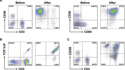
The viability of each type of immune cell in our culture system was found to exceed 95%, none of the cultured im-mune cells were found to be contaminated, and all of the preparations met the quality criteria. The percentage of NK (CD3CD56þ), gdT (CD3þVg9þ) and CIK (CD3þCD56þ) cells before and after induction was 8.35% (range, 3.97e18.42%) vs. 82.56% (range, 58.33e99.61%), 4.78% (range, 2.34e12.35%) vs. 80.63% (range, 55.72e98.21%) and 4.51% (range, 1.55e8.45%) vs. 34.52% (range, 26.65e58.23%), respectively (Table 2). Induction also resul-ted in a significant increase in the proportion of activaresul-ted NK cells (CD56þCD69þ). Representative results from a single patient are shown inFig. 1.
3.3. PFS and OS
Follow-up of all patients was ended on December, 2015, with a median follow-up time of 10.3 months (range, 3e55.2
months). PFS in the CIT group was superior to the control group (5 vs. 3.1 months, P ¼0.020) (Fig. 2A), and CIT as maintenance therapy significantly reduced the risk of ES-SCLC recurrence (hazard ratio [HR], 0.489, 95% confidence interval [CI], 0.264e0.909, P¼0.024). OS of the CIT group was also longer than that of the control group (13.3 vs. 8.2 months, P ¼ 0.044; HR, 0.528, 95% CI, 0.280e0.996, P¼0.048, respectively) (Fig. 2B). The 1-year and 2-year OS rate of the CIT and control group was 57.9% vs. 40.0% and 15% vs. 3.6%, respectively.
3.4. Potential factors influencing the outcome of CIT
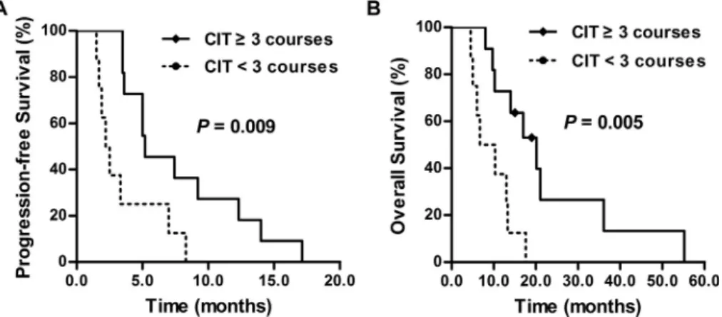
In the multivariate analysis, we found that sex, age, smoking index, ECOG performance status, chemotherapy course, radiotherapy and response to induction chemotherapy had no effect on the prognosis of ES-SCLC patients receiving CIT (P>0.05), but the CIT course had an effect both on the PFS and OS of CIT patients (P<0.05). We then investigated the influence of the CIT course on the prognosis of patients in the CIT group. The median number of CIT cycles was 3 (range, 1e6 cycles). Patients were divided into two subgroups: long-course group with CIT3 cycles and short-course group with CIT<3 cycles. The characteristics of the patients in these subgroups were well balanced (Table 3). The median PFS in the long-course group (n ¼ 11) was longer than that of the short-course group (n ¼8) (5.2 vs. 2.2 months, P ¼ 0.009; HR, 0.276; 95% CI, 0.098e0.779; P¼0.015) (Fig. 3A). The median OS of the long-course group was also significantly longer than that of the short-course group (20.1 vs. 6.7 months, P ¼ 0.005; HR, 0.217, 95% CI, 0.069e0.687, P ¼ 0.009) (Fig. 3B).
3.5. Response to second-line chemotherapy
A total of 14 patients accepted second-line therapy in the CIT group, and 15 patients received second-line therapy in the control group. The median second-line chemotherapy courses were 2 (range, 1e6) for both groups (P> 0.05). None of the patients in the two groups achieved CR after second-line treatment. Eight patients got a PR or SD with a CBR of 57.1% in the CIT group, and 7 patients got a PR or SD with a CBR of 46.7% in the control group, but there was no signif-icant difference between the groups (P¼0.573).
3.6. Side effects of CIT
Two patients had a transient fever after CIT infusion, but recovered within 2 h after administration of antipyretics or physical cooling. Three patients reported mild fatigue after CIT infusion. No other significant side effects were observed. 4. Discussion
ES-SCLC is a disease with poor prognosis. Treatment outcome has not improved significantly during the last decades despite great efforts have been made. Maintenance therapy has
Table 1
Clinical characteristics of patients in the CIT and control groups.
Clinical features CIT group Control group P-value Sex Male 18 22 0.127 Female 1 8 Age, years Median (range) 63 (54e79) 57 (36e74) 0.002 Smoking index Median (range) 400 (0e1200) 400 (0e2000) 0.983 ECOG 1 16 29 0.285 2 3 1 Chemotherapy course 6 6 1.000 Radiotherapy Intrathoracic radiation Yes 7 17 0.176 No 12 13 RT to symptomatic sites Yes 7 12 0.825 No 12 18 PCI Yes 2 1 0.551 No 17 29
Response to induction chemotherapy
CRþPR 14 22 0.978
SD 5 8
Abbreviations: CIT, cellular immunotherapy, ECOG, Eastern Cooperative Oncology Group; RT, radiotherapy; PCI, prophylactic cranial radiation; CR, complete remission; PR, partial remission, SD, stable disease.
Table 2
Summarized data of the percentage of NK,gdT and CIK cells before and after induction.
Immune cells Before (median, range) After (median, range) NK cells 8.35% (3.97e18.42%) 82.56% (58.33e99.61%)
gdT cells 4.78% (2.34e12.35%) 80.63% (55.72e98.21%) CIK cells 4.51% (1.55e8.45%) 34.52% (26.65e58.23%) Abbreviations: NK, natural killer; CIK, cytokine-induced killer.
been tried in several clinical trials with the aim to prevent the onset of recurrence and prolong survival. However, mainte-nance and/or consolidation using chemotherapy, cytokines and other biological agents failed to improve ES-SCLC outcomes
[5]. Molecularly targeted drugs have been used as mainte-nance therapy in ES-SCLC in recent years, but most of them did not improve the OS of ES-SCLC patients, such as sor-afenib, vandetanib, and imatinib [26e28]. Therefore, new therapeutic strategies are urgently needed to improve the outcome of this disease.
Immune escape in SCLC patients is closely associated with the recurrence of the disease, and may contribute to poor pa-tient survival [6,7,12e14]. Cancer cells employ multiple mechanisms to evade an immune response[29e31], and thus CIT with only one type of immune cells is unlikely to achieve an optimal anticancer effect. In our previous study, CIT as maintenance therapy with the combination of NK, gdT and CIK cells has exhibited a synergistic anticancer effect and
improved the outcome of SCLC patients, especially in ES-SCLC patients. The present study, with more patients enrolled and longer follow-up time, further confirmed the ef-ficacy and safety of combined CIT as maintenance therapy for ES-SCLC patients. Thus, CIT may be a novel treatment for ES-SCLC patients who respond to first-line therapy, and can provide a novel strategy for the devastating disease.
ES-SCLC is a highly malignant tumor associated with short survival and limited chemotherapy regimens, and hence there are fewer confounding factors for OS. In our study, OS was significantly longer after CIT in ES-SCLC patients. There are several possible reasons for the prolonged survival observed. Cancer stem cells (CSCs) have been reported to be responsible for cancer progression, metastasis, and the development of drug resistance [32,33]. They have also been shown to be susceptible to immunocyte-mediated toxicity, suggesting that CIT may be useful in eliminating minimal residual disease and thus reduce cancer recurrence [34e36]. Prolonged PFS and
Fig. 1.The percentage of NK,gdT and CIK cells before and after induction. Representative results from a single patient are shown. The percentage of NK cells (A),gdT cells (B) and CIK cells (C) before and after induction was 14.4% vs. 95.3%, 3.47% vs. 98.1% and 1.88% vs. 47.2%, respectively. CD56þCD69þcells were considered to be activated NK cells.
Fig. 2.Progression free survival (PFS) and overall survival (OS) in both groups. (A) PFS of the two groups. PFS in the CIT group was longer than the control group (5 vs. 3.1 months, P¼0.020). (B) OS of the two groups. OS in the CIT group was significantly longer than in the control group (13.3 vs. 8.2 months, P¼0.044).
lower risk of recurrence (HR¼0.489) might be responsible for longer OS of ES-SCLC patients in the CIT group. But one study exploring the relationship between PFS and OS in SCLC patients found no significant relationship between them [37], suggesting that OS might be affected by other factors, such as second or third-line therapy.
It has been reported that patients who received cancer vaccines responded better to subsequent chemotherapy than those who did not, suggesting that immunotherapy might sensitize cancer cells to cytotoxic drugs [38,39]. Indeed, pa-tients with CIT tended to have higher CBR than the control in our study, although the difference was not significant,
indicating that CIT might enhance the sensitivity of recurrent SCLC to second-line chemotherapy. It is noteworthy that only half of the patients in the control group received second-line chemotherapy, but most of the patients in the CIT group received second-line chemotherapy. The main reason why half of the patients in the control group didn't receive second-line chemotherapy was the low performance status. Only a very small number of patients didn't receive second-line chemo-therapy because they refused further treatment. Therefore, CIT might improve the quality of life of cancer patients, so that more patients have the opportunity to receive second-line chemotherapy after CIT maintenance therapy. This might be another reason that CIT could prolong OS. The underlying mechanism needs to be further explored.
Potential factors influencing the outcome of CIT were also evaluated in this study. Using a multivariate analysis, we found that sex, age, smoking history, ECOG performance status, chemotherapy course, radiotherapy and response to induction chemotherapy were not related to the efficacy of treatment. Patients receiving longer CIT course had longer PFS and OS. Both PFS and OS in patients who received more than 3 cycles of CIT was significantly longer than that of patients who received less than 3 cycles of CIT. OS was consistent with our previous report. In our previous study, PFS tended to be longer in patients who received more than 3 CIT cycles, but the different was not statistically significant. The difference be-tween the two studies may be due to the different sample size and follow-up time. Besides, some patients with early stage SCLC were included in the previous study. One study demonstrated that NSCLC patients who received more than 7 cycles of CIK cell treatment had a significantly better prog-nosis than those who received fewer cycles [40]. Taken together, our findings and those of a previous report [40]
suggest that a longer CIT course improved patient outcome. However, the optimal number of treatment cycle and the length of treatment course are yet to be determined. Even though the characteristics of the patients in these subgroups were well balanced, some patients stopped CIT treatment in the short course group because of disease progression. Therefore, the shorter PFS and OS might be affected not only
Table 3
Clinical characteristics of patients in two subgroups.
Clinical features Long-course Short-course P-value Sex Male 11 7 0.421 Female 0 1 Age, years Median (range) 63 (54e79) 66.5 (58e78) 0.422 Smoking index Median (range) 600 (0e1000) 150 (0e1200) 0.424 ECOG 1 10 6 0.546 2 1 2 Chemotherapy course 6 6 1.000 Radiotherapy Intrathoracic radiation Yes 4 3 1.000 No 7 5 RT to symptomatic sites Yes 3 4 0.377 No 8 4 PCI Yes 2 0 0.485 No 9 8
Response to induction chemotherapy
CRþPR 8 6 1.000
SD 3 2
Abbreviations: ECOG, Eastern Cooperative Oncology Group; RT, radio-therapy; PCI, prophylactic cranial radiation; CR, complete remission; PR, partial remission, SD, stable disease.
Fig. 3.The influence of cellular immunotherapy (CIT) course on extensive stage small cell lung cancer (ES-SCLC) patients'prognosis. (A) The median progression free survival (PFS) in the long-course group was longer than that of the short-course group (5.2 vs. 2.2 months, P¼0.009). (B) The median overall survival (OS) in the long-course group was significantly longer than that of patients in the short-course group (20.1 vs. 6.7 months, P¼0.005).
by fewer CIT cycles, but also by other factors, such as the worse disease status in this subgroup of patients.
Safety is an important factor determining the application of CIT. In this study, only 3 patients reported mild fatigue and 2 patients had a transient fever after infusion. All symptoms diminished after treatment. We did not observe any significant side effects after CIT. Thus, CIT was well tolerated and is a good candidate for maintenance therapy when side effects need to be minimized.
To assess of the impact of CIT on the immune system, we compared the number of immune cells (T, NK, NKT, B, Treg cells and monocytes) in the peripheral blood before and after CIT and did not find any significant change. There were relatively fewer Treg cells at some time points after CIT, but these differences were not significant. This might reflect the relatively few CIT courses and the small number of patients involved. However, this might indicate that response to CIT is not associated with the proportion of immune cells in the peripheral blood and this needs to be addressed in the further studies. This also reflects the challenge to find markers for assessing immune response.
In our study, ES-SCLC patients in the CIT group were on average older than those in the control group. However, the multivariate analysis showed that age had no effect on the prognosis of these patients. Therefore, age was no a con-founding factor, although it has been reported that effector cell function decreases in older people, and might be associated with reduced antitumor immunity in these patients[41].
One limitation of this study is that patients were allocated to each group according to the patients' therapeutic options. Further multi-center randomized clinical trials are needed to verify its efficacy.
5. Conclusions
CIT as a maintenance therapy after first-line treatment might prevent disease recurrence and prolong the survival of ES-SCLC patients with only minimal side effects. We also found that more CIT courses might improve treatment outcome. CIT might enhance the sensitivity of recurrent ES-SCLC to second-line chemotherapy. The exact mechanism through which CIT extends PFS and OS in ES-SCLC patients remains to be explored.
Authors'contributions
XD participated in the acquisition of data, analysis and interpretation of data and the drafting of the manuscript and its revision. HC participated in acquisition of data and analysis and interpretation of data. XC,YZ, KM, ZL, XW, LY, DL and HT participated in the acquisition of data. HJ, CN, JC and WH participated in preparation and analysis of immunocytes. HH helped statistical analysis. WL and JC participated in the conception, design and coordination of this study, acquisition of data, analysis and interpretation of data and helped to draft the manuscript or revise it critically. All authors read and approved the final manuscript.
Competing interests
The authors declare that they have no competing interests. Acknowledgments
This work was supported by the Jilin Provincial Science and Technology Department (Grants 20111807 and 20140414014 GH, and 20150101176 to JW.C.), the Platform Construction Project of Development and Reform Commis-sion of Jilin Province (Grant 2014N147 to JW.C.), the Bethune Program B of Jilin University (Grant 2012202 to JW.C.), Key Clinical Project of the Ministry of Health of the People's Re-public of China (Grant 2001133 to W.L), and National Major Scientific and Technological Special Project (Grant 2013ZX09102032 to JT.C).
References
[1]van Meerbeeck JP, Fennell DA, De Ruysscher DK. Small-cell lung cancer. Lancet 2011;378:1741e55.
[2]Jotte R, Conkling P, Reynolds C, Galsky MD, Klein L, Fitzgibbons JF, et al. Randomized phase II trial of single-agent amrubicin or topotecan as second-line treatment in patients with small-cell lung cancer sensitive to first-line platinum-based chemotherapy. J Clin Oncol 2011;29:287e93. [3]Schmittel A, Sebastian M, Fischer von Weikersthal L, Martus P,
Gauler TC, Kaufmann C, et al. A German multicenter, randomized phase III trial comparing irinotecan-carboplatin with etoposide-carboplatin as first-line therapy for extensive-disease small-cell lung cancer. Ann Oncol 2011;22:1798e804.
[4]Gerber DE, Schiller JH. Maintenance chemotherapy for advanced non-small-cell lung cancer: new life for an old idea. J Clin Oncol 2013;31: 1009e20.
[5]Rossi A, Garassino MC, Cinquini M, Sburlati P, Di Maio M, Farina G, et al. Maintenance or consolidation therapy in small-cell lung cancer: a systematic review and meta-analysis. Lung Cancer 2010;70:119e28. [6]Predina J, Eruslanov E, Judy B, Kapoor V, Cheng G, Wang LC, et al.
Changes in the local tumor microenvironment in recurrent cancers may explain the failure of vaccines after surgery. Proc Natl Acad Sci U S A 2013;110:E415e24.
[7]Shiao SL, Ganesan AP, Rugo HS, Coussens LM. Immune microenvi-ronments in solid tumors: new targets for therapy. Genes Dev 2011;25: 2559e72.
[8]Chiappori AA, Soliman H, Janssen WE, Antonia SJ, Gabrilovich DI. INGN-225: a dendritic cell-based p53 vaccine (Ad.p53-DC) in small cell lung cancer: observed association between immune response and enhanced chemotherapy effect. Expert Opin Biol Ther 2010;10:983e91. [9]Reck M, Bondarenko I, Luft A, Serwatowski P, Barlesi F, Chacko R, et al. Ipilimumab in combination with paclitaxel and carboplatin as first-line therapy in extensive-disease-small-cell lung cancer: results from a randomized, double-blind, multicenter phase 2 trial. Ann Oncol 2013;24: 75e83.
[10]Ott PA, Fernandez MEE, Hiret S, Kim DW, Moss RA, Winser T, et al. Pembrolizumab (MK-3475) in patients (pts) with extensive-stage small cell lung cancer (SCLC): preliminary safety and efficacy results from KEYNOTE-028. J Clin Oncol 2015;33 [suppl; abstr 7502].
[11]Antonia SJ, Bendell JC, Taylor MH, Calvo E, Jaeger D, De Braud FG, et al. Phase I/II study of nivolumab with or without ipilimumab for treatment of recurrent small cell lung cancer (SCLC): CA209e032. J Clin Oncol 2015;33 [suppl; abstr 7503].
[12]Koyama K, Kagamu H, Miura S, Hiura T, Miyabayashi T, Itoh R, et al. Reciprocal CD4þ T-cell balance of effector CD62Llow CD4þ and CD62LhighCD25þCD4þregulatory T cells in small cell lung cancer reflects disease stage. Clin Cancer Res 2008;14:6770e9.
[13] Wang W, Hodkinson P, McLaren F, MacKinnon A, Wallace W, Howie S, et al. Small cell lung cancer tumour cells induce regulatory T lympho-cytes, and patient survival correlates negatively with FOXP3þcells in tumour infiltrate. Int J Cancer 2012;131:E928e37.
[14] Afifi SS, Helal AM. CD11cþ and CD123þdendritic cell subsets in peripheral blood of lung cancer patients. Egypt J Immunol 2009;16: 9e15.
[15] Tsuchida T, Yamane H, Ochi N, Tabayashi T, Hiraki A, Nogami N, et al. Cytotoxicity of activated natural killer cells and expression of adhesion molecules in small-cell lung cancer. Anticancer Res 2012;32: 887e92.
[16] Ohira M, Nishida S, Tryphonopoulos P, Tekin A, Selvaggi G, Moon J, et al. Clinical-scale isolation of interleukin-2-stimulated liver natural killer cells for treatment of liver transplantation with hepatocellular carcinoma. Cell Transpl 2012;21:1397e406.
[17] Shi L, Zhou Q, Wu J, Ji M, Li G, Jiang J, et al. Efficacy of adjuvant immunotherapy with cytokine-induced killer cells in patients with locally advanced gastric cancer. Cancer Immunol Immunother 2012;61:2251e9. [18] Yu X, Zhao H, Liu L, Cao S, Ren B, Zhang N, et al. A randomized phase II study of autologous cytokine-induced killer cells in treatment of hepatocelluar carcinoma. J Clin Immunol 2014;34:194e203.
[19] Maniar A, Zhang X, Lin W, Gastman BR, Pauza CD, Strome SE, et al. Human gammadelta T lymphocytes induce robust NK cell-mediated antitumor cytotoxicity through CD137 engagement. Blood 2010;116: 1726e33.
[20] Cui J, Wang N, Zhao H, Jin H, Wang G, Niu C, et al. Combination of radiofrequency ablation and sequential cellular immunotherapy improves progression-free survival for patients with hepatocellular carcinoma. Int J Cancer 2014;134:342e51.
[21] Cui J, Li L, Wang C, Jin H, Yao C, Wang Y, et al. Combined cellular immunotherapy and chemotherapy improves clinical outcome in patients with gastric carcinoma. Cytotherapy 2015;17:979e88.
[22] Sato K, Kimura S, Segawa H, Yokota A, Matsumoto S, Kuroda J, et al. Cytotoxic effects of gammadelta T cells expanded ex vivo by a third generation bisphosphonate for cancer immunotherapy. Int J Cancer 2005; 116:94e9.
[23] Ding X, Cao H, Chen X, Jin H, Liu Z, Wang G, et al. Cellular immu-notherapy as maintenance therapy prolongs the survival of the patients with small cell lung cancer. J Transl Med 2015;13:158.
[24] NCCN clinical practice guidelines in oncology: small cell lung cancer version 2. 2009.http://www.nccn.org[accessed 15.01.09].
[25] Eisenhauer EA, Therasse P, Bogaerts J, Schwartz LH, Sargent D, Ford R, et al. New response evaluation criteria in solid tumours: revised RECIST guideline (version 1.1). Eur J Cancer 2009;45:228e47.
[26] Sharma N, Pennell N, Nickolich M, Halmos B, Ma P, Mekhail T, et al. Phase II trial of sorafenib in conjunction with chemotherapy and as maintenance therapy in extensive-stage small cell lung cancer. Invest New Drugs 2014;32:362e8.
[27] Arnold AM, Seymour L, Smylie M, Ding K, Ung Y, Findlay B, et al. Phase II study of vandetanib or placebo in small-cell lung cancer patients after complete or partial response to induction chemotherapy with or without radiation therapy: National Cancer Institute of Canada Clinical Trials Group Study BR.20. J Clin Oncol 2007;25:4278e84.
[28] Schneider BJ, Kalemkerian GP, Ramnath N, Kraut MJ, Wozniak AJ, Worden FP, et al. Phase II trial of imatinib maintenance therapy after irinotecan and cisplatin in patients with c-Kit-positive, extensive-stage small-cell lung cancer. Clin Lung Cancer 2010;11:223e7.
[29] Kim R, Emi M, Tanabe K. Cancer immunoediting from immune sur-veillance to immune escape. Immunology 2007;121:1e14.
[30] Whiteside TL. The tumor microenvironment and its role in promoting tumor growth. Oncogene 2008;27:5904e12.
[31] Facciabene A, Motz GT, Coukos G. T-regulatory cells: key players in tumor immune escape and angiogenesis. Cancer Res 2012;72:2162e71. [32] Fabian A, Vereb G, Szollosi J. The hitchhikers guide to cancer stem cell
theory: markers, pathways and therapy. Cytom A 2013;83:62e71. [33] Sarvi S, Mackinnon AC, Avlonitis N, Bradley M, Rintoul RC, Rassl DM,
et al. CD133þcancer stem-like cells in small cell lung cancer are highly tumorigenic and chemoresistant but sensitive to a novel neuropeptide antagonist. Cancer Res 2014;74:1554e65.
[34] Gammaitoni L, Giraudo L, Leuci V, Todorovic M, Mesiano G, Picciotto F, et al. Effective activity of cytokine-induced killer cells against autologous metastatic melanoma including cells with stemness features. Clin Cancer Res 2013;19:4347e58.
[35] Tallerico R, Todaro M, Di Franco S, Maccalli C, Garofalo C, Sottile R, et al. Human NK cells selective targeting of colon cancer-initiating cells: a role for natural cytotoxicity receptors and MHC class I molecules. J Immunol 2013;190:2381e90.
[36] Todaro M, D'Asaro M, Caccamo N, Iovino F, Francipane MG, Meraviglia S, et al. Efficient killing of human colon cancer stem cells by gammadelta T lymphocytes. J Immunol 2009;182:7287e96.
[37] Foster NR, Renfro LA, Schild SE, Redman MW, Wang XF, Dahlberg SE, et al. Multitrial evaluation of progression-free survival (PFS) as a sur-rogate endpoint for overall survival (OS) in previously untreated extensive-stage small cell lung cancer (ES-SCLC): an alliance-led analysis. J Clin Oncol 2013;31 [suppl; abstr 7510].
[38] Schlom J, Arlen PM, Gulley JL. Cancer vaccines: moving beyond current paradigms. Clin Cancer Res 2007;13:3776e82.
[39] Wheeler CJ, Das A, Liu G, Yu JS, Black KL. Clinical responsiveness of glioblastoma multiforme to chemotherapy after vaccination. Clin Cancer Res 2004;10:5316e26.
[40] Li R, Wang C, Liu L, Du C, Cao S, Yu J, et al. Autologous cytokine-induced killer cell immunotherapy in lung cancer: a phase II clinical study. Cancer Immunol Immunother 2012;61:2125e33.
[41] Kozlowska E, Biernacka M, Ciechomska M, Drela N. Age-related changes in the occurrence and characteristics of thymic CD4(þ) CD25(þ) T cells in mice. Immunology 2007;122:445e53.