RESEARCH
MicroRNA circulating in the early
aftermath of motor vehicle collision predict
persistent pain development and suggest a role
for microRNA in sex-specific pain differences
Sarah D. Linnstaedt
1,2*, Margaret G. Walker
1,2, Joel S. Parker
3, Eunice Yeh
1,2, Robert L. Sons
4, Erin Zimny
5,
Christopher Lewandowski
5, Phyllis L. Hendry
6, Kathia Damiron
7, Claire Pearson
8, Marc‑Anthony Velilla
9,
Brian J. O’Neil
10,11, Jeffrey Jones
12, Robert Swor
13, Robert Domeier
14, Scott Hammond
4and Samuel A. McLe
an
1,2,15Abstract
Background: Molecular mediators influencing the transition from acute to persistent musculoskeletal pain follow‑
ing common stress exposures such as motor vehicle collision (MVC) remain poorly understood. In this exploratory, proof of concept study, we compared circulating microRNA (miRNA) expression profiles in the early aftermath of MVC among individuals who did and did not subsequently develop persistent pain. Blood RNA samples were obtained from African American individuals (n = 53) who presented to the emergency department after MVC and were dis‑ charged to home after evaluation. The presence or absence of severe pain in the axial region, the most common and morbid region in which post‑MVC pain occurs, was assessed 6 weeks following MVC via standardized questionnaire. miRNA expression was determined using miRNA‑sequencing; nonparametric analyses were used to compare miRNA expression levels among individuals with and without persistent pain.
Results: Thirty‑two mature miRNA were differentially expressed (p < 0.05) in those with and without severe axial
pain at 6 weeks. miR‑135a‑5p, a regulator of the serotonin receptor that is known to be stress‑responsive, differed most significantly between groups (p = 3 × 10⁻⁴). This miRNA, and miR‑3613‑3p (p = 0.001) survived correction for
multiple testing (FDR = 0.15) in this small sample. Interestingly, differentially expressed miRNA were enriched for X chromosome location. In secondary analyses, the eight X chromosome miRNA were (a) more significantly associated with axial pain in women than men, (b) expressed more highly in the peripheral blood of women than men, and (c) predicted in pathway analyses (DIANA miRPath v 2.0) to regulate neuronal and neuroendocrine pathways previously implicated in various pain pathologies.
Conclusions: These results show that circulating miRNA predict persistent severe axial pain after MVC and suggest
that they may be involved in the pathogenesis of post‑traumatic musculoskeletal pain. However, further studies are needed to determine if these miRNA play a direct causal role.
Keywords: microRNA, Stress induced pain, Persistent axial pain, Motor vehicle collision, Sexual dimorphism,
microRNA, Motor vehicle collision, African Americans, Musculoskeletal pain
© 2015 Linnstaedt et al. This article is distributed under the terms of the Creative Commons Attribution 4.0 International License (http://creativecommons.org/licenses/by/4.0/), which permits unrestricted use, distribution, and reproduction in any medium, provided you give appropriate credit to the original author(s) and the source, provide a link to the Creative Commons license, and indicate if changes were made. The Creative Commons Public Domain Dedication waiver (http://creativecommons.org/ publicdomain/zero/1.0/) applies to the data made available in this article, unless otherwise stated.
Open Access
*Correspondence: [email protected]
2 Department of Anesthesiology, University of North Carolina, Medical School Wing C CB#7010, Chapel Hill, NC 27599‑7010, USA
Background
MicroRNA (miRNA) are small non-coding RNA mol-ecules that regulate gene expression by binding target mRNA. During the past decade, the study of miRNA has transformed understanding of the regulation of major biological pathways [1, 2] and advanced understanding of the pathogenesis of a number of common diseases (e.g. [3–5]). Substantial evidence suggests that miRNA may play a critical role in molecular pathways underpinning diverse pain conditions (e.g. [6–8]). Most of this evidence comes from animal studies; further human studies are needed which prospectively evaluate the potential role of miRNA in persistent pain development after potential triggering events.
One common potential triggering event for persistent pain is exposure to a motor vehicle collision (MVC). More than fifty million MVCs occur each year world-wide [9], and more than 4 million individuals present to US emergency departments (ED) each year for care after MVC [10, 11]. Ninety percent of individuals presenting to US EDs for care after MVC are discharged home after evaluation with little or no identifiable tissue injury [12]. A substantial proportion of these individuals develop musculoskeletal pain, most commonly in the axial region (neck, shoulders, and/or back) [13].
The molecular mechanisms responsible for axial pain (AP) development after MVC remain poorly understood. This lack of understanding is a major bar-rier to the development of more effective preventive interventions. If miRNA play an important role in the pathogenesis of post-MVC AP, then studies identifying differences in miRNA populations among those who do and do not subsequently develop these outcomes may advance understanding of post-MVC AP pathogenesis [14–16]. Such studies must be feasible and ethical, and should obtain miRNA samples from tissue relevant to disease pathogenesis and/or be representative of such tissue.
Several lines of evidence suggest that blood is not only a feasible source of miRNA for studies of post-MVC musculoskeletal pain pathogenesis, but also that blood miRNA studies may provide pathogenic insights [17–
19]. First, stress systems appear to be involved in the pathogenesis of persistent pain after MVC [20–23], and blood-borne stress and immune-related factors are an important component of the systemic stress response [24]. In addition, RNA expression patterns in blood, CNS, and endocrine tissues are strongly correlated [15,
25, 26], and a number of previous studies have linked miRNA expression in blood with neurologic disease outcomes [27–30]. Finally, despite challenges related to tissue-specific gene expression, the study of blood
miRNA expression has led to important new under-standing of diverse diseases, including pain conditions [27, 31, 32].
In this prospective study, we compared ED blood miRNA profiles among individuals who presented to the ED for evaluation after MVC. We hypothesized that ED blood miRNA profiles would differ among those who did and did not have severe persistent AP 6 weeks later. Results
Cohort
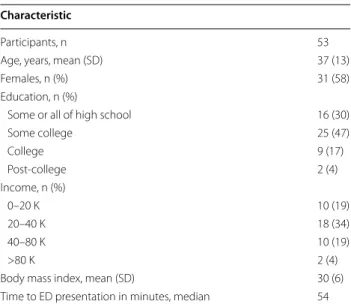
Characteristics of the study sample (n = 53) are shown in Table 1. Samples were drawn from a large prospec-tive cohort study of African Americans (R01AR060852); all participants were African American (AA) and nearly six in ten were female. Most were less than 40 years old, had some college education, made less than 40 K annu-ally, and were overweight (average BMI = 30). All indi-viduals in this study presented to the ED within 6 h of MVC and most arrived within 1 h. Only individuals who were discharged from the ED who reported no lacera-tions, avulsions, or major tissue injury were included. Additionally, participants all had a severity score of 1 on the Abbreviated Injury Scale (AIS) [33], indicating minimal anatomical injury. Six weeks following MVC, severe AP was present in 27/53 participants. Among these 27 individuals who developed severe AP, 16 (59 %) were women.
miRNA sequencing quality assessment
An average of 9 million sequencing reads were obtained per participant from blood samples obtained in the ED
Table 1 Study characteristics Characteristic
Participants, n 53
Age, years, mean (SD) 37 (13)
Females, n (%) 31 (58)
Education, n (%)
Some or all of high school 16 (30)
Some college 25 (47) College 9 (17) Post‑college 2 (4) Income, n (%) 0–20 K 10 (19) 20–40 K 18 (34) 40–80 K 10 (19) >80 K 2 (4)
Body mass index, mean (SD) 30 (6)
in the early aftermath of MVC. More than 95 % of these miRNA aligned with miRNA in miRBase, indicating that the majority of the sequencing reads were mature miRNAs (vs. degradation products, linker–linker con-taminants, etc.). Mature miRNA with an average of ≥300 sequencing reads across all 53 samples (n = 376 miRNA) were included in analyses. Relative proportions of several miRNA typically found in peripheral blood were very sim-ilar to those reported previously [15, 34] (data not shown).
Evaluation of ED miRNA expression levels among those who did and did not develop persistent AP 6 weeks following MVC
Thirty-two of 376 (9 %) miRNA detected in ED blood samples were differentially expressed at the p <0.05 level among those who did and did not report severe post-MVC AP at 6 weeks, with fold differences rang-ing from −3.71 to 2.49 (Table 2, sequencing read counts used for determination of mean fold differences for each miRNA are included in Additional file 1: Table S1). Nine of these 32 differentially expressed miRNA have previ-ously been associated with pain and/or stress system physiology in neurological tissue and/or blood (see ‘Ref’ column, Table 2). Two of the 32 differentially expressed miRNA, miR-135a-5p (p = 3 × 10⁻⁴) and miR-3613-3p (p = 0.001), met our pre-hoc significance level threshold for multiple testing of 0.15, corresponding to a p value cut-off of <0.003 [35].
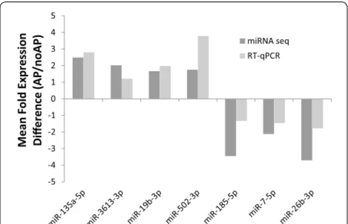
Validation of miRNA sequencing results using RT‑qPCR
Technical and qualitative validation of miRNA sequenc-ing results was performed on a random subsample of 7 of the 32 differentially expressed miRNA using reverse tran-scription quantitative-PCR (RT-qPCR) [36]. In each case, concordance between miRNA sequencing results and RT-qPCR results was observed for direction of differen-tial expression (i.e., positive or negative expression dif-ference, Fig. 1). Magnitude of direction of effect was also generally similar. The Spearman Correlation between the two methods was also calculated: r = 0.786 p = 0.036.
Differentially expressed miRNA were enriched for X chromosome location
Specific data regarding chromosomal origin and strand (sense or antisense) for the 32 differentially expressed miRNA is shown in Additional file 1: Table S1. The 32 miRNA predictive of severe post-MVC AP at 6 weeks were enriched for gene location on the X chromosome (Fig. 2) in comparison to all X chromosome miRNA identified in the sample (8/32 (25 %) vs. 28/376 (8 %), p = 0.038). This holds true despite the fact that approxi-mately 8 % of the detected blood miRNA originate from the X chromosome.
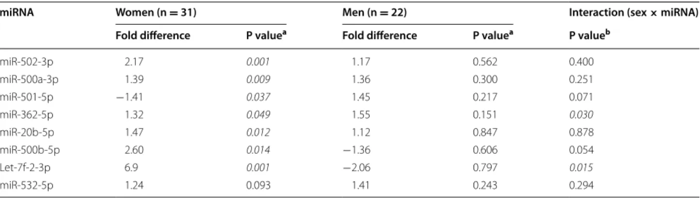
X chromosome miRNA identified in this study are more highly associated with the development of persistent AP following MVC in women than men
Because X chromosome gene expression can be sex-dependent [37], after we observed the X chromosome enrichment of differentially-expressed miRNA in our sample, we assessed for interactions between sex and the
Table 2 microRNA in whole blood circulating in the early aftermath of motor vehicle collision in African Americans that predict axial pain development 6 weeks after MVC trauma
a Mean fold difference was calculated by dividing the average sequencing read counts for individuals developing axial pain by the average sequencing read counts for individuals who recover
b p values were calculated using the Mann–Whitney U test. Italicized miRNA remained significant after correcting for multiple testing (FDR = 0.15) c Previous assoc = references describing a previously identified role for the miRNA in stress system biology (S), pain pathobiology (P), or neuropsychiatric disease (N)
microRNA Mean fold differencea p valueb Previous assocc
miR‑135a‑5p 2.49 3 × 10⁻⁴ S, N [47, 48] miR‑3613‑3p 2.02 0.001 miR‑19b‑3p 1.67 0.004 S, P [49, 51] miR‑502‑3p 1.75 0.004 miR‑500a‑3p 1.39 0.005 miR‑1296‑5p 1.99 0.006 S [50] miR‑454‑5p 1.58 0.010 miR‑99a‑5p 1.48 0.010 P [71] miR‑501‑5p −1.15 0.011 miR‑362‑5p 1.41 0.013 miR‑154‑5p 1.09 0.015 Let‑7a‑3p 1.48 0.020 S, P [50, 72] miR‑185‑5p −3.45 0.021 P [52] miR‑339‑5p 1.31 0.023 miR‑29c‑5p 1.67 0.023 miR‑4659b‑3p −2.19 0.023 miR‑15b‑5p −1.22 0.026 miR‑329‑3p 1.68 0.026 miR‑20b‑5p 1.35 0.029 S [73] miR‑500b‑5p 1.38 0.029 Let‑7f‑2‑3p 1.43 0.029 miR‑7‑5p −2.12 0.033 P [53, 73] miR‑378a 1.37 0.034 miR‑3130‑5p 1.91 0.034 miR‑532‑5p 1.31 0.036 miR‑345‑5p 1.62 0.037 miR‑16‑5p −2.70 0.043 miR‑18a‑3p 1.49 0.044 miR‑337‑3p −1.06 0.045 miR‑26b‑3p −3.71 0.046 P [54] miR‑26a‑5p −2.52 0.048 miR‑151b 1.33 0.048
effect on severe persistent AP of differentially expressed X chromosome miRNA. Even in the relatively small sam-ples of men and women assessed, sex × miRNA interac-tions were significant at the p <0.05 level for miR-362-5p and let-7f-2-3p, and were present at the trend level for
miR-501-5p and miR-500b-5p (Table 3, Additional file 1: Table S1). Much greater differences in expression accord-ing to pain outcome were observed in women (n = 31) vs. men (n = 22) (Table 3). Seven of the 8 X chromo-some miRNA were significantly associated with AP development in women, whereas none of the X chro-mosome miRNA were associated with AP development in men (Table 3). Additionally, after discovering signifi-cant sex × miRNA interactions for the X-chromosome miRNA described above, we assessed for additional interactions between participant sex and miRNA associ-ated with persistent AP. A significant sex × miRNA inter-action was present for two additional miRNA, miR-1296 (p = 0.017) and Let-7a-3p (p = 0.022) (see Additional file 1: Table S1 for results of all sex × miRNA interac-tions). These two miRNA are significantly associated with persistent severe AP in women (p < 0.001 and 0.001) but not in men (Additional file 2: Table S2).
X chromosome miRNA identified in this study are
expressed more highly in the blood in the early aftermath of MVC in women than men
Based on previous reports showing higher expression of some X chromosome genes in women than men (most notably in brain tissue [38] and possibly due to mecha-nisms such as escape from X chromosome inactivation [39]), we assessed whether the X chromosome miRNA identified in this study are expressed at higher levels in women than men. All eight miRNA were expressed at higher levels in women than in men developing severe AP, although only one was statistically significant (miR-502-3p, p = 0.017) (Table 4).
Evaluation of biologic pathways targeted by AP‑associated X chromosome miRNA
Using DIANA miRPath v 2.0 [40], we assessed for molecular pathways (KEGG pathways [41]) overrep-resented in predicted targeting by these 8 X chromo-some miRNA. The pathways with the highest number of gene transcripts targeted by the eight X chromo-some miRNA in Table 2 (i.e. most statistically significant enrichment) out of ~450 KEGG pathways that DIANA miRPath queries are shown in Table 5 (false discovery rates (FDRs) calculated via permutation testing [42]). These pathways include neuronal and neuroendocrine pathways such as the Long Term Potentiation path-way (p = 1.67 × 10⁻⁹, FDR ≤ 0.08), the Axon Guidance pathway (p = 1.32 × 10⁻⁷, FDR ≤ 0.22), Neurotrophin signaling (p = 7.35 × 10⁻⁷, FDR ≤ 0.22) and the Dopa-minergic synapse signaling pathway (p = 1.56 × 10⁻⁶, FDR ≤ 0.33). Substantial evidence supports an impor-tant role for these pathways in mediating physiologic responses to stress and the pathogenesis of acute and
-5 -4 -3 -2 -1 0 1 2 3 4 5 Mean Fold Expression Di ffe rence (A P/ noAP) miRNA seq RT-qPCR
Fig. 1 RT‑qPCR validation of microRNA that predict axial pain (AP)
development following motor vehicle collision (MVC). Dark grey bars represent expression differences calculated using mean microRNA sequencing counts in individuals developing AP divided by mean microRNA sequencing counts in individuals recovering after MVC.
Light grey bars represent expression differences in the same two
groups using mean cycle thresholds generated via RT‑qPCR. Spear‑ man Correlation assessing correlation of the two methods: r = 0.786, p = 0.036
Fig. 2 microRNA that predict axial pain (AP) development are tran‑
scribed from 14 different chromosomes. The percentage of miRNAs (out of the 32 associated with AP, Table 2) that originate from each of the 14 chromosomes are represented by pieces of the pie chart based on shading (lightest shading = 3 % and darkest shading = 25 %, with intermediate percentages having intermediate shading colors, see legend). The names/number of each chromosome are labeled inside each piece of the pie
persistent pain [43–45]. In addition to assessing X-linked miRNA targeted pathways, we also assessed which path-ways might be enriched in targeting by all 32 miRNA associated with persistent AP development. This data is provided in Additional file 3: Table S3.
Discussion
Persistent pain is a common and poorly understood sequela of traumatic/stressful events such as MVC [46]. The results of this study show that even in the relatively small study sample, circulating blood miRNA in the first hours after stress exposure differed significantly among AAs who did and did not have severe persistent MVC-related AP 6 weeks later. Study results also provide an example of the novel pathophysiologic insights that may be obtained, as enrichment of X chromosome miRNA
Table 3 Association of Emergency Department expression levels of microRNA (miRNA) from the X chromosome with per-sistent axial pain following motor vehicle collision in women vs. men and assessment of interaction between sex of an individual and miRNA
a p values were calculated using the Mann–Whitney U test
b p values for the interaction term were calculated using a logistic regression model adjusted for age and site. P values meeting a significance threshold of p <0.05 are italicized
miRNA Women (n = 31) Men (n = 22) Interaction (sex × miRNA)
Fold difference P valuea Fold difference P valuea P valueb
miR‑502‑3p 2.17 0.001 1.17 0.562 0.400 miR‑500a‑3p 1.39 0.009 1.36 0.300 0.251 miR‑501‑5p −1.41 0.037 1.45 0.217 0.071 miR‑362‑5p 1.32 0.049 1.55 0.151 0.030 miR‑20b‑5p 1.47 0.012 1.12 0.847 0.878 miR‑500b‑5p 2.60 0.014 −1.36 0.606 0.054 Let‑7f‑2‑3p 6.9 0.001 −2.06 0.797 0.015 miR‑532‑5p 1.24 0.093 1.41 0.243 0.294
Table 4 Emergency Department expression level dif-ferences of microRNA (miRNA) from the X chromosome in women vs. men developing persistent axial pain follow-ing motor vehicle collision
a Expression difference is the mean sequencing read counts of the specified miRNA in women who have axial pain at 6 weeks divided by the mean sequencing read counts of men who have axial pain at 6 weeks
miRNA Expression differencea (women/men) P value
miR‑502‑3p 1.80 0.017 miR‑500a‑3p 1.43 0.112 miR‑501‑5p 1.38 0.209 miR‑362‑5p 1.08 0.773 miR‑20b‑5p 1.60 0.126 miR‑500b‑5p 1.37 0.417 Let‑7f‑2‑3p 1.78 0.252 miR‑532‑5p 1.15 0.563
Table 5 DIANA miRPath predicted KEGG pathways enriched in targeting by X chromosome miRNA differentially regu-lated in the early aftermath of MVC trauma in AA individuals who develop AP following MVC vs. those who recover
* Denotes mRNA experimentally validated to interact with an miRNA from Table 2, as identified by TarBase v 7.0 KEGG pathway P value Example of predicted targets
Ubiquitin mediated proteolysis 2.15 × 10⁻¹⁰
Long‑term potentiation 1.67 × 10⁻⁹ PRKCA*, CAMK4, GRIA1, PPP3CC, KRAS, CALM2*, GRIA2, PPP3CA, GRM1, RPS6KA3*, EP300*, GNAQ*
Axon guidance 1.32 × 10⁻⁷ GSK3B*, ABLIM3, SEMA5A, EPHA5, ROCK2*, PAK7, ROBO2, SEMA3C*, SRGAP1*, PPP3CC, KRAS, EPHA7, PPP3CA, PTK2*, RASA1*, NFAT5*, EPHB4*, UNC5C, CFL2*, SEMA3D, SEMA3A*, PLXNC1*, EPHA4*, SEMA3E, UNC5D
ErbB signaling 7.04 × 10⁻⁷
Neurotrophin signaling 7.35 × 10⁻⁷ GSK3B, NTRK2*, CAMK4, CRK, SORT1*, NTRK3*, FRS2*, MAP3K1*, KRAS, CALM2*, JUN, MAPK8, SOS1*, RPS6KA3*, GAB1, AKT3*, CAMK2B, FOXO3, MAP2K1, PRDM4, RAP1B, MAP3K5
Insulin signaling 8.72 × 10⁻⁷
Dopaminergic synapse 1.56 × 10⁻⁶ FOS, GSK3B*, PRKCA*, PPP2R5E, PPP2R3A, PPP2R2C, CREB5, DRD1, GRIA1, PPP3CC, CALM2*, GRIA2, PPP3CA, MAPK8, PPP2R2A, SCN1A, PPP2R3C, GNAQ, PPP2CB, AKT3*, CAMK2B, PPP1CB, GRIA3
among differentially expressed miRNA, and sex differ-ences in the effect of these miRNA, suggest potential mechanisms contributing to sex differences in vulner-ability to persistent post-MVC pain. Supporting the role of these differentially expressed miRNA in persistent pain pathogenesis, these miRNA included a number of miRNA previously associated with the stress response and/or pain processing. However, further studies are needed to determine whether these miRNA play a causal role in the pathogenesis of pain post-MVC. Of note, study findings also support the legitimacy of persistent post-MVC musculoskeletal pain as a “real” disease out-come. This is important, as patients with post-MVC pain outcomes are highly stigmatized [47]. In addition, study findings also contribute to a growing literature indicating that stress-mediated changes in neurosensory processing play an important role in the pathogenesis of post-MVC outcomes.
We do not know whether miRNA predicting severe AP in the present study play a causal role in the develop-ment of these outcomes, or are markers for other cellular processes directly involved. If these miRNA play a causal role, mechanisms by which miRNA detected in blood may directly influence AP outcomes include: (1) miRNA expressed outside the central nervous system (CNS) and detected in the blood may cross the blood brain bar-rier [48, 49] to influence CNS processes, (2) miRNA expressed in the CNS may alter CNS transcription and also be released into the periphery (e.g., as part of a cellu-lar/systemic communication system [50, 51]), (3) miRNA expressed outside the CNS may influence extra-CNS processes involved in the pathogenesis of post-traumatic pain [52, 53] (animal model data suggest that systemic, extra-CNS processes may play an important role in the pathogenesis of stress-induced pain [54]). Future studies are needed to better understand the identified associa-tions between miRNA circulating in the immediate after-math of trauma and the development of persistent pain states. Such studies may provide new insights into the biology of chronic pain development.
A potential causal role of the miRNA identified in the present study in the pathogenesis of severe post-MVC axial pain is supported by the known role of several of these miRNA in pain and/or stress-related processes. For example, miR-135a-5p binds the mineralcorticoid recep-tor (NR3C2) [55], the serotonin transporter (SLC6A4) [56], and the serotonin receptor-1a (HTR1A) [56] tran-scripts, all of which can affect pain processing (e.g. [57–
59]), and miR-135a-5p has been shown to be expressed in pain-relevant tissues including the amygdala [60], 5-HT neurons [56], spinal cord [61], and pre-frontal cortex [62]. miR-19b-3p has been shown to be stress responsive in both the amygdala [63] and blood leukocytes [64], and
can regulate the adrenergic receptor β-1 (ADRB1) [63]. Other miRNA, such as miR-3613-3p, have not been stud-ied extensively; target prediction algorithms (e.g., Tar-getScan v7.0) predict that miR-3613-3p modulates the expression of pain-associated genes including GABRB3, GRIN3A, TRPV1, NPY1R, and SCN9A. Further examples of miRNA identified in this study that have prior asso-ciations with pain include miR-185-5p, miR-7-5p, and miR-26b-3p [61, 62, 65]. Equally important, many of the miRNA identified in this study are not currently known to be associated with stress or pain-related outcomes, suggesting that investigations such as the present study have the potential to lead to the identification of novel miRNA mediators.
An unexpected finding in the present study was that differentially expressed miRNA were enriched for miRNA located on the X chromosome. This finding holds true even accounting for the relatively high abundance of X chromosome miRNA expressed in the blood (8 %) compared to miRNA from other chromosomes. These X chromosomal miRNA were consistently expressed at higher levels in women than in men and appeared to con-tribute to persistent severe AP in women but not in men. These X chromosome miRNA were predicted to target pain-relevant transcripts from KEGG pathways known to be associated with various pain phenotypes, such as long term potentiation, neurotrophin signaling, and dopa-minergic signaling [43–45]. Pathophysiologic mecha-nisms by which these miRNA may contribute to severe post-MVC AP in women but not in men are currently unknown. Six of the eight X chromosome miRNA iden-tified to be associated with persistent severe AP in this study are transcribed as part of the miR-532-502 cluster of miRNA (a cluster which includes the expression of miR-532, -188, -500, -362, -501, -500b, -660, and -502). Upon examination of DNase hypersensitivity regions upstream of this cluster and upstream of miR-20b-5p and let-7f-2-3p, we did not find any obvious binding regions for sex hormone responsive transcription factors (TFs). However, let-7f-2-3p has been shown experimentally to be induced by estradiol and is in a dosage sensitive region of the X chromosome [66]. In addition to sex hormone responsive TFs, another mechanism that may contrib-ute to the observed sex differences in miRNA effect is X chromosome inactivation. This phenomenon is believed to influence the transcription of ~15 % of all X chromo-some genes [67, 68], and the results of at least two other studies suggest that miRNA genes can escape X chro-mosome inactivation. One study found that X chromo-some miRNA genes are over-expressed in the T cells of women with lupus [69], and another study found sex-biased miRNA expression in the neonatal brain [70]. Fur-ther studies are needed to assess for associations between
X chromosome miRNA expression and pain outcomes in men and women experiencing MVC, and to evaluate potential physiologic mechanisms by which sex differ-ences in the expression of these miRNA may occur.
Some limitations should be considered when inter-preting the results of this study. First, the sample size of this initial proof-of-concept study was relatively small. Future studies with much larger samples and greater power are needed. These studies should also adjust more stringently for multiple comparisons to reduce the probability of Type I error. Second, we did not adjust for potential confounders such as participant age, sex, or BMI in our primary analyses. However, adjusting for these factors in exploratory analyses did not dimin-ish our effect size estimates. Third, we were not able to adjust for the potential confounding influence of medi-cations administered in the ED and miRNA expression. We were able to evaluate chronic medication use in our cohort: no individuals in the study were taking opi-oids (due to exclusion criteria), 4 % of individuals were taking acetaminophen, and 13 % were taking NSAID. Adjusting for pre-MVC acetaminophen or NSAID use had negligible effect on effect size estimates for the association between miR-135a-5p and miR-3613-3p and severe AP development. Similarly, we were also unable to adjust for any potential confounding effect due to pre-MVC chronic illnesses, as comprehensive past med-ical history data on study participants was not available. Data from the emergency department record, for which past medical history data is often incomplete, indicated that the most prevalent chronic illnesses in the cohort were hypertension (25 %) and asthma (13 %). Hyperten-sion was not associated with the development of severe AP (p = 0.757), and adjusting for hypertension did not weaken the association between 135a-5p and miR-3613-3p and severe AP development. Similar results were obtained when assessing the potential effect of asthma. Fourth, miRNA expression differences between those who did and did not develop severe AP were eval-uated in the ED, in the early aftermath of MVC, and we do not know how the expression of these miRNA changed over time. However, we found that even in the very early aftermath of MVC, miRNA expression differed in those who did and did not subsequently develop persistent pain. Fifth, our study population was limited to African Americans, an understudied group that has been shown to experience an increased bur-den of adverse pain outcomes after trauma [71–74]. The generalizability of our findings to other ethnic groups is unknown. Sixth, larger sex specific strata are needed to fully understand miRNA expression differences associ-ated with persistent severe AP development in women
vs. men. Finally, pathway and gene target identification analyses were based on predicted binding rather than actual binding in biologic assays. However, predicted binding has been shown to have high concordance with actual binding, and predicted binding has the advantage of providing an unbiased assessment across the entire genome [75].
Conclusion
The results of this study show that two miRNA, miR-135a-5p and miR-3613-3p, predict persistent AP devel-opment after MVC. In addition, study results suggest that X chromosome miRNA contribute to persistent pain development after MVC stress exposure in women, and that such miRNA may contribute to sex differences in vulnerability to persistent pain after MVC. More broadly, the results of this study support the hypothesis that analyses of miRNA collected from blood in the early aftermath of trauma/stress exposure might provide new insights into mechanisms of persistent pain development. Further studies are needed in larger samples of individu-als experiencing MVC, both to validate current find-ings and to provide greater power to discover associated miRNA. Additionally, further experiments are needed to show whether the miRNA identified in this study play a causal role in persistent pain pathogenesis. The results of such studies may provide an important new window into these yet enigmatic processes.
Methods
Study design and setting
This prospective longitudinal study enrolled African American individuals who presented within 24 h of MVC to one of eight EDs in three states (Michigan, Pennsylva-nia, and Florida) between July 2012 and July 2013. The study only enrolled African Americans because of the pressing need for pain studies that focus on such under-studied, high risk groups [74, 76–79]. The study was approved by the institutional review boards of all par-ticipating hospitals. Each participant provided written informed consent before enrollment.
Participant eligibility criteria
Individuals ≥18 and ≤65 years of age presenting to the ED within 24 h of MVC who did not have frac-ture or other injury requiring hospital admission were screened for eligibility. Patients who were not alert and oriented were excluded, as were patients who did not self-identify as African American, pregnant patients, prisoners, patients unable to read and understand Eng-lish, or patients taking opioids above a total daily dose of 30 mg of oral morphine or equivalent.
Study procedures
Eligible and consenting participants provided a blood sample in the ED and completed an ED interview evalu-ation. Interview evaluations were performed by research assistants at the time of the ED visit using a web-based survey with explicit definitions of variables. Before enrolling patients in the ED, each research assistant com-pleted a study training module followed by an interview with a standardized mock ED patient. Comparison of mock ED patient data across research assistants dem-onstrated an error rate of 0.57 %. Injury characteristics and medications administered in the ED were obtained by data extraction from the ED medical record. Six weeks after the MVC, participants completed a follow-up inter-view by telephone, online, or via mail. Participants were compensated $75 for completing the ED protocol and $50 for completing the 6-week interview.
Participant demographics
Participant demographic characteristics (including age, gender, income, height, weight, and educational attain-ment) were obtained from the ED medical record and from participant self-report.
Pain assessments and outcome definitions
Severity of pain in each body region during the month prior to MVC was assessed at the time of ED evaluation using a 0–-10 Numeric Rating Scale (NRS) score [80]. Severity of pain due to MVC in each body region was assessed at the 6 week time point using this same method together with an assessment of the MVC-relatedness of the pain. Individuals reporting a pain severity score ≥7 in at least one axial body region (neck, upper back, lower back, left shoulder, right shoulder) were defined as hav-ing severe AP [81, 82]. Individuals reporting severe AP during the month prior to the MVC were excluded from analyses.
RNA collection and isolation
Research assistants collected blood samples in the ED at the time of enrollment using PAXgene RNA tubes. Total RNA (including miRNA) was isolated using the PAX-gene blood miRNA kit (Qiagen, Valencia, CA, USA) and stored at −80 °C until use. RNA concentration and purity were measured using a NanoDrop 1000 (Nanodrop Tech-nologies, Wilmington, DE).
Library preparation and miRNA sequencing
Template libraries for miRNA Sequencing were produced from 1.0 ug total RNA using an adaptation of published protocols [83]. Briefly, total RNA was sequentially ligated to a 3′ linker using T4 RNA ligase 2, polyacrylamide gel extracted to remove excess 3′ linker, then ligated via T4
RNA ligase 1 to an oligonucleotide adapter (sequences shown in Additional file 4: Table S4). The 5′ adapter contained a two nucleotide barcode for multiplexing libraries. RNA products were reverse transcribed and amplified by PCR. In order to purify the miRNA popu-lation, gel isolation was used to obtain template libraries with 15–40 nucleotide inserts. Twelve barcoded libraries were combined per lane and sequenced on a HiSeq 2000 (Illumina, San Diego, CA, USA).
Bioinformatics analysis and data normalization
Raw sequence reads were processed using a custom bioinformatics pipeline. Reads were de-multiplexed and barcode and adapter sequences removed. Mature miRNA sequences were obtained from miRbase v18.0 and genomic extensions were added before aligning with sequencing reads. Total read counts were generated including isomir and non-templated nucleotide addi-tion. Sequencing reads were normalized using quantile normalization. In order to avoid individual samples with lowly abundant or no miRNA expression, miRNA species with less than 300 reads across samples were dropped from analyses (adapted as described previously [28]).
RT‑qPCR validation
The miRNA RT-qPCR method used is based on the stem-loop method described by Chen et al. [36]. Stem-loop RT primers and TaqMan probes for each miRNA were obtained from Life Technologies (Carlsbad, CA, USA). MiRNA expression of each miRNA was normalized to RNU48 levels before determination of expression differ-ences. RT-qPCR validation was performed on a subset of the significant miRNA identified by miRNA sequencing (due to limited quantities of participant RNA).
Statistical analysis
Differences in ED miRNA expression between those who did and did not subsequently develop AP were quantified by dividing the mean expression levels in the two groups. Because miRNA distributions were non-normal [Kol-mogorov–Smirnov (K-S) test], expression levels of indi-vidual miRNA among indiindi-viduals with and without AP, including for women and men subgroup analyses, were compared using the Wilcoxon-Mann–Whitney (WMW) test. Logistic regression models were used to test for sex-miRNA interactions while adjusting for age and ED study site. To account for multiple testing, we used the meth-ods of False Discovery Rate (FDR) determination defined by Benjamini and Hochberg [35]. For this initial proof-of-concept discovery cohort, p value thresholds were set at p <0.05 and an FDR cut-off corresponding to 0.15. Statis-tical analyses were carried out using SPSS software ver-sion 21.0 or SAS University Edition.
Identification of biologic pathways targeted by differentially expressed miRNA
A web-based computational tool, DIANA miRPath v2.0, was used to identify molecular pathways overrepre-sented in predicted targeting by differentially expressed miRNA originating from the X chromosome [79]. Path-way enrichment was also performed for the full set of 32 miRNA identified in Table 2. DIANA miRPath uses its predictive binding algorithm, DIANA-microT-CDS, to define a list of potential targets for each miRNA, then assigns a Kyoto Encyclopedia of Genes and Genomes (KEGG) pathway [41] rank and significance level based on the relative number of targets in that pathway [79]. DIANA miRPath results have been validated and its pre-dictive binding algorithm has been shown to have high concordance with actual miRNA binding (e.g. [88, 90]). MiRNA that have been experimentally validated to bind to the predicted mRNA were identified using TarBase v2.0 [84]. The false discovery rates of the most highly ranked pathways were evaluated using permutation test-ing [42].
Abbreviations
MVC: motor vehicle collision; miRNA: microRNA; AP: axial pain; RT‑qPCR: reverse transcription‑quantitative polymerase chain reaction; ED: emergency department; AA: African American; KEGG: Kyoto Encyclopedia of Genes and Genomes; TF: transcription factor; NRS: numeric rating scale; FDR: false discovery rate.
Authors’ contributions
SL and SM conceived the manuscript, SL, MW, and RS isolated RNA from patient samples and prepared small RNA libraries for sequencing, SL and SH performed bioinformatics analyses, SL, JP and EY performed statistical analy‑ ses, EZ, CL, PH, KD, CP, MV, BO, JJ, RS, and RD assisted with study design and were responsible for data collection at individual ED sites, SL made figures and tables, SL and SM contributed to manuscript design and writing. All authors read and approved the final manuscript.
Author details
1 TRYUMPH Research Program, Chapel Hill, NC, USA. 2 Department of Anes‑ thesiology, University of North Carolina, Medical School Wing C CB#7010, Chapel Hill, NC 27599‑7010, USA. 3 Department of Genetics, Lineberger Comprehensive Cancer Center, University of North Carolina, Chapel Hill, NC, Additional files
Additional file 1: Table S1. microRNA in whole blood collected from African Americans in the early aftermath of Motor Vehicle Collision (MVC) that are predictive of Axial Pain vs. Recovery 6 weeks after MVC trauma. Additional file 2: Table S2. microRNA in whole blood collected from African American individuals in the early aftermath of Motor Vehicle Colli‑ sion (MVC) that are predictive of Axial Pain vs. Recovery 6 weeks after MVC trauma, as assessed independently in women and men.
Additional file 3: Table S3. Top 10 DIANA miRPath predicted KEGG pathways enriched in targeting by all 32 miRNA differentially regulated in the early aftermath of MVC trauma in AA individuals who develop AP following MVC vs. those who recover.
Additional file 4: Table S4. Oligonucleotide sequences of linker adapt‑ ers used in library preparation for miRNA sequencing.
USA. 4 Department of Cell Biology and Physiology, University of North Caro‑ lina, Chapel Hill, NC, USA. 5 Department of Emergency Medicine, Henry Ford Hospital, Detroit, MI, USA. 6 Department of Emergency Medicine, University of Florida College of Medicine‑Jacksonville, Gainesville, FL, USA. 7 Department of Emergency Medicine, Albert Einstein Medical Center, Philadelphia, PA, USA. 8 Department of Emergency Medicine, Detroit Receiving, Detroit, MI, USA. 9 Department of Emergency Medicine, Sinai Grace, Detroit, MI, USA. 10 Depart‑ ment of Emergency Medicine, Wayne State University, Detroit, MI, USA. 11 The Cardiovascular Research Institute, School of Medicine, Wayne State University, Detroit, MI, USA. 12 Department of Emergency Medicine, Spectrum Health But‑ terworth Campus, Grand Rapids, MI, USA. 13 Department of Emergency Medi‑ cine, William Beaumont Hospital, Troy, MI, USA. 14 Department of Emergency Medicine, St Joseph Mercy Health System, Ypsilanti, MI, USA. 15 Department of Emergency Medicine, University of North Carolina, Chapel Hill, NC, USA.
Acknowledgements
The authors would like to thank the study participants for taking part in this study.
Funding for this study was provided by the National Institute of Arthritis, Musculoskeletal, and Skin Diseases (R01AR060852 : Samuel A. McLean) and by the Mayday Fund. Neither of the above funding agencies had any role in the design and conduct of the study, in the collection, management, analysis and interpretation of the data, or in the preparation, review, or approval of the manuscript.
Scientific Meeting Presentation: presented at the American Pain Society meet‑ ing, May 2014 in Tampa, FL.
Competing interests
The authors declare that they have no competing interests. Received: 29 July 2015 Accepted: 13 October 2015
References
1. He L, Hannon GJ. MicroRNAs: small RNAs with a big role in gene regula‑ tion. Nat Rev Genet. 2004;5:522–31.
2. Bartel DP. MicroRNAs: target recognition and regulatory functions. Cell. 2009;136:215–33.
3. Calin GA, Croce CM. MicroRNA signatures in human cancers. Nat Rev Cancer. 2006;6:857–66.
4. Thum T, Gross C, Fiedler J, et al. MicroRNA‑21 contributes to myocardial disease by stimulating MAP kinase signalling in fibroblasts. Nature. 2008;456:980–4.
5. Kornfeld JW, Baitzel C, Konner AC, et al. Obesity‑induced overexpression of miR‑802 impairs glucose metabolism through silencing of Hnf1b. Nature. 2013;494:111–5.
6. Lutz BM, Bekker A, Tao YX. Noncoding RNAs: new players in chronic pain. Anesthesiology. 2014;121:409–17.
7. Bali KK, Kuner R. Noncoding RNAs: key molecules in understanding and treating pain. Trends in molecular medicine. 2014;20:437–48. 8. Andersen HH, Duroux M, Gazerani P. MicroRNAs as modulators and
biomarkers of inflammatory and neuropathic pain conditions. Neurobiol Dis. 2014;71:159–68.
9. Niska R, Bhuiya F, Xu J. National Hospital Ambulatory Medical Care Survey: 2007 emergency department summary. Natl Health Stat Rep. 2010;(26):1–31.
10. Bureau UC. Statistical abstract of the United States. Washington DC: US Census Bureau. 2012. http://www.census.gov/compendia/statab/. Accessed Jan 2014.
11. Tanskanen A, Hintikka J, Honkalampi K, Haatainen K, Koivumaa‑Hon‑ kanen H, Viinamaki H. Impact of multiple traumatic experiences on the persistence of depressive symptoms—a population‑based study. Nord J Psychiatry. 2004;58:459–64.
12. Platts‑Mills TF, Hunold KM, Esserman DA, Sloane PD, McLean SA. Motor vehicle collision‑related emergency department visits by older adults in the United States. Acad Emerg Med Off J Soc Acad Emerg Med. 2012;19:821–7.
13. Bortsov AV, Platts‑Mills TF, Peak DA, et al. Effect of pain location and duration on life function in the year after motor vehicle collision. Pain. 2014;155:1836–45.
14. Whitehead A, Crawford DL. Variation in tissue‑specific gene expression among natural populations. Genome Biol. 2005;6:R13.
15. Landgraf P, Rusu M, Sheridan R, et al. A mammalian microRNA expression atlas based on small RNA library sequencing. Cell. 2007;129:1401–14. 16. Sakai A, Suzuki H. Emerging roles of microRNAs in chronic pain. Neuro‑
chem Int. 2014;77:58–67.
17. Mitchell PS, Parkin RK, Kroh EM, et al. Circulating microRNAs as stable blood‑based markers for cancer detection. Proc Natl Acad Sci USA. 2008;105:10513–8.
18. Arroyo JD, Chevillet JR, Kroh EM, et al. Argonaute2 complexes carry a population of circulating microRNAs independent of vesicles in human plasma. Proc Natl Acad Sci USA. 2011;108:5003–8.
19. Bahn S, Chan MK. What can we learn about depression from gene expres‑ sion in peripheral tissues? Biol Psychiatry. 2015;77:207–9.
20. McLean SA. The potential contribution of stress systems to the transition to chronic whiplash‑associated disorders. Spine. 2011;36:S226–32. 21. McLean SA, Diatchenko L, Lee YM, et al. Catechol O‑methyltransferase
haplotype predicts immediate musculoskeletal neck pain and psycho‑ logical symptoms after motor vehicle collision. J Pain Off J Am Pain Soc. 2011;12:101–7.
22. Bortsov AV, Smith JE, Diatchenko L, et al. Polymorphisms in the glucocor‑ ticoid receptor co‑chaperone FKBP5 predict persistent musculoskeletal pain after traumatic stress exposure. Pain. 2013;154(8):1419–26. 23. Bortsov AV, Diatchenko L, McLean SA. Complex multilocus effects of cat‑
echol‑O‑methyltransferase haplotypes predict pain and pain interference 6 weeks after motor vehicle collision. Neuromol Med. 2014;16(1):83–93. 24. Desborough JP. The stress response to trauma and surgery. Br J Anaesth.
2000;85:109–17.
25. Sullivan PF, Fan C, Perou CM. Evaluating the comparability of gene expression in blood and brain. Am J Med Genet Part B Neuropsychiatr Genet Off Publ Int Soc Psychiatr Genet. 2006;141B:261–8.
26. Liu DZ, Tian Y, Ander BP, et al. Brain and blood microRNA expression profiling of ischemic stroke, intracerebral hemorrhage, and kainate seizures. J Cereb Blood Flow Metab Off J Int Soc Cereb Blood Flow Metab. 2010;30:92–101.
27. Orlova IA, Alexander GM, Qureshi RA, et al. MicroRNA modulation in complex regional pain syndrome. J Transl Med. 2011;9:195.
28. Leidinger P, Backes C, Deutscher S, et al. A blood based 12‑miRNA signa‑ ture of Alzheimer disease patients. Genome Biol. 2013;14:R78.
29. Keller A, Leidinger P, Steinmeyer F, et al. Comprehensive analysis of micro‑ RNA profiles in multiple sclerosis including next‑generation sequencing. Mult Scler. 2013.
30. Lai CY, Yu SL, Hsieh MH, et al. MicroRNA expression aberration as potential peripheral blood biomarkers for schizophrenia. PLoS One. 2011;6:e21635. 31. Schultz NA, Dehlendorff C, Jensen BV, et al. MicroRNA biomarkers in
whole blood for detection of pancreatic cancer. JAMA J Am Med Assoc. 2014;311:392–404.
32. Alevizos I, Illei GG. MicroRNAs as biomarkers in rheumatic diseases. Nat Rev Rheumatol. 2010;6:391–8.
33. Thomas A, Gennarelli EW. The Abbreviated Injury Scale 2005. In: AAAM. AAfAM, editor. Des Plaines, IL Update. 2008.
34. Chen X, Ba Y, Ma L, et al. Characterization of microRNAs in serum: a novel class of biomarkers for diagnosis of cancer and other diseases. Cell Res. 2008;18:997–1006.
35. Benjamini YHY. Controlling the false discovery rate—a practical and pow‑ erful approach to multiple testing. J Roy Stat Soc B Met. 1995;57:289–300. 36. Chen C, Ridzon DA, Broomer AJ, et al. Real‑time quantification of microR‑
NAs by stem‑loop RT‑PCR. Nucleic Acids Res. 2005;33:e179.
37. Xu J, Burgoyne PS, Arnold AP. Sex differences in sex chromosome gene expression in mouse brain. Hum Mol Genet. 2002;11:1409–19. 38. Nguyen DK, Disteche CM. Dosage compensation of the active X chromo‑
some in mammals. Nat Genet. 2006;38:47–53.
39. Morgan CP, Bale TL. Sex differences in microRNA regulation of gene expression: no smoke, just miRs. Biol Sex Differ. 2012;3:22.
40. Vlachos IS, Kostoulas N, Vergoulis T, et al. DIANA miRPath v. 2.0: investigat‑ ing the combinatorial effect of microRNAs in pathways. Nucleic Acids Res. 2012;40:W498–504.
41. Kanehisa M, Goto S, Sato Y, Furumichi M, Tanabe M. KEGG for integration and interpretation of large‑scale molecular data sets. Nucleic Acids Res. 2012;40:D109–14.
42. Tusher VG, Tibshirani R, Chu G. Significance analysis of microarrays applied to the ionizing radiation response. Proc Natl Acad Sci USA. 2001;98:5116–21.
43. Rygh LJ, Svendsen F, Fiska A, Haugan F, Hole K, Tjolsen A. Long‑term potentiation in spinal nociceptive systems—how acute pain may become chronic. Psychoneuroendocrinology. 2005;30:959–64. 44. Wood PB. Role of central dopamine in pain and analgesia. Expert Rev
Neurother. 2008;8:781–97.
45. Pezet S, McMahon SB. Neurotrophins: mediators and modulators of pain. Annu Rev Neurosci. 2006;29:507–38.
46. McLean SA, Ulirsch JC, Slade GD, et al. Incidence and predictors of neck and widespread pain after motor vehicle collision among US litigants and nonlitigants. Pain. 2014;155:309–21.
47. Carette S. Whiplash injury and chronic neck pain. N Engl J Med. 1994;330:1083–4.
48. Haqqani AS, Delaney CE, Tremblay TL, Sodja C, Sandhu JK, Stanimirovic DB. Method for isolation and molecular characterization of extracellular microvesicles released from brain endothelial cells. Fluids Barriers CNS. 2013;10:4.
49. Alvarez‑Erviti L, Seow Y, Yin H, Betts C, Lakhal S, Wood MJ. Delivery of siRNA to the mouse brain by systemic injection of targeted exosomes. Nat Biotechnol. 2011;29:341–5.
50. Pegtel DM, Peferoen L, Amor S. Extracellular vesicles as modulators of cell‑to‑cell communication in the healthy and diseased brain. Biological sciences: Philos Transact R Soc Lond Ser B; 2014. p. 369.
51. Pegtel DM, Cosmopoulos K, Thorley‑Lawson DA, et al. Functional delivery of viral miRNAs via exosomes. Proc Natl Acad Sci USA. 2010;107:6328–33. 52. Jung SH, Wang Y, Kim T, et al. Molecular mechanisms of repeated social
defeat‑induced glucocorticoid resistance: role of microRNA. Brain Behav Immun. 2015;44:195–206.
53. Wang WC, Juan AH, Panebra A, Liggett SB. MicroRNA let‑7 establishes expression of beta2‑adrenergic receptors and dynamically down‑ regulates agonist‑promoted down‑regulation. Proc Natl Acad Sci USA. 2011;108:6246–51.
54. Khasar SG, Burkham J, Dina OA, et al. Stress induces a switch of intracel‑ lular signaling in sensory neurons in a model of generalized pain. J Neurosci Off J Soc Neurosci. 2008;28:5721–30.
55. Sober S, Laan M, Annilo T. MicroRNAs miR‑124 and miR‑135a are potential regulators of the mineralocorticoid receptor gene (NR3C2) expression. Biochem Biophys Res Commun. 2010;391:727–32.
56. Issler O, Haramati S, Paul ED, et al. MicroRNA 135 is essential for chronic stress resiliency, antidepressant efficacy, and intact serotonergic activity. Neuron. 2014;83:344–60.
57. Hains BC, Everhart AW, Fullwood SD, Hulsebosch CE. Changes in seroto‑ nin, serotonin transporter expression and serotonin denervation super‑ sensitivity: involvement in chronic central pain after spinal hemisection in the rat. Exp Neurol. 2002;175:347–62.
58. Lucas JJ, Mellstrom B, Colado MI, Naranjo JR. Molecular mechanisms of pain: serotonin1A receptor agonists trigger transactivation by c‑fos of the prodynorphin gene in spinal cord neurons. Neuron. 1993;10:599–611. 59. Myers B, Greenwood‑Van Meerveld B. Divergent effects of amygdala glu‑
cocorticoid and mineralocorticoid receptors in the regulation of visceral and somatic pain. American journal of physiology Gastrointestinal and liver physiology. 2010;298:G295–303.
60. Mannironi C, Camon J, De Vito F, et al. Acute stress alters amygdala micro‑ RNA miR‑135a and miR‑124 expression: inferences for corticosteroid dependent stress response. PLoS One. 2013;8:e73385.
61. Genda Y, Arai M, Ishikawa M, Tanaka S, Okabe T, Sakamoto A. micro‑ RNA changes in the dorsal horn of the spinal cord of rats with chronic constriction injury: a TaqMan(R) low density array study. Int J Mol Med. 2013;31:129–37.
62. Pohl KW, Yeol J‑F, Ongl WY. MicroRNA changes in the mouse prefrontal cortex after inflammatory pain. Eur J Pain. 2012;15:801–12.
63. Volk N, Paul ED, Haramati S, et al. MicroRNA‑19b associates with Ago2 in the amygdala following chronic stress and regulates the adrenergic receptor beta 1. J Neurosci Off J Soc Neurosci. 2014;34:15070–82.
64. Muhie S, Hammamieh R, Cummings C, Yang D, Jett M. Transcriptome characterization of immune suppression from battlefield‑like stress. Genes Immun. 2013;14:19–34.
65. Sakai A, Saitow F, Miyake N, Miyake K, Shimada T, Suzuki H. miR‑7a alleviates the maintenance of neuropathic pain through regulation of neuronal excitability. Brain J Neurol. 2013;136:2738–50.
66. Bhat‑Nakshatri P, Wang G, Collins NR, et al. Estradiol‑regulated microR‑ NAs control estradiol response in breast cancer cells. Nucleic Acids Res. 2009;37:4850–61.
67. Carrel L, Willard HF. X‑inactivation profile reveals extensive variability in X‑linked gene expression in females. Nature. 2005;434:400–4. 68. Wutz A. Gene silencing in X‑chromosome inactivation: advances in
understanding facultative heterochromatin formation. Nat Rev Genet. 2011;12:542–53.
69. Hewagama A, Gorelik G, Patel D, et al. Overexpression of X‑linked genes in T cells from women with lupus. J Autoimmun. 2013;41:60–71. 70. Morgan CP, Bale TL. Early prenatal stress epigenetically programs dysmas‑
culinization in second‑generation offspring via the paternal lineage. J Neurosci Off J Soc Neurosci. 2011;31:11748–55.
71. Lawlis GF, Achterberg J, Kenner L, Kopetz K. Ethnic and sex differences in response to clinical and induced pain in chronic spinal pain patients. Spine. 1984;9:751–4.
72. Edwards RR, Fillingim RB. Ethnic differences in thermal pain responses. Psychosom Med. 1999;61:346–54.
73. Campbell CMER. Ethnic differences in pain and pain management. Pain Manage. 2012;2:219–30.
74. Campbell CM, Edwards RR, Fillingim RB. Ethnic differences in responses to multiple experimental pain stimuli. Pain. 2005;113:20–6.
75. Lewis BP, Shih IH, Jones‑Rhoades MW, Bartel DP, Burge CB. Prediction of mammalian microRNA targets. Cell. 2003;115:787–98.
76. Focus on pain. Nature Neurosci. 2014;17:145.
77. Denk F, McMahon SB, Tracey I. Pain vulnerability: a neurobiological per‑ spective. Nat Neurosci. 2014;17:192–200.
78. Rahim‑Williams FB, Riley JL 3rd, Herrera D, Campbell CM, Hastie BA, Fill‑ ingim RB. Ethnic identity predicts experimental pain sensitivity in African Americans and Hispanics. Pain. 2007;129:177–84.
79. McBeth J, Jones K. Epidemiology of chronic musculoskeletal pain. Best Prac Res Clin Rheumatol. 2007;21:403–25.
80. Wolfe F. Pain extent and diagnosis: development and validation of the regional pain scale in 12,799 patients with rheumatic disease. J Rheuma‑ tol. 2003;30:369–78.
81. Krebs EE, Carey TS, Weinberger M. Accuracy of the pain numeric rating scale as a screening test in primary care. J Gen Intern Med. 2007;22:1453–8.
82. Fejer R, Jordan A, Hartvigsen J. Categorising the severity of neck pain: establishment of cut‑points for use in clinical and epidemiological research. Pain. 2005;119:176–82.
83. Pfeffer S, Sewer A, Lagos‑Quintana M, et al. Identification of microRNAs of the herpesvirus family. Nat Methods. 2005;2:269–76.
84. Vlachos IS, Paraskevopoulou MD, Karagkouni D, et al. DIANA‑TarBase v7.0: indexing more than half a million experimentally supported miRNA:mRNA interactions. Nucleic Acids Res. 2015;43:D153–9.
Submit your next manuscript to BioMed Central and take full advantage of:
• Convenient online submission • Thorough peer review
• No space constraints or color figure charges • Immediate publication on acceptance
• Inclusion in PubMed, CAS, Scopus and Google Scholar • Research which is freely available for redistribution
Submit your manuscript at www.biomedcentral.com/submit