Detection of damage on single- or
double-stranded DNA in a population exposed to
arsenic in drinking water
J. Jiménez-Villarreal1,3, D.I. Rivas-Armendariz1, C.P. Pineda-Belmontes1,
N.D. Betancourt-Martínez1, M.A. Macías-Corral2, A.J. Guerra-Alanis1,
M.S Niño-Castañeda1 and J. Morán-Martínez1
1Departamento de Biología Celular y Ultraestructura,
Centro de Investigación Biomédica, Facultad de Medicina, Universidad Autónoma de Coahuila, Torreón, Coahuila, México
2CONACYT-CIMAV Unidad Durango, Durango, México
3Departamento de Investigación, Escuela de Medicina, Unidad Norte,
Universidad Autónoma de Coahuila, Piedras Negras, Coahuila, México
Corresponding author: J. Morán-Martínez E-mail. [email protected]
Genet. Mol. Res. 16 (2): gmr16029241 Received September 12, 2016
Accepted April 10, 2017 Published May 18, 2017
DOI http://dx.doi.org/10.4238/gmr16029241
Copyright © 2017 The Authors. This is an open-access article distributed under the terms of the Creative Commons Attribution ShareAlike (CC BY-SA) 4.0 License.
the exposed population had low nutritional consumption compared to the control group (P < 0.05). Furthermore, the water consumed by the exposed group had As concentration of 14.3 ± 8.4 mg/L, whereas the As level in the water consumed by the control group was 7.7 ± 3.5 mg/L. Analysis shows that the frequency of double strand break (DSB) fragmentation was higher in the population exposed to higher levels of As compared to that of the control group. These results suggest a possible association between the concentration of As in drinking water and lifestyle variables, with increasing fragmentation of DSBs in the exposed population.
Key words: Arsenic; Two-tailed comet assay; Single-stranded DNA breaks; Double-stranded DNA breaks
INTRODUCTION
The evaluation of environmental exposure to As via water is important due to its impact on human health. Biomonitoring can quantify the extent of exposure and the response of the human body to environmental As exposure (Chanpiwat et al., 2015). DNA fragmentation is an important parameter in determining the genotoxicity of environmental contaminants. The comet assay is used to evaluate DNA fragmentation; two types of comets, alkaline and neutral, were developed, respectively, by Singh et al. (1988) and Olive et al. (1991). However, these methods are unable to differentiate between SSB and DSB in the same cell. The presence of DSB and SSB fragmentation in the same cell may have important clinical implications, and thus, the identification and evaluation of these two types of damages is important at the cellular level. The two-tailed comet assay (TTC) is a new standardized technique capable of evaluating DSBs and SSBs in the same cell. TTC is a rapid, sensitive, and reliable method for the characterization of DNA damage in genotoxic assessment (Enciso et al., 2009). The relationship between exposure to As and extent of DNA damage is another important issue that requires investigation. Therefore, the objective of this study is to determine the relationship between the concentration of As in drinking water of an exposed population and the extent of SSB and DSB, and compare it with the results of a control group.
MATERIAL AND METHODS
Study area
The study was conducted in the community of Ejido El Lequeitio, located in the Municipality of Francisco I. Madero, Coahuila, Mexico (25°46'31''N, 103°16'23''W). The study area was categorized as contaminated by arsenic, the levels of which were higher than the permissible exposure limits. Based on studies of genotoxicity of exposure (Tian et al, 2001; Bartolotta et al, 2011), the experimental design included exposed and control subjects in 188 participants.
Data collection
Assessment of arsenic exposure
The exposure assessment in the study area was conducted by collecting household water samples. Arsenic concentration was determined according to the instructions of the Wag-WE10500 Wagtech Digital technique Arsenator® (Erlanger, KY, USA) commercial portable kit, which has been validated by other studies (Kinniburgh and Kosmus, 2002; Safarzadeh-Amiri et al., 2011). The Arsenator® was calibrated in triplicate before use with standard solutions containing 0, 10, 25, 50, and 75 µg/L As.
Genotoxicity assessment
The genotoxic evaluation study was performed on peripheral blood leukocytes of subjects exposed to arsenic and residing for more than 2 years in the study area; extraction was by venipuncture into a BD 4 mL Vacutainer® (7.2 mg of k
2E/k2 EDTA). The leukocyte samples were processed according to the TTC technique developed by Enciso et al. (2009) with some modifications in the incubation time of the samples and electrophoresis conditions, especially, in the volume and concentration of the solutions. DNA was stained with GelGreenTM (Nucleic Acid Gel Stains, Biotium®, Fremont, CA USA) in phosphate buffered saline (PBS). Comets were visually analyzed and the images were processed as explained below; The DNA fragmentation index (DFI) was obtained by counting 500 comets per slide and cells were classified as without damage (no DNA migration) or damaged (DNA migration). The tails of comets oriented in the X-axis represent double strand breaks (DSBs) and tails of comets oriented in the Y-axis represent single strand breaks (SSBs). Comets were classified into seven types according to the length of the tail (fragmented DNA): 1, without damage; 2, low level of SSB damage; 3, high level of SSB damage; 4, low level of DSB damage; 5, high level of DSB damage; 6, low level SSB and low level DSB; 7, high level of SSB and high level DSB. The TTCs were observed with a fluorescence microscope (LX 400, LABOMED, Germany) and images were captured with fluorescent high resolution camera (IVU 7000, Jenopik, Germany). The ImageJ V.1.8.0 software (Collins, 2007) was used for the visual classification of TTC.
Statistical analysis
Results are reported as means ± standard deviation, and categorical variables are expressed in percentage and were analyzed by the chi square test. For the t-test, two independent samples were used to observe the differences between groups, and the Mann Whitney U-test was used for differences between groups of categorical variables with non-normal distribution. The Pearson correlation analysis was used for the association of variables, bivariate analysis for the association of variables with genotoxic effect, and multivariate analysis for the modifying effect variables associated with genotoxic damage. P < 0.05 was considered to be statistically significant for the analyses.
RESULTS
Analysis of arsenic exposure
test subjects was above the current permissible limit (WHO, 2004). The exposed group was statistically different from the control group (P < 0.05). The exposure range of arsenic in water was 11.2-17.4 µg/L for the exposed group, whereas it was 6.4-9.1 µg/L for the control group. The average time for exposure was 4 ± 0.11 years for the test group, whereas it was 3 ± 0.07 years for the control group.
Sociodemographic analyses
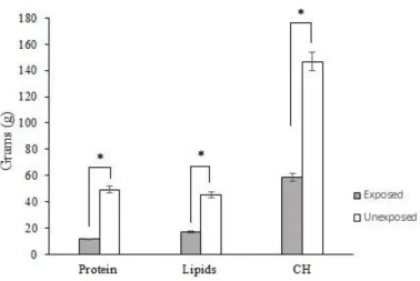
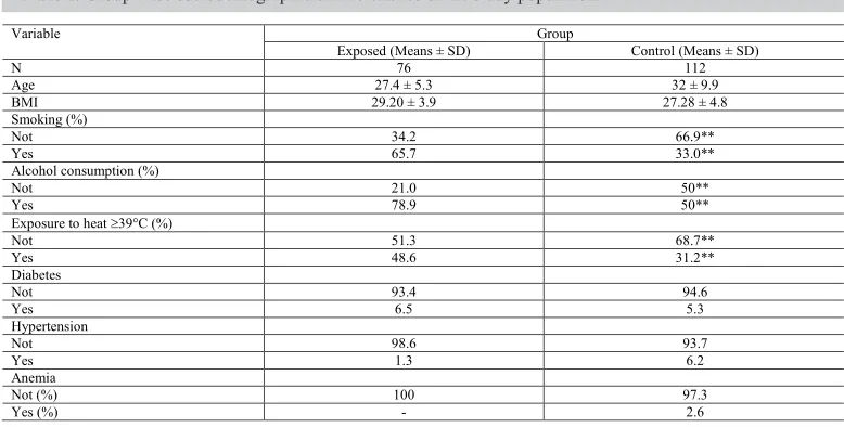
Analysis of sociodemographic variables showed that the groups varied only in lifestyle factors and not in the occurrence of chronic diseases. There was no statistical difference between the groups in terms of presence of diabetes, hypertension, and anemia, which might affect the health of participants. The sociodemographic and lifestyle characteristics of the participant population are shown in Table 1. The main lifestyle variables were associated with the development of chronic diseases and other conditions. Evaluation of the nutritional intake (grams) of protein, lipids, and carbohydrates (CH) between the evaluated groups showed that nutritional status was significantly different between the exposed group and the control group (Figure 1). The differences in the nutritional intake of protein, lipids, and CH between the groups may be due to socioeconomic differences as the studied area is classified as a rural area.
Genotoxicity analysis
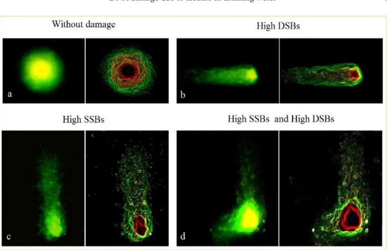
We analyzed the leukocytes of subjects exposed to 11.2-17.4 µg/L As in drinking water (exposed group) or 6.4-9.1 µg/L of As (control group) using the two-tailed comet assay. Fluorescence microscopic assessment of DNA fragmentation showed different types of damage. The observed comets were classified as mentioned in Materials and Methods. As shown in Figure 2, comets were stained with GelGreenTM (left-hand side images) and were contrasted with an electronic filter (Find EDGES) for assessing fragmentation (right-hand side images). Cells without DNA damage (Figure 2a), with high DSB (Figure 2b), high SSB (Figure 2c), and DSB and SSB damage in the same cell (Figure 2d) were observed. The result of the visual classification and enumeration of comets with DNA fragmentation is shown in Figure 3. In all categories, the fragmentation observed in the DNA of the exposed group was significantly and statistically different from that of the control group; however, comets registered a higher number of cells in the “high DSB” and “low SSB” categories in the exposed group compared to those in the control group (P < 0.05). Furthermore, the results show nonparametric distribution in the frequency of DSB and SSB in both groups. Figure 3 shows the number of damaged cells in the exposed group.
The DNA fragmentation index (Table 2), shows difference in the percentage of DSB between the two groups; however, there was no difference in SSB-DFI between the two groups. This suggests that exposure to As through drinking water is associated with lifestyle variables that increase the possibilities of DSB.
Correlation of biologically relevant variables with DSB and SSB damage were considered at P value < 0.05. Only the variable “exposure ≥39°C” was associated with SSB DNA fragmentation in the bivariate regression analysis (Table 3). The interactions of lifestyle variables with arsenic exposure were associated with increased DNA fragmentation, and all the variables analyzed were associated with DSB DNA fragmentation except for basal metabolic index (BMI).
Variable Group
Exposed (Means ± SD) Control (Means ± SD)
N 76 112
Age 27.4 ± 5.3 32 ± 9.9
BMI 29.20 ± 3.9 27.28 ± 4.8
Smoking (%)
Not 34.2 66.9**
Yes 65.7 33.0**
Alcohol consumption (%)
Not 21.0 50**
Yes 78.9 50**
Exposure to heat 39°C (%)
Not 51.3 68.7**
Yes 48.6 31.2**
Diabetes
Not 93.4 94.6
Yes 6.5 5.3
Hypertension
Not 98.6 93.7
Yes 1.3 6.2
Anemia
Not (%) 100 97.3
Yes (%) - 2.6
Table 1. Group-wise sociodemographic characteristics of the study population.
Figure 2. Representation of comets evaluated in different categories. a. Basal DNA structure in good condition; b. migration of DNA-DSB; c. SSB damage; d. DNA fragmentation in DSB and SSB in the same cell. Cells stained With GelGreenTM (right-side images) and with electronic filter (left-side images) at 100X magnification.
The association coefficient of the variable “exposure to As” showed interaction with lifestyle variables (P < 0.05) associated with the development of DSBs and SSBs. However, a positive association (P < 0.05) was observed when the exposure to As was adjusted with lifestyle variables that showed associations with the development of DSB fragmentation but not with SSB (Table 4).
Table 2. Fragmentation index of SSB and DSB.
DNA Damage (%) Exposed (Means ± SD) Control (Means ± SD)
DSB-DFI 32.5 ± 5.7 19.7 ± 4.7*
SSB-DFI 30.3 ± 6.1 31.7 ± 9.9
DSB-DFI = double strand breaks-DNA fragmentation index. SSB-DFI = single strand breaks-DNA fragmentation
index. *Statistically significant P < 0.05; t-test, 2 independent samples.
Table 3. r2 values of correlation and bivariate regression analysis of variables associated with DNA damage
in DSB and SSB.
Correlation analysis Bivariate regression analysis
DSB SSB DSB SSB
Variable r2 P r2 P Coef.. Std. Err. P Coef.. Std. Err. P
Exposed to As 0.77 <0.001 -0.08 <0.27 1.92 0.11 <0.001 -0.02 0.19 <0.27
Age -0.17 <0.01 0.05 <0.04 -0.16 0.06 <0.001 0.05 0.07 <0.46
BMI 0.08 <0.24 -0.01 <0.81 0.14 0.12 <0.24 -0.03 0.13 <0.8
Smoking 0.02 <0.001 0.01 <0.85 3.97 1.16 <0.001 0.23 1.26 <0.80
Alcohol consumption 0.32 <0.001 0.005 <0.94 5.47 1.15 <0.001 0.09 1.3 <0.93 Exposure to heat 39°C 0.11 <0.01 0.16 <0.01 2.96 1.20 <0.015 3.01 1.28 <0.02
Table 4. Multivariate regression analysis and its relationship with increased DNA fragmentation in DSB and SSB.
aMultivariate regression analysis adjusted for variables such as age, smoking, alcohol consumption, exposure to ≥39°C. Coef., coefficient; Std. error, standard error.
DSB SSB
Coef. Std. error P Coef. Std. error P
As exposure (crude) 1.92 0.11 <0.001 -0.021 0.19 <0.27
As exposurea (adjusted) 1.87 0.13 <0.001 -0.33 0.21 <0.123
Limit of detection of the TTC assay
DNA damage in the exposed group and control group indicated that the TTC test can detect DNA fragmentation in the aqueous arsenic range used in our study. The arsenic content in water consumed by both groups is above 1 µM, the arsenic concentration that has been reported to initiate DNA damage.
DISCUSSION
adenosine diphosphate poly-ribose polymerase-1 (PARP-1), thereby increasing SSB and DSB fragmentation (Qin et al., 2008). Sampling of peripheral blood via venipuncture is the best way to evaluate the genotoxic effects of arsenic in exposed population in a minimally invasive way (Basu et al., 2005; Palus et al., 2005; Vuyyuri et al., 2006; Banerjee et al., 2008; Jasso-Pineda et al., 2012). The time of exposure to arsenic is an important parameter for determination of genotoxic damage. Certain studies have demonstrated an association of exposure time to arsenic with DNA damage by evaluation of peripheral blood. This time was estimated to be 5 years in Bengal, (Basu et al., 2005), 18 years in Poland (Palus et al., 2005), 3 years in the study by Vuyyuri et al. (2006), 10 years in the study of Banerjee et al. (2008), and San Luis Potosi, Mexico (Jasso-Pineda et al. 2012) evaluated 6 years of exposition of arsenic. Basu et al. (2005) reported 59.74% ± 10.54 DNA damage and Palus et al. (2005) showed tail moment of 13.2 x 10⁻³ for DNA fragmentation. Vuyyuri et al. (2006) showed an increase of 14.95 ± 0.21 mm in the tail length of fragmented comets, whereas Banerjee et al. (2008) showed an Olive tail moment of 2.76 ± 1.39 of damaged DNA, and Jasso-Pineda et al. (2012) reported significant damage while evaluating tail moment of 5.2 ± 0.6, 3.5 ± 0.4, and 2.5 ± 0.4. Our results are similar to the published results regarding DNA fragmentation upon environmental exposure to arsenic. We observed 32.5 ± 5.7% DSB-DFI and 30.3 ± 6.1% SSB-DFI in our subjects.
The parameters used for evaluating DNA fragmentation vary among studies; however, the comet assay is commonly used for assessing DNA damage in populations that are environmentally exposed to arsenic. There are two types of comet assays: 1) the neutral comet assay (Olive et al., 1991), in which DNA migrates under neutral conditions, for identification of double-stranded DNA breaks (DSB), and 2) the alkaline comet assay, in which DNA is mobilized under alkaline conditions for DNA denaturation. The latter technique detects both SSB and DSB, without distinguishing between the two (Singh et al., 1988).
kinetics (Jackson and Bartek, 2009). Therefore, further research is required for determining the biological impact of SSBs and DSBs on the health of the exposed population. Here, we established TTC as a method for detecting DNA fragmentation upon exposure to arsenic in water in a concentration range of 6.4-9.1 µg/L.
In conclusion, a continuous analysis of the concentration of arsenic in water can reduce arsenic exposure, and TTC is a sensitive, reliable, and rapid methodology for determining the extent of genotoxic damage in exposed people. Frequent analysis of DNA fragmentation might promote lifestyle change and thereby alter the interaction of these variables with arsenic toxicity, which is associated with high levels of DSB. In summary, we showed that As is an environmental toxin associated with DNA damage in exposed people. We believe that our results would clarify the type of DNA fragmentation induced by As exposure through drinking water using the TTC technique. The limitations of this study are the inability to analyze the concentration of arsenic (or its metabolic by-products) in biological samples such as blood and urine for establishing a relationship between individual concentrations in the study subjects and their level of personal genotoxicity.
Conflicts of interest
The authors declare no conflict of interests.
ACKNOWLEDGMENTS
We thank Dr. G. Hankings of the West Virginia State University, for critical reading of the manuscript and CONACyT for grant #378398.
REFERENCES
Abernathy CO, Thomas DJ and Calderon RL (2003). Health effects and risk assessment of arsenic. J. Nutr. 133 (Suppl 1): 1536S-1538S.
Aitken RJ and Krausz C (2001). Oxidative stress, DNA damage and the Y chromosome. Reproduction 122: 497-506. http://dx.doi.org/10.1530/rep.0.1220497
Akter T, Jhohura FT, Akter F, Chowdhury TR, et al. (2016). Water Quality Index for measuring drinking water quality in rural Bangladesh: a cross-sectional study. J. Health Popul. Nutr. 35: 4. http://dx.doi.org/10.1186/s41043-016-0041-5 Banerjee M, Sarma N, Biswas R, Roy J, et al. (2008). DNA repair deficiency leads to susceptibility to develop
arsenic-induced premalignant skin lesions. Int. J. Cancer 123: 283-287. http://dx.doi.org/10.1002/ijc.23478
Bartolotta SA, Pacskowski MG, Hick A and Carballo MA (2011). Micronuclei assay in exfoliated buccal cells from individuals exposed to arsenic in Argentina. Arch. Environ. Contam. Toxicol. 61: 337-343. http://dx.doi.org/10.1007/ s00244-010-9607-1
Basu A, Som A, Ghoshal S, Mondal L, et al. (2005). Assessment of DNA damage in peripheral blood lymphocytes of individuals susceptible to arsenic induced toxicity in West Bengal, India. Toxicol. Lett. 159: 100-112. http://dx.doi. org/10.1016/j.toxlet.2005.05.001
Chanpiwat P, Himeno S and Sthiannopkao S (2015). Arsenic and other metals’ presence in biomarkers of Cambodians in arsenic contaminated areas. Int. J. Environ. Res. Public Health 12: 14285-14300. http://dx.doi.org/10.3390/ ijerph121114285
Chowdhury UK, Biswas BK, Chowdhury TR, Samanta G, et al. (2000). Groundwater arsenic contamination in Bangladesh and West Bengal, India. Environ. Health Perspect. 108: 393-397. http://dx.doi.org/10.1289/ehp.00108393
Collins TJ (2007). ImageJ for microscopy. Biotechniques 43 (Suppl): 25-30. http://dx.doi.org/10.2144/000112517 Dauphiné DC, Smith AH, Yuan Y, Balmes JR, et al. (2013). Case-control study of arsenic in drinking water and lung
Dong Z (2002). The molecular mechanisms of arsenic-induced cell transformation and apoptosis. Environ. Health Perspect. 110 (Suppl 5): 757-759. http://dx.doi.org/10.1289/ehp.02110s5757
Enciso M, Sarasa J, Agarwal A, Fernández JL, et al. (2009). A two-tailed Comet assay for assessing DNA damage in spermatozoa. Reprod. Biomed. Online 18: 609-616. http://dx.doi.org/10.1016/S1472-6483(10)60003-X
Faita F, Cori L, Bianchi F and Andreassi MG (2013). Arsenic-induced genotoxicity and genetic susceptibility to arsenic-related pathologies. Int. J. Environ. Res. Public Health 10: 1527-1546. http://dx.doi.org/10.3390/ijerph10041527 Gidron Y, Russ K, Tissarchondou H and Warner J (2006). The relation between psychological factors and DNA-damage:
a critical review. Biol. Psychol. 72: 291-304. http://dx.doi.org/10.1016/j.biopsycho.2005.11.011
Hartwig A, Pelzer A, Asmuss M and Bürkle A (2003). Very low concentrations of arsenite suppress poly(ADP-ribosyl) ation in mammalian cells. Int. J. Cancer 104: 1-6. http://dx.doi.org/10.1002/ijc.10911
Hoque MA and Butler AP (2015). Medical hydrogeology of Asian deltas: status of groundwater toxicants and nutrients, and implications for human health. Int. J. Environ. Res. Public Health 13: 81. http://dx.doi.org/10.3390/ijerph13010081 IARC - International Agency for Research on Cancer (2002). Some drinking-water disinfectants and contaminants,
including arsenic. In Monographs on the Evaluation of Carcinogenic Risks to Humans; Lyon, France, Vol. 84. Islam LN, Nabi AH, Rahman MM, Khan MA, et al. (2004). Association of clinical complications with nutritional status
and the prevalence of leukopenia among arsenic patients in Bangladesh. Int. J. Environ. Res. Public Health 1: 74-82. http://dx.doi.org/10.3390/ijerph2004020074
Jackson SP and Bartek J (2009). The DNA-damage response in human biology and disease. Nature 461: 1071-1078. http:// dx.doi.org/10.1038/nature08467
Jasso-Pineda Y, Díaz-Barriga F, Calderón J, Yáñez L, et al. (2012). DNA damage and decreased DNA repair in peripheral blood mononuclear cells in individuals exposed to arsenic and lead in a mining site. Biol. Trace Elem. Res. 146: 141-149. http://dx.doi.org/10.1007/s12011-011-9237-0
Khalequzzaman M, Faruque FS and Mitra AK (2005). Assessment of arsenic contamination of groundwater and health problems in Bangladesh. Int. J. Environ. Res. Public Health 2: 204-213. http://dx.doi.org/10.3390/ijerph2005020002 Kinniburgh DG and Kosmus W (2002). Arsenic contamination in groundwater: some analytical considerations. Talanta
58: 165-180. http://dx.doi.org/10.1016/S0039-9140(02)00265-5
Mostafa MG and Cherry N (2015). Arsenic in drinking water, transition cell cancer and chronic cystitis in rural Bangladesh.
Int. J. Environ. Res. Public Health 12: 13739-13749. http://dx.doi.org/10.3390/ijerph121113739
Murray MP and Sharmin R (2015). Groundwater arsenic and education attainment in Bangladesh. J. Health Popul. Nutr.
33: 20. http://dx.doi.org/10.1186/s41043-015-0029-6
Nava RC and Méndez AM (2011). Efectos neurotóxicos de metales pesados (cadmio, plomo, arsénico y talio). Arch. Neurocien. (Mex) 16: 140-147.
Norma Oficial Mexicana NOM-008-SSA3-2010 Para el tratamiento integral del sobrepeso y la obesidade, 1-8.
Norma Oficial Mexicana NOM-043-SSA2-2012 Servicios básicos de salud. Promoción y educación para la salud en
materia alimentaria, 1-79.
Norma Oficial Mexicana NOM-127-SSA1-1994 Salud ambiental, agua para uso y consumo humano-límites permisibles
de calidad y tratamientos a que debe someterse el agua para su potabilización, 1-8.
Olive PL, Wlodek D and Banáth JP (1991). DNA double-strand breaks measured in individual cells subjected to gel electrophoresis. Cancer Res. 51: 4671-4676.
Palus J, Lewinska D, Dziubaltowska E, Stepnik M, et al. (2005). DNA damage in leukocytes of workers occupationally exposed to arsenic in copper smelters. Environ. Mol. Mutagen. 46: 81-87. http://dx.doi.org/10.1002/em.20132 Qin XJ, Hudson LG, Liu W, Timmins GS, et al. (2008). Low concentration of arsenite exacerbates UVR-induced DNA
strand breaks by inhibiting PARP-1 activity. Toxicol. Appl. Pharmacol. 232: 41-50. http://dx.doi.org/10.1016/j. taap.2008.05.019
Ratnaike RN (2003). Acute and chronic arsenic toxicity. Postgrad. Med. J. 79: 391-396. http://dx.doi.org/10.1136/ pmj.79.933.391
Ribas-Maynou J, García-Peiró A, Abad C, Amengual MJ, et al. (2012). Alkaline and neutral Comet assay profiles of sperm
DNA damage in clinical groups. Hum. Reprod. 27: 652-658. http://dx.doi.org/10.1093/humrep/der461
Rossiello F, Herbig U, Longhese MP, Fumagalli M, et al. (2014). Irreparable telomeric DNA damage and persistent DDR signalling as a shared causative mechanism of cellular senescence and ageing. Curr. Opin. Genet. Dev. 26: 89-95. http://dx.doi.org/10.1016/j.gde.2014.06.009
Safarzadeh-Amiri A, Fowlie P, Kazi AI, Siraj S, et al. (2011). Validation of analysis of arsenic in water samples using Wagtech Digital Arsenator. Sci. Total Environ. 409: 2662-2667. http://dx.doi.org/10.1016/j.scitotenv.2011.03.016 Shibata T, Meng C, Umoren J and West H (2016). Risk assessment of arsenic in rice cereal and other dietary sources for
Singh NP, McCoy MT, Tice RR and Schneider EL (1988). A simple technique for quantitation of low levels of DNA damage in individual cells. Exp. Cell Res. 175: 184-191. http://dx.doi.org/10.1016/0014-4827(88)90265-0
Tian D, Ma H, Feng Z, Xia Y, et al. (2001). Analyses of micronuclei in exfoliated epithelial cells from individuals chronically exposed to arsenic via drinking water in inner Mongolia, China. J. Toxicol. Environ. Health A 64: 473-484. http://dx.doi.org/10.1080/152873901753215939
Vuyyuri SB, Ishaq M, Kuppala D, Grover P, et al. (2006). Evaluation of micronucleus frequencies and DNA damage in glass workers exposed to arsenic. Environ. Mol. Mutagen. 47: 562-570. http://dx.doi.org/10.1002/em.20229 WHO (2004) Guideline for drinking water quality, background document for development arsenic in drinking-water
Geneva: 1: 41.