*
Corresponding author: Farzaneh Ghanavati Behbahan, Plant Protection Dept., University of Zabol, Zabol,
herbmed.skums.ac.ir
INTRODUCTION
Over the recent decade, gold nanoparticles (NPs) have attracted much interest as a novel platform for various applications such as nanobiotechnology and biomedicine because of convenient surface bioconjugation with molecular probes and remarkable plasmon- resonance optical
properties. Moreover, Nanoparticles (NPs) have attracted great interest in their development as potential antibacterial drugs.1 It has been reported that biophysical interactions occur between NPs and bacteria including biosorption, NPs breakdown or aggregation, and cellular uptake with effects
Origin
al
ar
tic
le
Antimicrobial activities of Gold nanoparticles against
Salmonella
typhimurium
Farzaneh Ghanavati Behbahan1*, Mohammad Salari1, Seyedeh Razeyeh Mousavi2, Razeyeh Rezaei1
1
Plant Protection Dept., University of Zabol, Zabol, I.R. Iran; 2Horticulture and Landscape Dept., University of Zabol, Zabol, I.R. Iran.
Received: 12/Dec/2015 Accepted: 24/Apr/2016
ABSTRACT
Background and aims: One of the major problems in hospitals is resistant pathogenic bacteria to antimicrobial substances. The problem of increased costs of treatment failure and mortality rates is increasing.The aim of this study was to evaluate the antimicrobial activity of gold nanoparticles has been on Salmonella typhimurium.
Methods: This cross-sectional study was performed and Salmonella typhimurium
bacteria were isolated from poultry. Gold nanoparticles for business were purchased. Minimum inhibitory concentrations of gold nanoparticles in different concentrations by dilution in the wells were determined on bacteria. Susceptibility to several antibioticswas evaluated by Kirby-Bauer disk diffusion method.
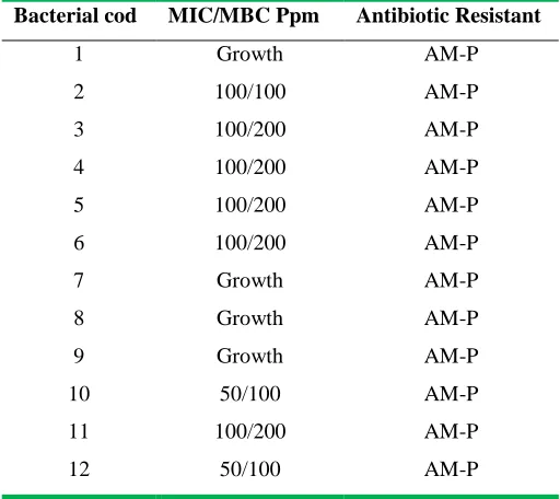
Results: The result of gold nanoparticles showed the highest MIC (the minimum inhibitory concentration) was 100 ppm concentration that 6 strains of them were inhibited by this concentration. The lowest MIC was 50 ppm concentration that 1 strain of
Salmonella was inhibited. The highest and lowest MBC value of extractwas 200 and 100 ppm, respectively.
Conclusions: The results showed that gold nanoparticles have good inhibitory effect on all studied bacteria.
including membrane damage and toxicity.2,3 The success of gold as catalyst is a consequence of the manipulation of this metal at the nanometric size, mainly stabilizing nanoparticles in different inorganic supports such as silica, alumina, zeolites. Use of gold in the killing of bacteria has been focused to some treatments of arthritis.4 Medical applications of gold include the use of sulphur-gold compounds as anti-inflammatory.5 The aim of the present study was to investigate antimicrobial activities of Gold nanoparticles against
Salmonella typhimurium.
METHODS
This current cross-sectional study was performed and all strains were isolated at different times during 2013-2014 from contaminated birds. Samples were diluted and/or homogenized in TSB medium, and isolates obtained by Salmonella selective enrichment in Rappaport-Vassiliadis (RV) medium after 24 h incubation at 43°C.
The susceptibility of all antibiotics was evaluated using disc diffusion method on Muller-Hinton agar as recommended by CLSI.6 Thefollowed procedures were briefly described here. Bacteria plates were grown overnight on blood agar. Nutrient agar and colony suspension was preparedequivalent to a 0.5×10⁷ McFarland Standard using the sterile saline water McFarland standard. Suspension (100 μl) was spread over the media plate and antibiotic disc was transferred aseptically on the surface of inoculated media plate. Isolated plates were tested with different antibiotics and their concentration was shown in parenthesis viz. ampicillin and penicillin.
The broth microdilution method was used to determine Minimum Inhibitory Concentration. All tests were performed in Mueller Hinton broth supplemented with Tween 80 at a final concentration of 0.5% (v/v). Briefly, serial doubling dilutions of the extract were prepared in a 96-well microtiter plate ranged from 6.25 ppm to 100 ppm. 10 μl of indicator and 10 μl of Mueller Hinton Broth were added to each well. Finally, 10 μl of bacterial suspension (106 CFU/ml) was added to each well to achieve a concentration of 104 CFU/ml. The plates were wrapped loosely with cling film to ensure that the bacteria did not get dehydrated. The plates were prepared in triplicates, and then they were placed in an incubator at 37°C for 18-24 hours. The color change was then assessed visually. The lowest concentration at which the color change occurred was taken as the MIC value. The average of 3 values was calculated providing the MIC value for the tested extract. The MIC was defined as the lowest concentration of the extract at which the microorganism did not demonstrate the visible growth. The microorganism growth was indicated by turbidity.
All experiments and measurement were repeated at least three times. Statistical analyses were performed using SPSS and Excel 2010 software. All experimental results were analyzed using mean descriptive statistics and correlation-coefficient. A value of P<0.05 was regarded as statistically significant.
RESULTS
concentration) was 100 ppm concentration that 6 strains of them were inhibited by this concentration. The lowest MIC was 50 ppm concentration that 1 strain of Salmonella
was inhibited. The highest and lowest Minimum Bacterial concentration (MBC) value of extract was 200 and 100 ppm, respectively (Table 1).
Table 1: Minimum inhibitory concentration and Minimum Bacterial concentration of gold nanoparticles against Salmonella typhimurium
Antibiotic Resistant MIC/MBC Ppm
Bacterial cod
AM-P Growth
1
AM-P 100/100
2
AM-P 100/200
3
AM-P 100/200
4
AM-P 100/200
5
AM-P 100/200
6
AM-P Growth
7
AM-P Growth
8
AM-P Growth
9
AM-P 50/100
10
AM-P 100/200
11
AM-P 50/100
12
DISCUSSION
The use of nanotechnology in medicine can fix this problem in the future to help mankind. Because nanoparticles, nanotechnology is the foundation, so their use in medicine, new prospects in the fight against pathogenic bacteria has opened.7,8
The study of Mubarak Ali showed that the leaf extract of menthol is very good bioreductant for the synthesis of silver and gold nanoparticles and synthesized nanoparticles active against clinically isolated human pathogens, Staphylococcus aureus and Escherichia coli.9 The study of Burygin, revealed that the antibiotic concentration in the wells decreased by twofold dilutions from 2.25 to 0.56 mg/mL.
It can be seen that the gentamicin–NP mixture retarded bacterial growth to a degree comparable to that demonstrated by the free antibiotic. When the free antibiotic and its mixture with NPs were diluted twofold, the diameter of the zone of culture-growth inhibition was reduced to the same extent in both cases. To obtain reliable statistical data, we ran five independent experiments, with three replicates per experiment.10 The study of Zhou showed the effects of gold and silver NPs on BCG and
applied in bacterial culture. The growth of E. coli was monitored through colony forming units (CFU). The mechanism of interaction between NPs and bacteria was analyzed through bacterial thin sections followed by TEM and scanning electron microscopy. Antibacterial effects on BCG were observed by recording fluorescent protein expression levels.11 The study of Lima detected the gold nanoparticles were supported onto clinoptilolite, mordenite and faujasite zeolites. Content of gold in materials varied between 2.3 and 2.8 wt%. The size, dispersion and roughness of gold nanoparticles were highly dependent on the zeolite support. The faujasite support was the support where the 5 nm nanoparticles were highly dispersed. The efficiency of gold-zeolites as bactericides of Escherichia coli and Salmonella typhi was determined by the zeolite support.12
The effectiveness of gold nanoparticles with and without stabilizing agents in the study of Prema was tested against both Gram positive and Gram negative bacteria such as
Staphylococcus aureus, Streptococcus
epidermis, Bacillus cereus, Vibrio vulnificus,
Pseudomonas aeruginosa, Escherichia coli, Flexibacter sp. and Klebsiella pneumoniae. Gold nanoparticles exhibited the highest antibacterial sensitivity to all the test strains used in this study. The maximum inhibition zone (30mm) was obtained against E. coli, K. pneumoniae and V. vulnificus for chitosan capped gold nanoparticles which are followed by CMC and starch.13
The study of Zawrah, TEM revealed small size of gold nanoparticles (range 9-19 nm) trapped by the biofilm released by
S. Typhimurium that easily attached to the
surface of cell membrane and drastically disturbed its proper function like respiration and permeability. Interaction between S. Typhimurium and ciprofloxacin coated with gold nanoparticles revealed that the cell wall was loosened and separated from the membrane or disrupted with complete absence of flagella.14
CONFLICT OF INTEREST
The authors declare no conflict of interest.
ACKNOWLEDGEMENT
We would like to thank all individuals who collaborated and helped us to complete this project.
REFERENCES
1. Sondi I, Salopek-Sondi B. Silver nanoparticles as antimicrobial agent: a case study on E. coli as a model for Gram-negative bacteria. J Colloid Interface Sci. 2004; 275(1): 177-82.
2. Priester JH, Stoimenov PK, Mielke RE, Webb SM, Ehrhardt C, Zhang JP, et al. Effects of soluble cadmium salts versus CdSe quantum dots on the growth of planktonic Pseudomonas aeruginosa. Environ Sci Technol. 2009; 43(7): 2589-94. 3. Brayner R, Ferrari-Iliou R, Brivois N, Djediat S, Benedetti MF, Fievet F. Toxicological impact studies based on Escherichia coli bacteria in ultrafine ZnO nanoparticles colloidal medium. Nano Lett. 2006; 6(4): 866-70.
How to cite the article: Ghanavati Behbahan F, Salari M, Mousavi SR, Rezaei R. Antimicrobial activities of Gold nanoparticles against Salmonella typhimurium. Adv Herb Med. 2016; 2(1): 26-30.
complexes. Met Based Drugs. 1994; 1(5-6): 395-404.
5. Wright V. Oral gold for rheumatoid arthritis. Br Med J. 1984; 289(6449): 858-9. 6. Clinical and Laboratory Standards Institute. Performance standards for antimicrobial susceptibility testing (2002). 16 th International supplement. CLSI; 1997. 7. Chen WY, Lin JY, Chen WJ, Luo L, Wei-Guang Diau E, Chen YC. Functional gold nanoclusters as antimicrobial agents for antibiotic-resistant bacteria. Nanomedicine. 2010; 5(5): 755-64.
8. Grace AN, Pandian K. Antibacterial efficacy of aminoglycosidic antibiotics protected gold nanoparticles: A brief study. Colloids Surf A Physicochem Eng Asp. 2007; 297(1): 63-70.
9. MubarakAli D, Thajuddin N, Jeganathan K, Gunasekaran M. Plant extract mediated synthesis of silver and gold nanoparticles and its antibacterial activity against clinically isolated pathogens. Colloids Surf B Biointerfaces. 2011; 85(2): 360-5.
10. Burygin GL, Khlebtsov BN, Shantrokha AN, Dykman LA, Bogatyrev VA, Khlebtsov NG. On the Enhanced Antibacterial Activity of Antibiotics Mixed with Gold Nanoparticles. Nanoscale Res Lett. 2009; 4(8): 794-801.
11. Zhou Y, Kong Y, Kundu S, Cirillo JD, Liang H. Antibacterial activities of gold and silver nanoparticles against Escherichia coli and bacillus Calmette-Guerin. J Nanobiotechnol. 2012; 10: 19.
12. Lima E, Guerra R, Lara V, Guzman A. Gold nanoparticles as efficient antimicrobial agents for Escherichia coli and Salmonella typhi. Chem Cent J. 2013; 7(1): 11.
13. Prema P, Thanyapanpiyan S. In-vitro antibacterial activity of gold nanoparticles capped with polysaccharide stabilizing agents. Int J Pharm Pharm Sci. 2013; 5(1): 310-314.