Reversible External Control of Gene Expression
in an NRl Knock-in Mouse
Mohammed A.K. Nassar B.Sc. (Hons)
A thesis presented for the degree of
Doctor of Philosophy in the
University of London
Wellcome Laboratory for Molecular Pharmacology
Department of Pharmacology
University College London
ProQuest Number: U641857
All rights reserved
INFORMATION TO ALL USERS
The quality of this reproduction is dependent upon the quality of the copy submitted.
In the unlikely event that the author did not send a complete manuscript and there are missing pages, these will be noted. Also, if material had to be removed,
a note will indicate the deletion.
uest.
ProQuest U641857
Published by ProQuest LLC(2015). Copyright of the Dissertation is held by the Author.
All rights reserved.
This work is protected against unauthorized copying under Title 17, United States Code. Microform Edition © ProQuest LLC.
ProQuest LLC
789 East Eisenhower Parkway P.O. Box 1346
ABSTRACT
The NMDA type of glutamate receptors plays a crucial role in the induction of long
term synaptic potentiation (LTP) which is believed to be one of the molecular basis
underlying learning and memory. I have generated a new mouse model for the study
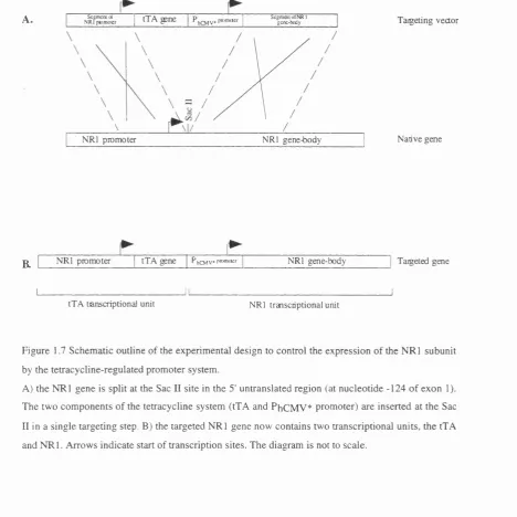
of the NMDA receptor function in vivo. This model should allow external (non invasive) reversible control over the native expression of the NRl gene, the subunit
common to all NMDA receptors. My strategy is based on modifying the N Rl gene
so that its expression is mediated through the tetracycline-regulated promoter
system. This new strategy replaces the NRl promoter with the PhCMV* promoter and
uses the N R l promoter to drive the expression of the tetracycline-sensitive transactivator (tTA).
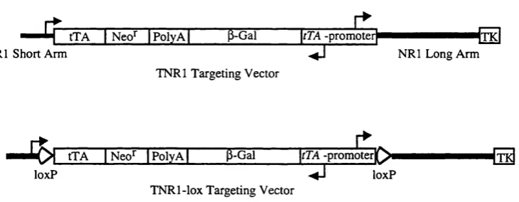
The N R l gene was modified in the E14.1 embryonic stem (ES) cell line by
homologous recombination with a single targeting vector. A -22 kb targeting vector was designed and cloned to achieve a tetracycline regulatable expression of NRl and hence is termed TN Rl. Eleven chimeric mice have been produced from 4 of the
TNRl-targeted ES clones. One chimera transmitted the ES genome to 7 offspring of
which 2 harbour the targeted NRl allele.
Furthermore, a variant of the model was generated where the inserted elements of the tetracycline-regulated promoter system can be deleted in tissues expressing Cre,
a site-specific recombinase. As a result, the expression of NRl gene retains a native tetracycline-independent pattern in Cre expressing tissues. This should allow the
control of expression of N R l in all tissues but those where the loss of NMDA
receptor activity causes fatality. A second targeting vector identical to TN Rl but
contains two loxP sites, the site for Cre excision, was cloned and is termed TN Rl- lox. Nine chimeric mice were obtained from 3 TNR1 -lox-targeted ES clones. Two
chimeras transmitted the ES genome to 5 offspring of which non harbour the targeted N Rl allele. Breeding of those chimeras is continued to obtained offspring
CONTENTS
ACKNOWLEDGEMENTS. 5
DEDICATION. 5
LIST OF FIGURES AND TABLES. 6
INTRODUCTION. 9
1.1 Classification of glutamate receptors 11
1.2 The intriguing properties of NMDA receptor 11
1.3 Physiological significance of NMDA receptor 12
1.4 Molecular diversity of the NMDA receptor 14
1.5 Expression of NMDA receptor subunits mRNAs 18
1.6 Functional heterogeneity of NMDA receptor 20
1.7 Transmembrane topology of NMDA receptor subunits 22
1.8 Molecular determinants of Ca^+ permeability and Mg^+ block 24
1.9 Agonists binding sites 24
1.10 Studies of NMDA receptor in vivo 24
1.11 Gene targeting 26
1.12 Gene knockout 29
1.13 Limitations of the gene knockout approach 33
1.14 Cre-mediated knockout 34
1.15 Gene regulation by inducible promoter systems 37
1.16 The tetracycline-regulated promoter system 38
MATERIALS AND METHODS. 50
2.1 Materials 51
2.2 Methods 54
A. Molecular biology methods 54
B. Embryonic stem cell culture 62
C. Transgenic methods 67
RESULTS. 71
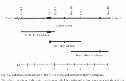
3.1 Subcloning and mapping of mouse genomic N Rl sequences. 73
3.2 Cloning of the ES cell targeting and the PCR-test vectors. 77
3.3 Designing primers and optimisation of the screening PCR. 86
3.4 Transfection of the TNRl and TNRl-lox targeting vectors into
ES cells. 90
3.5 Confirmation of PCR-positive ES clones through Southern
blots. 94
3.6 Generation of chimeric mice from confirmed ES clones. 107
3.7 Breeding of chimeras. I l l
DISCUSSION. 115
4.1 A new NMDA mouse model 116
4.2 A Strategy for external control of expression of N Rl 117
4.3 Suitability of the tetracycline-regulated promoter system 119
4.4 Discussion of results 120
4.5 Future work 122
ACKNOWLEDGEMENTS
I would like to thank the Wellcome Trust for funding this work.
Many thanks to Dr. Ralf Scheopfer and Prof. David Colquhoun, my supervisors, for
three years of advise and support.
I am also in debt to Matthias Kneussel, Alexander Annala and Philip Chen for their
advice and help throughout the project.
Special thanks to Tony Langford at the LMP and the staff of UCL Biological
Services for their patience and sincere help in dealing with my never ending "urgent"
requests.
DEDICATION
LISTS OF FIGURES AND TABLES
FIGURESINTRODUCTION
1.1 Schematic outline of the structure of the eight splice variants of
the N R l subunit. 18
1.2 Schematic representation of the transmembrane topology model
of glutamate receptors subunits. 23
1.3 Knockout of a gene of interest gene through homologous
recombination with a targeting vector. 27
1.4. Cre mediated deletion and inversion of loxP flanked DNA. 35 1.5. Schematic representation of the tetracycline-regulated
promoter system. 40
1.6. Schematic representation of the rtTA version of the
tetracycline-regulated promoter system. 43
1.7 Schematic outline of the experimental design to control the
expression of the N Rl subunit by the tetracycline-regulated
promoter system. 47
1.8 Co-regulation of the p-galactosidase reporter gene and N R l
subunit by the bi-directional PhCMV* promoter. 48
1.9. Cre mediated deletion of the components of the tetracycline
system restores direct control of NRl promoter over expression of
NRl subunit. 49
RESULTS
3.1. Southern Blot analysis of a 129SvJ-mouse genomic ^-library
clone (No. 36.1) showing signals for oligonucleotide probes to
exon 4. 74
3.3. The position of cutting sites for 17 restriction enzymes mapped
in the genomic NRl sequences covered by the three subclones. 76
3.4. Schematic diagram of the TNRl and TN Rl-lox targeting
vectors outlining their components. 78
3.5. Dividing the components of each targeting vector between
three intermediate constructs. 80
3.6A-B. Assembly of the TN Rl targeting vector from its
intermediate constructs in three steps. 82
3.6C. Assembly of the TNRl targeting vector from its intermediate
constructs in three steps. 83
3.7. Cloning PCR-test 2 vector. 85
3.8 The product of PCR performed on various amounts of PCR-test
2 vector DNA as template. 87
3.9 The product of PCR performed on serial dilution of an ES
clone transfected with PCR-test 2 vector. 89
3.10. Modification of an N Rl allele as a result of homologous
recombination between the native NRl allele and a targeting
vector. 91
3.11 The PCR screen of pools of transfected ES cells with TN R
l-lox vector. 92
3.12. Confirmation of correct homologous recombination in
targeted ES cells by Southern blot analysis. 95
3.13. Southern blot of PCR positive TN Rl-ES clones digested with
EcoRV-Hindm and labelled with probe A. 97
3.14. Southern blot of PCR positive TNRl-lox-ES clones digested
with EcoRV-Hindin and labelled with probe A. 98
3.15. Southern blot of PCR positive ES clones digested with
3.16. Southern blot of PCR positive ES clones digested with
H indni and labelled with probe B 102
3.17. Southern blot of PCR positive ES clones digested with Nde I
and labelled with probe E. 104
3.18. Southern blot of PCR positive ES clones digested with Nde I
and labelled with probe D. 106
3.19. Chimera C/96-010 obtained from ES clone TNRl 6. 110
3.20. Testing for germ-line transmission of the targeted N Rl allele
by mating chimeras with C57BL/6 mice. 112
3.21. Southern blot of EcoRV digest of genomic DNA isolated
from tail biopsies labelled with probe A. 114
TABLES RESULTS
3.1. PCR screen of transfected ES colonies with TN Rl targeting
vector for homologous recombination. 92
3.2. PCR screen of transfected ES colonies with T N R l-lox
targeting vector for homologous recombination. 92
3.3. Results of injection of ES clones into host blastocysts to
generate chimeric mice. 108
3.4. List of sex and serial number of obtained chimeras. 109
3.5 Results of screening agouti FI from chimeras C/96-017,
C/97-022 and C/97-026 with Southern blots for the transmission of the
INTRODUCTION
OverviewThis thesis describes the generation of a transgenic mouse in which the formation of
the NMDA receptor is controlled by external non-invasive means. This mouse serves
as a new model for the study of the function of the NMDA receptor in vivo.
I begin by a brief introduction of the NMDA receptor discussing its physiological
significance, molecular and functional diversity. Published investigations of the
function of the NMDA receptor in vivo is based on observing the phenotype resulting from the loss of NMDA receptor activity. These studies used strategies that
targeted the NMDA receptor at two levels, the protein level by using NMDA
receptor antagonists and the DNA level by knockout of N R l, the common subunit to
all NMDA receptors. I discuss the limitations of using NMDA receptor antagonists
for in vivo studies. This is followed by a brief description of the principles of gene targeting, the method used to generate knockout mice. Then I discuss the results of
knockouts studies of the cloned NMDA receptor subunits. There are significant
limitations to the knockout approach and in the light of these limitations the use of
the CxdloxP recombinase system to generate a cell- or tissue-specific NMDA knockout is described.
As discussed later the use of the tetracycline-regulated promoter system to
genetically control the formation of the NMDA receptor circumvents the limitations
of the conventional and CidloxP mediated knockout approaches. The aim of thesis is to generate a new mouse model where the formation of the NMDA receptor is
reversibly controlled by external non-invasive means. Also, the external control can
be refined to exclude a particular cell or tissue type. Finally I detail my experimental
design to control the N Rl subunit expression by the tetracycline-regulated promoter
1.1 Classification of glutamate receptors
Glutamate activates ionotropic and metabotropic receptors in the vertebrate nervous
system. The ionotropic receptors constitute the major fast component of excitatory
synaptic transmission in the CNS. Ionotropic glutamate receptors have been
classified into three families (Watkins et a l, 1981) based on their pharmacology to the agonists a-amino-3-hydroxy-5-methyliosxazolepropoinic acid (AMPA), kainic
acid and N-methyl-D-aspartic acid (NMDA), and the antagonists 2-amino-5-
phosphonopentanoic acid (AP5) (Davies et a l , 1981) and 6-cyano-7- nitroquinoxaline-2,3-dione (CNQX). NMDA-activated glutamate receptors are
sensitive to block by AP5 but not CNQX. In contrast, kainate- and AMPA-activated
glutamate receptors are sensitive to block by CNQX but not AP5. Kainate and
AMPA receptors are collectively referred to as non-NMDA.
1.2 The intriguing properties of NMDA receptor
NMDA receptor channels differ in fundamental ways from the non-NMDA and
other ligand-gated ion channels. NMDA receptor channels are gated in a unique
manner both by ligand and voltage. NMDA receptor channels require glycine as a
co-agonist in addition to glutamate (or NMDA) (Kleckner and Dingledine, 1988).
The voltage dependence is due to block of the ion channel by Mg^+ at normal resting
potentials (McBain and Mayer, 1994; Nowak et a l, 1984). The NMDA receptor channel is highly permeable to Ca^+ (Ascher and Nowak, 1986; MacDermott et a l ,
1986). Furthermore, excitatory postsynaptic currents mediated by NMDA receptor
channels are characterised by a prolonged time course relative to that mediated by
the non-NMDA receptor channels (Dale and Roberts, 1985). In addition, the
function of the NMDA receptor channel is modulated by many endogenous
compounds and second messenger systems (for a review see Collingridge and
1.3 Physiological significance of NMDA receptor
NMDA receptors are considered to play an important role in activity-dependent
synaptic plasticity. Due to the voltage-dependent block by Mg^+, NMDA receptor
channels are activated only when the presence of glutamate in a synapse is paired
temporally with depolarisation in the postsynaptic cell. The postsynaptic
depolarisation alleviates the Mg^+ block. Thus the NMDA receptor acts as a co
incidence detector of pre- and postsynaptic depolarisations. The activated NMDA
receptor channel is permeable to Ca^+ (Ascher and Nowak, 1986; MacDermott et a l, 1986) which triggers biochemical processes that result in enhancement of synaptic
efficacy.
1.3.1 Developmental plasticitv
A variety of studies have implicated NMDA receptor activation in the experience-
dependent rewiring of the brain that occurs during development (McDonald and
Johnston, 1990). In the developing visual system, chronic infusion of AP5 into kitten
visual cortex blocks the physiologically determined ocular dominance shift normally
associated with monocular deprivation (Kleinschmidt et a i , 1987). In Xenopus laevis, NMDA plays a role in the experience dependent establishment of matching binocular tectal maps (Scherer and Udin, 1989; Scherer and Udin, 1991) and the eye-
specific segregation of retinotectal projections in surgically produced 3-eyed
tadpoles (Cline et a i, 1987).
In the cerebellum, in vivo application of AP5 prevents the regression of supernumerary climbing fibre synapses in Purkinje cells (Rabacchi et a l , 1992) It was reported that NMDA receptor antagonists reduce granule cell migration
(Komuro and Rakic, 1993).
In the superior colliculus, application of NMDA receptor antagonists interferes with
postnatal weeks (Simon et a l , 1992). NMDA receptor activation may also play a role in early postnatal olfactory learning in rat (Lincoln et al, 1988).
Mice lacking NMDA receptor activity die shortly after birth indicating an important
role in development (Forrest et a l, 1994; Li et a l, 1994). Furthermore, mutant mice fail to refine the whisker-related neural pattern in the brainstem trigeminal complex
(Forrest et a l , 1994; Li et a l, 1994). This suggests the involvement of the NMDA receptor in the refinement of periphery-related neural patterns in the mammalian
brain (Li et a l, 1994).
1.3.2 Svnaptic plasticitv and learning
The prerequisite postsynaptic depolarisation necessary for the alleviation of the
block by Mg^+ enables the NMDA receptor to act as coincidence detector of pre-
and postsynaptic depolarisations. In experimental setups, tetanic stimulation
provides a greater and longer-lasting postsynaptic depolarisation which alleviates the
block by Mg^+ of NMDA receptors. Tetanic stimulation in the hippocampus induces
long-term potentiation (LTP), a persistent and dominant form of activity-dependent
synaptic enhancement of synaptic efficacy (Bliss and Collingridge, 1993).
Hippocampal LTP includes NMDA receptor-dependent (Schaffer collaterals-CAl
synapse) and NMDA-independent (Mossy fibre-CA3 synapse) forms (Bliss and
Collingridge, 1993). Hippocampal LTP is believed to provide a physiological model
for one of the molecular mechanisms underlying learning and memory (Bliss and
Collingridge, 1993).
Adm inistration of AP5 reversibly prevented the induction of LTP in the
hippocampal CA l layer (Collingridge et a l, 1983). Chronic intraventricular infusion of AP5 resulted in impairment of both hippocampal LTP and spatial learning in rat
1.3.3 Neuronal cell death
Glutamate is implicated in acute neuronal cell death as a result of various types of
insults to the nervous system, including anoxia, hypoglycaemia, seizure and
mechanical trauma (Choi, 1988; Choi, 1990; Meldrum and Garthwaite, 1990).
Damage occurs through excessive activation of NMDA receptors, triggering
mechanisms leading to neuronal cell death. Excessive activation of NMDA receptors
may also be involved in chronic neurodegenerative diseases such as Alzheimer's and
Huntingdon's (Meldrum and Garthwaite, 1990).
In accord with this, antisense oligonucleotides to the NRl subunit of the NMDA
receptor prevent the neurotoxicity elicited by NMDA in vitro and reduce the volume of focal ischaemic infarction caused by occlusion of the middle cerebral artery in rat
(Wahlestedt et a i, 1993).
1.4 Molecular diversity of the NMDA receptor
Molecular cloning studies identified multiple cDNAs coding for NMDA receptor
channel subunits. These cDNAs were classified into two gene families, NMDARl
(NRl) and NMDAR2 (NR2). Highly active NMDA receptor channels are formed in vitro by co-expression of members of the N Rl and NR2 families. The cloned NMDA receptor subunits are characterised by a distinct distribution pattern,
functional properties and regulation.
1.4.1 Cloning of the N Rl subunit
The first cDNA of an NMDA receptor subunit (NMDARl) was isolated from rat
forebrain poly(A)+ RNA by expression cloning in Xenopus oocytes (Moriyoshi et a l , 1991; Nakanishi et a l, 1992). The largest open reading frame in the isolated clone predicted a 938 amino acids polypeptide that has about 20% sequence identity
currents (-10 nA) in the Xenopus oocytes expression system. These currents have electrophysiological and pharmacological properties characteristic of the NMDA
receptor (Moriyoshi et a l, 1991).
The mouse homologue to NRl was independently isolated from a forebrain cDNA
library using the mouse GluRA and GluRB non-NMDA receptor subunits cDNAs as
probes under low-stringency conditions (Yamazaki et a l, 1992). The isolated mouse clone, designated as Çl, codes for a polypeptide with >99% amino acid identity to
that of N Rl with only 2 out of 938 amino acid substitutions (Yamazaki et a l, 1992). The human homologue to N R l, designated as hNM DARl, was isolated using a
probe from N R l (Karp et a l, 1993). The predicted amino acid sequence from the human cDNA has 99% amino acid identity to rat sequence with 3 amino acids
substitutions in the signal peptide region and 4 amino acids substitutions in the N-
terminal region (Karp et a l, 1993). The Drosophila melanogaster homologue to N R l, named DNMDARl, shows 46% amino acid identity to N R l (Ultsch et al,
1993).
1.4.2 Cloning of the NR2 subunits
To date, four members of the NMDAR2 (NR2) family of NMDA receptor subunits,
designated as NR2A-D for rat or 81-4 for mouse, were identified by molecular
cloning. The NR2A-D, e l and e4 subunits were cloned through PCR mediated
amplification of brain cDNA using degenerate primers to conserved amino acid
sequences between ionotropic glutamate receptor subunits (Ikeda et a l , 1992; Ishii et a l , 1993; Meguro et a l , 1992; Monyer et a l , 1992). Whereas the e2 and e3 subunits were cloned by probes generated from e l (Ikeda et a l, 1992).
The amino acid identity between the NR2 subunits and other ionotropic glutamate
receptor subunits is only 11-18%. A large C-terminal region is a prominent feature
of NR2 subunits which distinguishes them from other ionotropic glutamate subunits
overall amino acid identity among NR2 subunits is about 50%. The identity is
highest around the putative transmembrane domains (80-90%), moderate in the N-
terminal region (45-60%) and lowest in the C-terminal region (20-30%)(Ikeda et a l, t992; Ishii et a i , 1993). In general, the size and amino acid sequences more closely resemble each other between NR2A and NR2B than between NR2C and NR2D
(Ishii et a l, 1993).
Expression of one or two members of the NR2 family in the X enopus oocyte expression system does not result in functional NMDA receptor channels. However,
co-expression of NRl and an NR2 subunit resulted in NMDA-activated currents that
are 100 times larger than those obtained from homomeric expression of N R l (Ishii et a l, 1993; Meguro e t a l , 1992; Monyer era/., 1992).
1.4.3 Splice variants of NMDA receptor subunits
Shortly after cloning the first N R l cDNA and as work to identify other NMDA
cDNAs was underway, several laboratories reported cloning of a family of N Rl
splice variants. NR2A-C subunits do not have any splice variants whereas NR2D
exists in two isoforms (Ishii et a l, 1993).
Seven functional isoforms of the cloned N Rl subunit generated by different splicing
were isolated independently in different laboratories from rat (Durand et a l, 1992; Hollmann et a l , 1993; Nakanishi, 1992; Sugihara et a l, 1992) and mouse (Yamazaki et a l , 1992). These isoforms were generated by an insertion at the N- terminal region, deletions at two different C-terminal regions, or combinations of the
insertion and deletions.
Hollmann et al (1993) analysed the structure of genomic rat NRl gene and mapped the intron-exon boundaries. His analysis revealed that the N-terminal variant is
generated by alternative use of exon 5 (63 bp long, after nucleotide 516).
Furthermore, three different deletions in the C-terminal region of N R l arise from
The first deletion is generated by alternative use of exon 21 (111 bp long, nucleotide
2536-2646). The second deletion is generated by alternative use of a splice-acceptor
site in exon 22 resulting in a 356 bp deletion (nucleotide 2647-3002). The second
deletion removes the original stop codon of N R l, leading to the use of a second
previously out-of-frame stop codon occurring at nucleotide 3068 in the 3'
untranslated region. This switch to an alternative stop codon converts 66 bp of non
coding sequence of N Rl to coding sequence. The third deletion is a combination of
the first and second deletions (467 bp, nucleotides 2536-3002). All nucleotide
positions are numbered starting from the first nucleotide of the first codon of the
mature protein (Hollmann et a l, 1993).
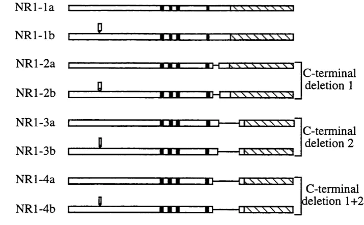
Several nomenclature systems were suggested to describe the N R l splice variants
but that of Hollmann et al (1993) is widely accepted. The size of the C-terminal deletion is described by a number (from 1-4). The N R l splice variant with no
deletions has the number 1 (NRl-1), splice variants with the first deletion has the
number 2 (NRl-2), splice variant with the second deletion has the number 3 (NR 1-3)
and splice variant with the third deletion has the number 4 (NR 1-4). The absence or
presence of the N-terminal insertion in each of the C-terminal variants is denoted by
the letter a and b, respectively. The stmcture of the eight splice variants of the N R l
subunit are schematically outlined in figure 1.1. The human homologue for N R l-la ,
N R l-2a and N R l-4a have been isolated (Foldes et a l, 1993). Also, the isolation of two human splice variants containing the N-terminal insert, N R l-3b and N R l-4b,
N R l-la
N R l-lb
N Rl-2a
NRl-2b
N R l-3a
N Rl-3b
N R l-4a
N Rl-4b
j J 1 ,
r r r
■
----“n ■
XXE XTT TTT TXT
Tnrr
C-terminal deletion 1C-terminal deletion 2
I W W W I
C-terminal deletion 1+2
Figure 1.1 Schematic outline o f the structure of the eight splice variants of the N R l subunit.
The structures shown start as in the mature protein. N R l-la is equivalent to the N R l of (Moriyoshi et
a i , 1991). Black boxes represent the four membrane domains. Stippled bars represent the 3'
untranslated region o f the cDNA clone. The presence of the N-terminal insertion (exon 5) is indicated
by an open box in N R l-#b variants. The different terminal deletions are indicated by a line.
C-terminal deletion 1 involves exon 21. C-C-terminal deletion 2 involves exon 22 and an alternative stop
codon in the 3' untranslated region.
1.5 Expression of NMDA receptor subunits mRNAs
1.5.1 Expression in the brain
The N Rl subunit is ubiquitously expressed in the brain throughout development.
Only the NR2B and NR2D subunit mRNAs are expressed in the embryonic brain.
NR2B is distributed widely, in contrast to NR2D subunit mRNA which is
predominately found in the diencephalon and the brainstem. Transcripts encoding
the NR2A and NR2C are not detectable during the entire embryonic period (Monyer
During the first two weeks after birth, the expression of the NR2 subunits changes
drastically. The NR2A subunit mRNA starts to appear in the whole brain with the
highest level of expression occurring in the cerebral cortex, the hippocampal
formation and the cerebellar granule cells (Wenzel et a l, 1997). The NR2B subunit mRNA becomes restricted to the forebrain with high levels of expression observed
in the cerebral cortex, the hippocampal formation, the septum, the caudate-putamen,
the olfactory bulb and the thalamus. The NR2D subunit mRNA expression is greatly
decreased with low levels found in the thalamus, the brainstem, the olfactory bulb
and non-pyramidal neurones in the hippocampus (Monyer et a l, 1994). The NR2C subunit mRNA appears strongly in the cerebellum with weak expression detected in
the thalamus, the olfactory bulb and non-pyramidal neurones in the hippocampus
(Monyer et a l, 1994; Watanabe et a l, 1994d; Zhong et a l, 1995).
1.5.2 Expression in the spinal cord
At embryonic stages. The N R l, NR2B and NR2D subunit mRNAs are expressed
widely in the spinal cord. In addition, restricted expression of the NR2A subunit
mRNA is reported in the developing ventral horn (Watanabe et a l, 1994c). Reports on the expression patterns in the adult spinal cord are controversial. The N R l, NR2A
and NR2B subunit mRNAs are the predominant transcripts in the cervical cord
(Watanabe et a l, 1994c). Whereas the N R l, NR2C and the NR2D subunit mRNAs are found in the rat lumbar cord (Tolle et al, 1993).
1.5.3 Expression of the splice variants
The distribution of N R l splice variants shows regional and developmental
heterogeneity (Laurie and Seeburg, 1994; Luque et a l, 1994; Zhong et a l, 1995; Zukin and Bennett, 1995). The relative abundance of the variants is about 67% for
N R l-la , 18% for variants with the COOH deletions but without the NH2 insertion
(2a, 3a and 4a) and 15% for variants with the NH2 insertion (lb , 2b, 3b and 4b)
1.6 Functional heterogeneity of NMDA receptor
Functional NMDA receptors from homomeric expression of N Rl subunit has only
been observed in the Xenopus oocyte expression system (Moriyoshi et aL, 1991). Homomeric expression of the N Rl subunit does not yield functional NMDA
receptor channels in mammalian cell lines (Monyer et a l, 1992; Sucher et a l , 1993). Active NMDA receptor channels are only produced when the N Rl subunit is
expressed together with at least one of the four NR2 subunits. In accord with this,
most brain regions express both N Rl and NR2 subunits mRNAs. No functional
NMDA receptor channels have been reported for mature cerebellar Purkinje cells
which express the NRl subunit mRNA but none of the NR2 subunits (Monyer et a i, 1994; Watanabe e t a l , 1993; Watanabe e t a i , 1994c).
Heteromeric assembly of the NMDA receptor channel subunits is also suggested
from biochemical and immunoprécipitation studies (Chazot and Stephenson, 1997;
Luo et a l , 1997; Sheng et a i , 1994; Tingley et a i , 1993). Anti-NRl subunit antibodies immunoprecipitated not only a =120 kDa protein (N R l) but also
significant amounts of the NR2A and NR2B subunits from the rat cerebral cortex. In
addition, the anti-NR2A subunit efficiently co-immunoprecipitated the NR2B
subunit and conversely, the anti-NR2B subunit antibodies co-immunoprecipitated
the NR2A subunit.
Each of the NR2 subunits confer variable electrophysiological and pharmacological
properties upon the NMDA receptor channel. These include offset decay time
constant, the affinities for agonists, sensitivity for competitive and non-competitive
antagonists (Ishii et a i, 1993; Kutsuwada e t a i , 1992; McBain and Mayer, 1994; Monyer et a i, 1992). The functional diversity of native NMDA receptors channels, therefore, is determined by the differential association of the N Rl subunit with the
NR2 subunits.
The Ca^+ permeability of the four NR1-NR2 subunit configurations are comparable
block by extracellular Mg^+ is observed for different NR1-NR2 subunit
configurations (Kuner and Schoepfer, 1996). At physiological concentrations of 1
mM extracellular Mg^+ the NR2A and NR2B channels are characterised by a
stronger sensitivity to Mg^+ block than NR2C and NR2D channels (Kuner and
Schoepfer, 1996; Monyer gr a/., 1992).
The excitatory postsynaptic potential mediated by NMDA receptor channels displays
a prolonged time course relative to that mediated by non-NMDA receptor channels
(Dale and Roberts, 1985). The offset decay time constant of recombinant N R l-
NR2A channels (118 ms) is approximately 3-4 times faster than that of either N R l-
NR2B (400 ms) or NR1-NR2C (382 ms) channels. NR1-NR2D channels are
characterised by a particularly long offset decay time constant of 4.8 s, which is 10-
40 fold longer than that for the other subunit combination (Monyer et a l, 1994).
Differential RNA splicing of the N R l subunits alters the physiological and
pharmacological properties of NMDA receptor channels (Zukin and Bennett, 1995).
Recombinant NMDA receptors containing NRl variants lacking exon 5 (N-terminal
insertion) show marked potentiation by poly amines (Durand et a l, 1993; Durand et a l, 1992). Whereas recombinant NMDA receptors containing N Rl variants lacking exon 5 show high sensitivity to inhibition by protons (IC5 0 40 nM, pH 7.4)
(Traynelis et a l, 1995). Recombinant NMDA receptors containing N R l variants lacking exon 22 (the second deletion) showed a marked potentiation by protein
kinase C (PKC) (Durand et al, 1993; Durand et a l, 1992). It has been suggested that NMDA receptor complexes can contain more than one N Rl splice variant (Blahos
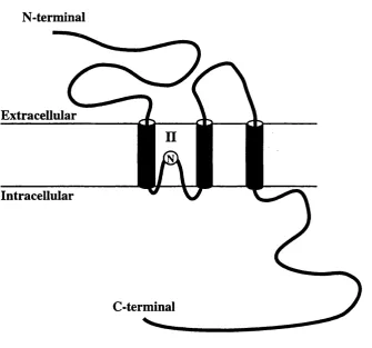
1.7 Transmembrane topology of NMDA receptor subunits
Cloned glutamate receptor channel subunits posses four hydrophobic segments that
were initially believed to form four transmembrane domains homologous to other
ligand-gated ion channels (Moriyoshi et a l, 1991). However, phosphopeptide analysis suggested that the C-terminal region of the N Rl subunit is phosphorylated
in vivo (Tingley et al, 1993). In addition, the C-terminal region of the NR2A subunit is responsible for the activation of the NR1/NR2A channel by a protein kinase C
activator, 12-0-tetradecanyoly-phorbol 13-acetate (TPA) (Mori gf a l, 1993). These findings are in contradiction to the initial predication since they suggest an
extracellular rather than intracellular location of the C-terminal region.
Furthermore, mutational analysis of the glycine-binding and redox modulation sites
of the N R l subunit suggests that the region between segments 3 and 4 exist
extracellularly (Kuryatov et a l , 1994; Sullivan et a l, 1994). It was found that the exchange of the first 150 amino acids upstream of the M l and the putative
extracellular M3-M4 loop were required to switch agonist selectivity between the
AMPA and kainate receptor (Stern Bach et a l, 1994). In addition, site-directed mutagenesis has identified amino acid residues found on NR2 subunits that control
glutamate potency in recombinant NMDA receptors (Anson et a l, 1998; Laube et a l , 1997). The identified residues are located in the NR2 subunit at positions homologous to those on the NRl subunit that control glycine potency.
These findings can be explained by a three transmembrane model as proposed for
kainate binding proteins and the GluRl subunit based on the analysis of engineered
N-glycosylation sites (Hollmann et a l, 1993; Wo et a l , 1995; Wo and Oswald, 1994; Wo and Oswald, 1995), in which the putative channel-lining segment M2,
loops into the membrane without traversing it.
This model was further supported by recent work where the residues contributing to
the channel pore were mapped by the cysteine-substitution method. The amino acid
individually substituted with a cystein residue. Residues contributing to the channel
pore were then identified by their ability to interact with sulfhydry 1-specific reagents.
This work suggested that the M2 segment forms a channel-lining loop originating
and ending on the cytoplasmic side of the channel (Kuner et a l, 1996). The current model of transmembrane topology of glutamate receptor subunits is schematically
represented in figure 1.2.
N-terminal
Extracellular
Intracellular
C-terminal
Figure 1.2 Schematic representation o f the transmembrane topology model o f glutamate receptors
subunits. The three transmembrane domains are represented by filled cylinders. The membrane
region 2 forms a loop originating and ending on the cytoplasmic side. The position o f the asparagine
(N) crucial for Ca^'*' permeability and sensitivity to block by Mg^'*' of NM DA receptor (Bumashev et
1.8 Molecular determinant of Ca^+ permeability and Mg^+ block
Site-directed mutogenesis revealed that arginine at position 586 in the putative
membrane segment M2 of the GluR2 subunit determines the Ca^+ permeability of
the AMPA receptor (Burnashev et a l, 1992a; Hume et a l, 1991; Mishina et al, 1991). The corresponding position of the NMDA receptor channel subunits, position
598 for rat and mouse N R l (Moriyoshi et a l, 1991; Yamazaki et a l , 1992), is occupied by asparagine. Site-directed mutogenesis revealed that this asparagene
critically controls the Ca^+ permeability and sensitivity to block by Mg^+ of NMDA
receptor channels (Bumashev et al, 1992b; Mori et a l, 1992; Sakurada et a l, 1993).
1.9 Agonists binding sites
Site-directed mutagenesis revealed that the glycine binding site is controlled by
residues on the N Rl subunit (Hirai et a l, 1996; Kuryatov et a l , 1994; Wafford et a l, 1995; Williams et a l, 1996). These residues are located in two areas termed SI and S2, SI is N-terminal of the putative TM I and S2 is C-terminal of the TM III
(Stem Bach et a l, 1994). Site-directed mutogenesis also revealed that the glutamate binding site is controlled by residues in the NR2 subunit. These residues are
conserved throughout the NR2 subunits and exist in similar areas to those of the
glycine binding site (Anson et a l, 1998; Laube et a l, 1997).
1.10 Studies of NMDA receptor in vivo
Synaptic plasticity, represented by LTP in the hippocampus, has been thought to
underlie leaming and memory (Bliss and Collingridge, 1993), but the experimental
evidence is limited. The role of NMDA receptor channel-dependent synaptic
plasticity in cognitive processes has been examined by two independent approaches.
The first approach relies on blocking the function of NMDA receptors using
competitive antagonists and channel blockers (e.g. AP5 and MK801 respectively).
The other approach involves manipulation of candidate genes in transgenic mice
Chronic intraventricular infusion of AP5 resulted in impairment of both hippocampal
LTP and spatial learning in rats (Davis et a l, 1992; Morris, 1989; Morris et a l, 1986). However, there are important limitations to the use of NMDA receptor
antagonist to study the function of NMDA receptors in vivo. The administered antagonist should be reasonably permeable to cross the blood-brain-barrier, non
toxic and specific. AP5 does not cross the blood-brain-barrier and thus is
administered through cannula inserted in the ventricles. This invasive method is
inappropriate for use with small animals, e.g. young mice and rats.
MK801 is a non-competitive use-dependent blocker of the NMDA receptor channel
(Cotman and Iversen, 1987) that readily crosses the blood brain barrier and therefore
can be administered by intraperitoneal injection. Systemic administration of MK801
fail to block LTP in vivo or impair spatial memory in the rat (Halliwell and Morris, 1987). Furthermore, MK801 did not induce a delay-related impairment in spatial
working memory in rats as observed with intrventricular administration of AP5
(Tonkiss and Rawlins, 1991).
CGP-37849 is an analogue of AP5 designed for systemic administeration (Fagg et a l , 1990). Systemic administerstion of CGP-37849 produced non-specific impariment in a working memory task, an effect that does not reseble those of AP5
administration and lesions of the hippocampus (Gutnikov and Rawlins, 1996). This
implyies that other neural mechanisms are equally or more sensitive to NMDA
blockade (Gutnikov and Rawlins, 1996).
As learning is not observed directly but inferred from behaviour, antagonists should
not create side effects that could interfere or contribute to the observed behavioural
changes. It was suggested that systemic administration of NMDA antagonists may
affect a broad range of anatomical structures and interfere with neural mechanisms
of motor performance (Gutnikov and Rawlins, 1996). Indeed, disruption of
sensorimoter functions is the most obvious behavioural effect of NMDA antagonists.
Carey, 1994; Hargreaves and Cain, 1992). Also observed are alteration in receptor
gene expression and release of neurotransmitter in the motor system (Healy and
Meador Woodruff, 1996a; Healy and Meador Woodruff, 1996b). Furthermore,
normal coding and transmission of information in the visual, somatosensery,
auditory and pain modalities require NMDA receptor activity (Binns and Salt,
1996a; Binns and Salt, 1996b; Fox e t a l , 1990; Sillito e ta l, 1990).
1.11 Gene targeting
Methods of reverse genetics involve regulating the activity of endogenous proteins
by controlling their genetic expression, hence the name. Genetic modifications in the
mouse genome greatly benefited from the development of two techniques in the
1980s (Bronson and Smithies, 1994). These are the mouse embryonic stem (ES) cell
system and gene targeting techniques.
ES cells, isolated from mouse blastocysts (day 3.5 embryos) or teratocarcinomas, are
an undifferentiated and pluripotent cell type. ES cells can be maintained in vitro and remain undifferentiated under suitable culture conditions (Evans and Kaufman,
1981). ES cells can be reintroduced into the blastocyst stage of mouse embryos.
Introduced ES cells differentiate into all tissue types including gonads.
Desirable modifications of the mouse genome are carried out in ES cells through
gene targeting. Gene targeting uses homologous recombination between DNA to
alter a gene of interest in a predetermined way (Capecchi, 1989). The desired genetic
modification is engineered in a targeting vector in vitro. The targeting vector is then introduced in ES cells by a suitable method of transfection. Homologous
recombination between the targeting vector and ES genome incorporates the
modification into the ES genome as outlined in figure 1.3.
A gene targeting vector consists of three functional components; a designed genetic
modification (mutation, deletion or insertion), selection markers and two stretches of
homology are derived from the target gene and flank the other two components. The
targeting vector's arms of homology and their endogenous ES counterparts are the
substrate of homologous recombination in ES cells. Thus the arms of homology
determine the specific site at which the targeting vector is integrated.
A.
B. + Selection Gene Selection Gene Vector Sequences
Select for + Selection Gene and against - Selection Gene
+ Selection Gene
Ï ' n B ~ i
Figure 1.3. Knockout of a gene of interest gene through homologous recombination with a targeting vector.
A) structure of a hypothetical target gene in ES cells. Black boxes represent exons. B) structure o f the
linearised targeting vector. The vector contains a positive selection marker flanked by regions of
homology (marked by dashed lines) to the target gene. The vector also contains a negative selection
marker and the vector backbone. Genetic crossover events (the X), within the homologous regions of
the target gene and the targeting vector, result in the insertion of the positive selection marker and
deletion of some exons in the target gene. C) structure of the correct product of homologous
recombination. Insertion of the positive selection marker and deletions of exons disrupts the target
The positive selection marker allows the selection of ES cells where the targeting
vector has integrated in the ES genome. Independent of homologous recombination,
the targeting vector can also randomly integrate into the ES genome. Therefore, not
all the selected ES cells possess the desired genetic modification. Unlike random
integration, correct homologous recombination does not result in the integration of
the negative selection marker in the ES genome. Therefore, the combination of
positive and negative selection of transfected ES cells enrich for clones harbouring
the correctly targeted locus of interest (Capecchi, 1989).
Since random integration of the targeting vector occurs at a higher probability
(>90%) than the correct homologous recombination. An additional screening scheme
has to be devised to differentiate between correct homologous recombination and
random integration events.
Correctly modified ES cells are then used to generate chimeric mice. ES cells
carrying the desired modification are introduced into the blastocyst stage of mouse
embryos. Blastocysts are then implanted into surrogate mice. ES cells are potentially
capable of differentiating and contributing to all tissue types in embryos including
gonads. The most frequently used ES cell/blastocyst combination is ES cell lines
from the 129 mouse strain and blastocysts from the C57BL/6 strain. Mice derived
from both blastocyst and ES cells are identified from patches of agouti colour (129
derived) in their black coat (C57BL/6 derived). ES contribution to the gonads of
chimeric mice allows germ-line transmission of the genetic modification to their
offspring (Bradley e t a l , 1984).
From all the above, the major advantages of using ES cells in genetic modifications
includes the ease to culture, it is readily manipulated by homologous recombination
Pronuclear injection is another method of generating genetically modified mice. In
this method, a DNA fragment known as the transgene is injected in fertilised mouse
eggs where it may integrate in the genome. The injected eggs are implanted into
surrogate mice. Resulting mice are then screened for the presence of the injected
transgene. The transgene integration is random, therefore, this method is suitable for
introducing dominant negative and gain of function mutants of a gene o f interest.
This method can not be used to generate site specific modifications (e.g. Knockouts).
1.12 Gene knockout
The first and most frequent application of gene targeting is gene knockout. Gene
knockout is a genetic modification of a gene of interest which results in its
disruption. The modification is achieved through homologous recombination
between the gene of interest and a "replacement" targeting vector (Thomas et a l , 1992). Correct integration of the vector replaces part of the gene of interest with the
positive selection marker, figure 1.3. Thus the product of the gene of interest is
irreversibly lost in all tissue types and at all developmental stages. Therefore, gene
knockout may reveal the effect of the complete loss of the disrupted gene on the
development and normal physiology of the mutant animal.
The approach of gene knockout greatly contributed to the understanding of
biological functions of many proteins in vivo. The following is a brief description of the result of gene knockout studies of the NMDA receptor subunits.
1.12.1 NR 1 subunit knockout
Mutant mice defective in the NRl subunit of the NMDA receptor channel died 10-
20 hours after birth (Forrest et a l , 1994; Li et a l, 1994). Pathological evidence suggested that respiratory failure was the ultimate cause of death (Forrest et a l,
1994). New bom mutant mice were not feeding, as indicated by the absence of milk
a l, 1994). Additionally, mutant mice were severely ataxic and could not support their body weight on their hindlimbs (Li et a l , 1994). However, the overall neuroanatomy of the mutant mice appeared normal (Forrest et a l, 1994).
The development of the whisker-related pattern in the brainstem trigeminal complex
(BSTC) was examined in N Rl mutant mice (Li et a l, 1994). Whiskers and sinus hairs on the snout of mice are arranged in a discrete array. Peripheral and central
processes of the trigeminal ganglion connect the whisker pad with the BSTC.
Central axon arbors of trigeminal ganglion cells and their postsynaptic target
neurones in the BSTC replicate the topographic arrangement of the whiskers.
In the N Rl mutant mice, the pathfinding, initial targeting and crude topographic
projection of the trigeminal ganglion central axons in the BSTC were unaffected (Li
et a l, 1994). However, the refinement of the crude topographic projections into a representation of the whisker-related pattern failed (Li et a l, 1994). These results suggest that the development of the whisker-related patterns is dependent on NMDA
receptor channel activity. This is in contrast to studies where the role of neuronal
activity in general, and NMDA receptor channel activity in particular, in the
development of the whisker-related pattern has been studied using activity blockers,
such as TTX and AP5 respectively (Chiaia et a l, 1992; Henderson et a l , 1992; Schlaggar et a l, 1993). This contradiction further highlights the limitations of the use of functional blockers for study of NMDA receptors in vivo.
1.12.2 NR2A subunit knockout
Mutant mice defective in the NR2A subunit of the NMDA receptor channel showed
significant reduction of the NMDA receptor current (about 50%) and LTP (about
30%) at the hippocampal CA l synapse (Sakimura et a l, 1995) The mice also showed moderate deficiency in spatial learning in the Morris water-maze task. The
lack of the NR2A subunit did not appreciably affect the growth and mating of the
only postnatally and thus may exert little effect on development (Sakimura et a l, 1995).
The fact that LTP was not completely abolished by the knockout of the NR2A
subunit implies that the NR2B subunit plays a role in NMDA-dependent LTP in the
CA l region of the hippocampus. Both subunit mRNAs are expressed in the adult
hippocampus and share similar functional properties such as Mg2+ blockage and
modulation (Meguro et a l, 1992; Monyer et a l, 1992).
1.12.3 NR2B subunit knockout
Mutant mice defective in the NR2B subunit of the NMDA receptor channel died
shortly after birth. Similar to N R l knockout, mutant mice lacked the suckling
response but survive by hand feeding (for up to 6 days in one case)(Kutsuwada et a l, 1996). This suggests that the primary cause of death is the lack of nutrition caused by the defect in suckling response, not due to respiration abnormality as
proposed for the N Rl knockout mice (Forrest et a l, 1994; Li et a l, 1994).
No significant histological difference was observed in the brain structure or
development between the mutant and wildtype mice at PO and after 2 days of hand
feeding (Kutsuwada et a l, 1996). Similar to N R l knockout, NR2B knockout hindered the formation of the Whisker-related neuronal barrelette structure and the
clustering of primary sensory afferent terminals in the brainstem trigeminal complex
(Kutsuwada et a l, 1996).
Although the NR2D subunit mRNA is expressed in the BSTC, the NR2D subunit did
not compensate for NR2B. It is not known whether this is for quantitative,
qualitative differences or both (Kutsuwada et a l, 1996). Another possible explanation is the existence of a difference in the inter-synaptic composition of
NMDA receptor channels in a single neurone in the BSTC. Evidence for such a case
has been recently presented for neurones of the hippocampal CA3 region which
In the CA l region of the hippocampus of P0-P3 knockout mice, synaptic NMDA
responses were abolished, indicating the absence of functional synaptic NMDA
receptors (Kutsuwada et a l, 1996). Unlike NMDA receptor channel-dependent LTD, LTP in neonatal wildtype mice (P0-P3) can not be induced in the hippocampus, in
accordance with previous reports (Izumi and Zorumski, 1995; Kutsuwada et a l, 1996). LTD in NR2B knockout mice was abolished (Kutsuwada et a l, 1996). The result of the study of the NR2B knockout suggests that NMDA receptors containing
the NR2B subunit play an essential role in both neuronal pattern formation and
synaptic plasticity (Kutsuwada et a l, 1996).
1.12.4 NR2C subunit knockout
Mutant mice defective in the NR2C subunit of the NMDA receptor channel
displayed normal fertility and developed normally without obvious disturbances of
health, motor activity or behavioural problems (Ebralidze et a l, 1996). However, examining single NMDA receptor channels in granule cells of the cerebellum, where
NR2C mRNA is abundant, revelled the disappearance of the low-conductance
NMDA receptor channels ( < 3 7 pS) normally expressed in mature cells (Ebralidze
et a l, 1996). In addition, the non-NMDA receptor component of the EPSC was significantly smaller in mutant mice. The net result of the elimination of the NR2C
subunit is a composite EPSC which is similar to that of immature wildtype granule
cells (D'Angelo e ta l, 1993).
1.12.5 NR2D subunit knockout
Mutant mice defective in the NR2D subunit of the NMDA receptor channel grew
and mated normally. Furthermore, they exhibited no obvious histological
abnormalities in the various brain regions and develop whisker-related neuronal
patterns in the BSTC where they are expressed. However, they exhibited reduced
1.13 Limitations of the gene knockout approach
The NR2 subunits, with distinct patterns of expression and functional properties, are
major determinants of the NMDA receptor channel diversity. Therefore, the
molecular composition and functional properties of NMDA receptor channels differ
depending on brain regions and developmental stages. The results of NR2 subunit
knockouts suggest that their molecular and functional diversity may underlie the
various physiological roles of the NMDA receptor channel.
However, the gene knockout approach suffers from significant limitations. First, the
onset of gene knockout can not be temporally controlled. When the gene of interest
is crucial for development its knockout may lead to severe developmental defects or
premature death. Thus, nothing can be learnt about the gene's role in adults. This is
certainly the case for the NRl and NR2B knockouts.
The second limitation is that the knockout can not be spatially controlled. When the
gene of interest is widely distributed in the body or region of the body, a "global"
knockout does not provide information about the gene's role in specific regions or
cell types. Therefore, global gene knockout makes it difficult to attribute abnormal
phenotypes to a particular type of cells or tissues. As an example, the lethality of the
NR2B subunit knockout revealed its role in developing the suckling response
(Kutsuwada et a l, 1996). Nevertheless, it hindered the study of the physiological significance of the dramatic changes in the NR2 subunit mRNA expression during
the first 3 postnatal weeks (Watanabe et a l, 1994d). A cerebellar granule cell- specific knockout of the NR2B subunit may provide the answer.
One of the most significant limitations is the fact that the knockout is irreversible.
Reversible knockouts would allow to study the effect of the loss of the gene of
The fourth limitation is that gene knockout allows the loss of the gene of interest to
be studied at two levels only. These are 50% and 0% in the heterozygous and
homozygous knockout mice respectively (Bronson and Smithies, 1994).
1.14 Cre-mediated knockout
One way to partially overcome some of the limitations of the "conventional" gene
knockout approach above is to combine it with a conservative site-specific
recombination system to create "conditional" knockouts. Unlike homologous
recombination, conservative site-specific recombination depends on extremely short
and specific regions of homology. One type of site-specific recombination is
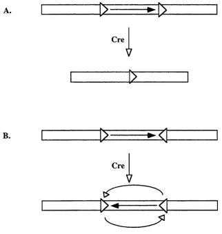
mediated by Cre recombinase, an integrase from bacteriophage P I, which catalyses
recombination between two loxP sites in the absence of any additional co-factors (Sternberg and Hamilton, 1981). A loxP site consists of two 13 bp inverted repeats separated by an 8 bp spacer. These repeats are the recognition and binding sites for
Cre.
In the presence of Cre, the intervening DNA between two loxP sites positioned head to tail is excised along with one loxP site, figure 1.4a. Alternatively, repetitive inversion of the intervening DNA occurs if the loxP sites are positioned head to head, figure 1.4b. Cre-mediated recombination has been demonstrated to excise
exogenous DNA in eukaryotic cells in vitro and in vivo (Byrne and Ruddle, 1989; Gu et a i, 1994; Ou et a i, 1993). Since the LoxP site of the PI is 34 bp in size, the natural occurrence of this exact sequence is unlikely in any eukaryotic genome.
However, related sequences may exist in eukaryotic genomes that could recombine
at low efficiency (<1 x 10$) with an authentic loxP site (Sauer, 1992).
gene can be activated, inactivated or altered by crossing the resulting transgenic
mouse line with a second transgenic line expressing the Cre recombinase. Under the
control of a tissue specific promoter, Cre recombinase can be used to manipulate the
flanked gene in a specific tissue or cell type during development or in adults. Gu et al. (1994) were the first to demonstrate that CvdloxP system can be used to excise DNA flanked by loxP sites in a tissue- and developmentally restricted fashion in vivo. Using the C idloxP system, gene knockouts can be temporally and spatially controlled to avoid stages and tissues where the gene under investigation is crucial
for development.
A.
Cre
V
B.
Cre
V
Figure 1.4. Cre mediated deletion and inversion of loxP flanked DNA.
A) Deletion o f flanked DNA when two loxP sites (open arrowheads) are positioned head to tail. B)
Repetitive inversion o f flanked D NA when two loxP sites are positioned head to head. The
1.14.1 Cre mediated subregional and cell type restricted N Rl knockout
The CvdloxP system was used to generate an NRl knockout in the pyramidal cells of the CA l layer of the hippocampus (Tsien et a l, 1996a; Tsien et a l, 1996b). Two loxP sites in a head-to-tail orientation were introduced in the N R l gene by homologous recombination. One loxP site was introduced in intron 10 while a second was introduced 3' of exon 22, thus flanking exons 11-22 (Tsien et a l, 1996b). In this way the introduced loxP sites did not interfere with the expression and correct processing of N R l RNA transcripts (Tsien et a l, 1996b). In the presence of Cre, however, the loxP flanked region was deleted. This resulted in production of truncated N R l transcripts lacking the coding sequence for the putative membrane
and C-terminal domains (Tsien e ta l, 1996b).
Tsein et a l (1996a) generated several Cre expressing mouse lines by pronuclear injection of a Cre-expression cassette. Cre expression was driven by the aCaMKII
promoter which is characterised by a postnatal and forebrain-restricted pattern. One
Cre line showed a strong Cre activity in the CAl region of the hippocampus and
little activity elsewhere in the brain (Tsien et a l, 1996a). Mice lacking the N Rl subunit in the CA l layer were obtained by crossing the CA 1-specific Cre mouse
with the modified N Rl mouse.
The CA l-restricted N R l knockout mouse grows to adulthood without obvious
pathologies and behavioural abnormalities (Tsien et a l, 1996b). However, the CA l- restricted N R l knockout mouse suffered some impairment o f spatial memory as
tested by the Morris water maze task. The NMDA-mediated EPSCs and LTP in the
CAl layer were absent (Tsien et a l, 1996b). These results suggested a role for the NMDA-dependent LTP of CAl layer in acquisition of spatial memories.
Although Cre mediated deletions allowed the generation of temporally and spatially
refined gene knockouts, this strategy suffers from some limitations. First the strategy
relies on the availability of a suitable promoter with the desirable temporal and
1.15 Gene regulation by inducible promoter systems
Gene knockout and transgenic mice expressing constitutively active or dominant-
negative molecules can provide insights into the physiological roles of a gene of
interest. However, interpretations of the phenotypes of such mice are often limited
by several factors. For example, compensation for the hypo- or hyperactivity of the
altered gene by others. Lethality that could result from manipulating many genes is
another example (Spencer, 1996). Therefore, the study of the function of a particular
gene in complex multicellular organisms would greatly benefit from inducible
promoter systems which allow stringent temporal and spatial control of the gene's
expression. Ideally, not only would such systems allow for "On and o f f levels of
gene expression but also permit limited expression at intermediate levels. This is
especially desirable when the regulated gene product is cytotoxic, essential for life or
when its effect during development is to be studied.
1.15.1 Eukarvotic inducible promoter svstems
Control of gene expression using various inducible eukaryotic promoters responsive
to heavy metal ions (Searle et a l, 1985), heat shock (Yoshida et a l, 1995) and steroids hormones (Israel and Kaufman, 1989) has been described. These systems
suffer from major disadvantages, the "leakiness" of expression due to residual
activity of the uninduced promoter (e.g. heavy metal ions), low induction ratios (10-
20 fold) and the pleiotropic effects caused by the lack of specificity of the inducing
agents themselves (e.g. temperature and hormones). Thus conclusions concerning
the physiological consequences of the product of the controlled gene are difficult to
interpret (for review see Spencer, 1996; Yarranton, 1992).
1.15.2 Bacterial inducible promoter svstems
Approaches to adapt bacterial transcriptional control systems to eukaryotic cells are
successful alternatives to eukaryotic inducible systems. A major advantage of these
Bacterial inducible systems rely on the interaction of a bacterial protein with its
DNA binding sequence (known as the operator). The operator sequence is unlikely
to occur in promoter sequences of eukaryotic genes, thus non-specific activation or
repression, which may cause pleiotropic effects, is unlikely to occur.
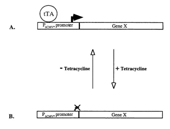
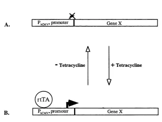
1.16 The tetracycline-regulated promoter system
Gossen and Bujard (1992) developed an eukaryotic inducible system based on the
regulatory elements of the TniO-specified tetracycline-resistance operon of E. Coli. The E. Coli regulatory system consists of a specific operator sequence (teiO) and a specific repressor protein (tetR) that binds to tetO with high specificity (Gossen and Bujard, 1992). In E. Coli, the presence of the antibiotic tetracycline results in failure of the tetR to bind to telO and relieve of the transcriptional repression over the tetracycline-resistance gene.
Gossen and Bujard (1992) adapted the system for eukaryotic cells by converting the
repressor tetR into a transcriptional activator. The tetR was fused with the activating domain of VP 16 which is essential for transcription of the immediate early viral
genes in herpes simples virus (HSV). The hybrid protein was called tetracycline-
controlled transactivator (tTA).
To produce a promoter responsive to tTA, the enhancer region of the human
cytomegalovirus immediate-early (hCMV IE) promoter-enhancer was deleted. As a
result, the generated minimal promoter has no or little intrinsic transcriptional
activity (Gossen and Bujard, 1992). Multiple telO sequences were then inserted upstream of the minimal promoter. Subsequent to binding of the tetR domain of the
tTA to tetO in the minimal promoter, the viral activation domain of the tTA activates transcription from the otherwise silent promoter. Tetracycline interferes with tTA
binding to tetO and thus inhibits the activation of the minimal promoter. The tetracycline-regulated promoter system is schematically outlined in figure 1.5.
highest level of expression upon induction but virtually no expression when low
levels of tetracycline are present (Gossen and Bujard, 1992). The tXA-responsive
promoter was named P h C M V * - l , hereafter it will be referred to as the P h C M V *
promoter.
The usefulness of the tetracycline-regulated promoter system depends on three
factors: the intrinsic (or background) activity of the P h C M V * promoter, the efficiency
of activation of the P h C M V * promoter by the tTA; and the extent and efficiency of
de-activation by tetracycline. In the Hela cell line, the P h C M V * promoter has very
little background activity. Expression of tTA, however, induced the activity of the
luciferase reporter gene under the control of the P h C M V * promoter by up to five
orders of magnitude. Tetracycline concentrations of 1 p.g/ml reduced the activity of
the luciferase reporter gene to background levels. This is 10 times lower than the
cytotoxic concentration of tetracycline (Gossen and Bujard, 1992). When
tetracycline is introduced in culture medium, the activity of the luciferase reporter
gene in Hela cells was reduced by >50-fold after 12 hours. On the other hand, when
tetracycline is removed from the medium, the activity of the luciferase reporter gene