REGULATION OF MMP-9
IN
HUMAN OVARIAN CANCER
THOMAS MATTHIAS LEBER
A thesis submitted for the degree of Doctor of Philosophy
University College London
May 1998
Imperial Cancer Research Fund
Lincoln’s Inn Fields
P roQ uest Num ber: U 642 1 1 0
All rights reserved
IN F O R M A T IO N T O ALL U S E R S
T h e quality of this reproduction is d ep en d en t upon the quality of the copy subm itted.
In the unlikely event that the author did not send a com plete m anuscript
and there are missing pages, these will be noted. Also, if m aterial had to be rem oved, a note will indicate the deletion.
uest.
P roQ uest U 6 4 2 1 1 0
Published by P roQ uest LL C (2015). Copyright of the Dissertation is held by the Author.
All rights reserved.
This work is protected against unauthorized copying under Title 17, United States C ode.
Microform Edition © P roQ uest LLC.
ProQ uest LLC
789 East E isenhow er P arkw ay
Abstract
Matrix metailoproteases (M M P s ) are a family of structurally and functionally related
endopeptidases. T h e y have a zinc ion at their active site and are released as inactive
pro-forms. Activation enables M M P s to degrade components of the extracellular matrix
and this been associated with tumour growth and metastasis. T h e aim of this thesis w a s
to establish the pattern of M M P s and tissue inhibitor of metalloproteinases (T IM P s )
present in human ovarian cancer and to investigate further one M M P , M M P -9 .
M M P /T IM P gene expression w as assessed by R T -P C R in biopsies of normal, benign
and malignant human ovary and compared to ovarian cancer cell lines and xenograft
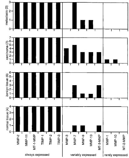
models of human ovarian cancer. Expression of M M P -2 , -11, M T -1 -M M P , T IM P -1 , -2
and -3 w as detectable in all tissue sam ples. M M P -1 , -1 0 and M T -2 -M M P w ere rarely
detectable and M M P -3 , -7, -9, -1 3 and M T -3 -M M P w ere variably expressed.
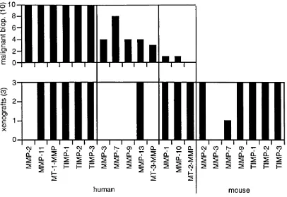
M M P /T IM P expression in the malignant biopsies was similar to that in ovarian cancer cell
lines and xenograft models of human ovarian cancer.
M M P -9 production w as further investigated in an in vitro model of tumour cell macrophage interaction. In human ovarian cancer M M P -9 localises to infiltrating
macrophages but little is known of its regulation. Co-culture of ovarian cancer cells
(P E 0 1 ) and a monocytic cell line (T H P -1 ) led to production of 9 2kD a p ro M M P -9. P E 0 1
conditioned medium (C M ) also stimulated TH P -1 cells or isolated peripheral blood
monocytes to produce proM M P -9. Expression of T IM P -1 , how ever, remained
unaffected. T h e metalloprotease stimulating factor (M M P S F ) present in C M w as not
T N F -a , but acted in a synergistic fashion with autocrine m onocyte-derived T N F -a to
stimulate release of monocytic proM M P-9. M M P S F was further characterised.
In all experim ents only proM M P -9 was released. Cell extracts of TH P-1 cells contained a
92 and 85kD a form of M M P -9 . N-terminal sequencing revealed that both proteins
contained the full pro-peptide and w ere therefore proteolytically inactive pro-forms of
Acknowledgements
I would like to acknowledge the help and advise I obtained throughout this thesis from all
members of the Biological Therapies Laboratory. In particular I would like to thank Dr.
Frances R. Balkwill for including me in her team here at the IC R F. I very much appreciate
her support, the space she gave me to develop m y own thoughts and the time she
spent to discuss our view s. Furthermore, I would like to thank my co-P hD s Dr. Rupert
Negus and Sergio Dias for their help and friendship over the past years. I will miss the
conversations w e had over the “swift ones down T h e G eorge” on Friday nights. I also
would like to mention Dr. AI Stewart for his advise during the characterisation of M M P S F
and his always encouraging “Hang in, man! Hang ini”.
Contents
1. Introduction 12
1 . 1 O varian C an cer 12
1.1.1 T h e ovary and ovarian tumours 12
1.1.2 Clinical description of ovarian cancer - stage 12
1 .1.3 Tum our histology 13
1.1.4 Risk factors associated with epithelial ovarian cancer 14
1.1.5 Current treatment of ovarian cancer 15
1.2 Matrix M etailoproteases - An overview 16
1.2.1 Tissue architecture and M M P s 16
1.2.2 M M P s in tumour growth and invasion 17
1.2.3 The M M P family 18
1.2.4 The protein structure of matrix metailoproteases 18
1.2.5 Regulation of matrix metailoproteases 22
1.2.6 Synthetic inhibitors of matrix metailoproteases 24
1.3 M M P -9 25
1.3.1 Protein structure of M M P -9 25
1.3.2 Role of M M P -9 in tumours growth and invasion 26
1.3.3 Regulation of gene expression 27
1.3.4 Activation of M M P -9 27
1.3.5 Regulation of M M P -9 activity by T IM P s 28
1.4 Aims of thesis 29
2 . P C R -screen for M M P s and T IM P s in biopsies of human ovarian cancer 30
2.1 Introduction 30
2 . 2 Material and Methods 32
2 .3 Results 38
2.3.1 Specificity of P C R primers 38
2.3 .2 M M P /T IM P expression in biopsies of the normal, benign and malignant
hum an ovary 41
2 .3 .3 Comparison of the M M P /T IM P expression pattern in malignant human
biopsies with cell lines 44
2 .3 .4 Comparison of the M M P /T IM P expression pattern in malignant human biopsies with xenograft models of human ovarian cancer 44
2 .4 Discussion 47
3. Optimisation of the zym ography technique for the quantitation of M M P -9 51
3.1 Introduction 51
3 .2 Material and Methods 53
3 .3 Results 56
3.3.1 Tim e course of zymogram staining 56
3 .3.2 Intergel reproducibility of linear range and detection limit 59 3.3 .3 Intragel variation of proM M P -9 quantitation 60
4. Regulation of monocytic M M P -9 production in human ovarian cancer 63
4.1 Introduction 63
4 .2 Material and Methods 65
4 .3 Results 71
4.3.1 Analysis of proM M P -9 production in co-cultures of ovarian cancer cells
and monocytic cells 71
4 .3 .2 Cell ratio experiments of the ovarian cancer cell line P E 0 1 and the
monocytic cell line T H P -1 . 72
4 .3 .3 Co-culture experiments without direct cell-cell contact 73 4 .3 .4 Tum our cell derived C M induced proM M P -9 production by the monocytic
cell line TH P-1 73
4 .3 .5 Tum our cell derived C M induced proM M P -9 release from isolated
peripheral blood monocytes 74
4 .3 .6 Analysis of M M P -9 and TIM P -1 gene expression and protein 75 4 .3 .7 T N F -a stimulated p roM M P -9 production by TH P-1 cells in a dose
dependent manner 77
4 .3 .8 Antibodies to T N F -a blocked pro M M P -9 production in both co-cultures
and C M stimulated THP-1 cells 78
4 .3 .9 The compound B B -2116 blocked T N F -a release and p ro M M P -9
production in both co-cultures and C M stimulated TH P-1 cells 79 4 .3 .1 0 Analysis of T N F -a gene expression and protein in TH P-1 cells 79 4.3.11 T N F -a acts in a synergistic fashion with P E 0 1 derived C M 81 4 .3 .1 2 Immunoprécipitation of T N F -a from C M did not alter C M ’s ability to
induce proM M P-9 from THP-1 cells 82
4 .3 .1 3 Production of C M in presence of B B -2 1 16 did not alter C M ’s capacity to
induce proM M P-9 from THP-1 cells. 83
4 .3 .1 4 Heating of C M reduced its pro M M P -9 inducing activity 84
4 .4 Discussion 85
5. Characterisation of M M P S F 89
5.1 Introduction 89
5 .2 Materials and Methods 90
5 .3 Results 93
5.3.1 Binding studies 93
5 .3 .2 Partial characterisation of W G A binding proM M P -9 inducing activity
(W G A -M M P S F ) 94
5 .3 .3 Partial characterisation of Q -Sepharose binding proM M P -9 inducing
activity (Q -M M P S F ) 96
5 .3 .4 Binding and elution of M M P S F to M onoQ 99 5 .3 .5 Gel filtration of Q -M M P S F obtained from M onoQ at pH 7 .5 100 5 .3 .6 Re-binding of M M P S F to M onoQ and elution at pH 6.5 1 0 2
5 .3 .7 Concentration of Q -M M P S F obtained at pH 6 .5 in fractions 4-11 103 5 .3 .8 Gel filtration of M M P S F from step 4 and Lectin blot 104
5 .4 Discussion 106
6 . Characterisation of a 85kD a form of M M P -9 110
6.1 Introduction 110
6 .2 M aterials and Methods 1 1 1
6.3.1 LPS stimulates proM M P -9 release in a dose dependent m anner 114 6.3 .2 Detection of cell associated gelatinolytic activity in LPS stimulated TH P-1
cells 115
6 .3 .3 W estern blotting confirmed the 9 2kD a and the 8 5kD a band to be a forms
of M M P -9 116
6 . 3 . 4 1rito n -X -114 extractable forms of M M P -9 could be pelleted at 100 OOOg 116 6.3 .5 Optimisation of cell culture conditions to obtain maximal yield of cell
associated M M P -9 forms 117
6.3.6 Purification of proM M P -9 (92kD a) from cell culture supernatant of LPS
stimulated THP-1 cells 118
6.3 .7 Purification and N-terminal sequencing of cell associated forms of M M P -9 119
6 .4 Discussion 121
7. Sum m ary and future plans 124
8 . List of publications 127
List of figures
Fig. 1.1 Domain structure of the M M P family 21
Fig. 1.2 Domain structure of proM M P -9 and sites of activation 26
Fig. 2.1 Restriction digests of P C R products 38
Fig. 2 .2 S equence comparison of P C R products with a data base 40
Fig. 2 .3 A garose gel of R T -P C R products 41
Fig. 2 .4 M M P /T IM P expression in tissue biopsies 42
Fig. 2 .5 M M P expression in malignant and non-malignant tissue biopsies 43
Fig. 2.6 M M P /T IM P expression in malignant biopsies and ovarian cancer cell lines 44
Fig. 2 .7 M M P /T IM P expression in malignant tissue biopsies and xenograft models of
human ovarian cancer 45
Fig. 3.1 Tim e course of zymogram staining 56
Fig. 3 .2 Quantitation of the time course of zymogram staining 58
Fig. 3.3 Inter gel reproducibility of the linear range for proM M P -9 59
Fig. 4.1 Co-culture of the monocytic THP-1 cell line and the ovarian cancer cell line
P E 0 1 71
Fig. 4 .2 Quantitation of proM M P -9 release in co-cultures of TH P-1 and P E 0 1 71
Fig. 4 .3 Effect of the P E Q 1/TH P -1 cell ratio on the proM M P -9 production 72
Fig. 4 .4 Role of cell cell contact on proM M P-9 production in co-cultures 73
Fig. 4 .5 Dose dependent production of p roM M P -9 by C M stimulated TH P -1 cells 74
Fig. 4 .6 C M stimulation of isolated peripheral blood monocytes 74
Fig. 4 .7 Kinetics of proM M P -9, T N F -a and TIM P-1 gene expression in unstimulated
and C M stimulated THP-1 cells 75
Fig. 4 .8 Tim e course of M M P -9 production in unstimulated and C M stimulated THP-1
cells 76
Fig. 4 .9 Dose dependent production of pro M M P -9 by T N F -a stimulated TH P -1 cells 77
Fig. 4 .1 0 Effect of an anti T N F -a antibody on proM M P -9 release 78
Fig. 4.11 Effect of B B -2 1 16 on release of T N F -a and proM M P -9 80
Fig. 4 .1 2 Synergistic action of C M with an agonistic polyclonal antibody specific for
the p55 T N F -a receptor 81
Fig. 4 .1 3 Th e pro M M P -9 release stimulating activity in C M can not be precipitated
with an anti T N F -a antibody 82
Fig. 4 .1 4 R elease of the p ro M M P -9 inducing activity present in C M is not inhibited by
B B -2 1 1 6 83
Fig. 4 .1 5 W arm ing of C M abrogates proM M P -9 induction from TH P-1 cells 84
Fig. 4 .1 6 Model of monocytic proM M P-9 production 8 6
Fig. 5.1 Binding of M M P S F to w heat germ agglutinin (W G A ) beads 93
Fig. 5 .2 Binding of M M P S F with Q -S ep h aro se 93
Fig. 5 .4 Lectin blot of sam ples obtained by gel filtration 95
Fig. 5 .5 Optimisation of the salt concentration required for the elution of Q -M M P S F 96
Fig. 5.6 Optimisation of the amount of Q -Sepharose required for maximal concentration
of Q -M M P S F 97
Fig. 5 .7 Analysis of Q -S ep h aro se eluate by S D S -P A G E and silver staining 97
Fig. 5 .8 Extensive washing of Q -S epharose beads did not reduce the am ount of
contaminating protein co-eluted with Q -M M P S F 98
Fig. 5 .9 Large scale enrichment of Q -M M P S F from C M with Q -S epharose 99
Fig. 5 .1 0 Binding Q -M M P S F to M onoQ at pH 7 .5 1 0 0
Fig. 5.11 Gel filtration of eluate obtained from M onoQ 101
Fig. 5 .1 2 Sizing of p roM M P -9 inducing activity present in fractions 2 4 -2 8 101
Fig. 5 .1 3 Elution of proM M P -9 inducing activity from M onoQ at pH 6 .5 1 0 2
Fig. 5 .1 4 Concentration of fractions 4-11 eluted from M onoQ at pH 6 .5 103
Fig. 5 .1 5 Analysis of proteins by S D S -P A G E and silver staining 104
Fig. 5 .1 6 Lectin blot of Q -M M P S F eluted at 80m M N aC I in histidine buffer at pH 6 .5 105
Fig. 6.1 M M P -9 production in response to endotoxin 114
Fig. 6.2 Gelatinolytic activity in extracts of LPS stimulated TH P -1 cells 115
Fig. 6.3 Detection of M M P -9 by western blotting in culture medium and in the aqueous
phase T X -1 14 extracts of LPS stimulated TH P -1 cells 116
Fig. 6.4 Subcellular fractionation of THP-1 cells by differential centrifugation 117
Fig. 6.5 Qptimisation of the production of cell associated forms of M M P -9 118
Fig. 6 . 6 S D S -P A G E of M M P -9 purified from cell culture supernatant 119
List of tables
T ab le 1.1 Definition of clinical stages, their incidences and associated 5-y e a r survival
rates 13
T ab le 1.2 Incidence and survival rates of histological subtypes of ovarian cancer 14
T ab le 1.3 Matrix metailoproteases and their substrates 19
T ab le 2.1 Tissue biopsies included in P C R screen for human M M P s and T IM P s 32
Tab le 2 . 2 P C R primers for a panel of human and murine M M P s and T IM P s 39
Abbreviations
A PM A aminophenyl mercuric acetate
B S A bovine serum albumin
C M conditioned medium
D T T dithiothreitol
E G F epidermal growth factor
E LISA enzym e linked immunosorbent assay
F C S foetal calf serum
FG F fibroblast growth factor
F P L C /H P L C fast protein liquid chromatography/high pressure liquid chromatography
IFN interferon
IL interleukin
M molar (mol per litre)
M -C S F macrophage colony stimulating factor
M M P matrix metalloproteinase
M M PI (synthetic) matrix metalloproteinase inhibitor
M M P S F matrix metalloproteinase stimulating factor
PAF platelet activating factor
PA G E polyacrylamide gel electrophoresis
P B S phosphate buffered saline
P D G F platelet derived growth factor
PHA phytohemagglutinin or phaseolus vulgaris agglutinin
PVDF polyvinylidene fluoride
S D S sodium dodecyl sulphate
T E A triethanolamine
T G F -a transforming growth factor-alpha
T IM P tissue inhibitor of matrix metalloproteinases
T N F -a tumour necrosis factor-alpha
1. Introduction
1.1 Ovarian Cancer
1.1.1 The ovary a n d ovarian tumours
T h e ovary consists of a central medulla surrounded by a cortex which in turn is
surrounded by a monolayer of epithelium. T h ese epithelial cells stem from the coelomic
endoderm and are separated from the stromal cells by basem ent membrane. T h e cortex,
which consists of dense stroma, ovarian follicles and corpora lutea, is the functional part
of the ovary with respect to oocyte production (1).
O varian cancer is the sixth most common cancer among wom en in the U S (2). T h e
empirical lifetime risk of developing ovarian cancer is 1:70 and the median age at
diagnosis is 62 years (3). T h e incidence of ovarian cancer is highest in the highly
industrialised countries, in particular W estern and Northern Europe and North Am erica (3).
O varian tumours can arise from any component of the ovary but most (6 5 -7 0 % ) are
derived from the epithelial layer (4). This type of tumour usually affects wom en older
than 20 years and its incidence increases with age peaking at approxim ately 5 0 cases
per 1 0 0 0 0 0 wom en in the age group 7 0 -7 5 (2 ). T h e second most common ovarian
tumours (15-20% ) arise from germ cells present in the ovary. In contrast to the epithelial
tumours, germ cell tumours tend to affect wom en of young age (0 -2 5 years). Finally,
tumours can also arise from the cortex stroma (5 -1 0 % ) or m ay be derived from
m étastasés (5% ) of a non ovarian primary tumour (4).
1.1.2 Clinical description o f ovarian cancer - stage
More than 5 0 % of patients present with advanced stage disease (stage lll-IV, T a b le
1 .1) characterised by m etastasis beyond the pelvic cavity (5). S tage at presentation is
strongly correlated to the 5 -y e a r survival rate (Table 1.1) and therefore cure. Late
presentation is thought to be the main reason for the poor prognosis (Table 1.1). Th e
cellular and molecular changes associated with ovarian cancer are poorly understood.
there has been no significant improvement in overall survival for patients with advanced
stage disease.
Stage incidence 5-year survival rate
Clinical description
Stage 1 la lb Ic 26.1% 85-100% 85-100% 85-100%
Growth limited to the ovaries.
Growth limited to one ovary; no ascites. No tumour on external surface; intact capsule.
Growth limited to both ovaries; no ascites. No tumour on external surface; intact capsule.
Tumour either stage la or lb, but with ascites or positive peritoneal washings.
Stage II lia lib lie 15.4% 40-50% 40-50% 40-50%
Growth involving one or both ovaries with pelvic extension. Extension and/or métastasés to the uterus and/or fallopian tubes. Extension to other pelvic tissues including peritoneum and uterus. Tumour either stage lia or lib, but with ascites or positive peritoneal washings.
Stage III
Ilia Nib NIC 39.1% 40-50% 20% 5-10%
Growth involving one or both ovaries with intraperitoneal métastasés outside the pelvis and/or positive retroperitoneal nodes. Tumour limited to the true pelvis with histologically proven malignant extension to the small bowel or omentum.
Tumour grossly limited to the true pelvis with negative nodes but with histologically confirmed microscopic seeding of abdominal peritoneal surfaces.
Tumour involving one or both ovaries with histologically confirmed implants of abdominal peritoneal surfaces, none exceeding 2cm in diameter. Nodes are negative.
Abdominal implants greater than 2cm in diameter. Nodes are negative.
Stage IV 16.3% 5-10% Growth involving one or both ovaries with distant métastasés. Pleural effusion must have positive cytology. Parenchymal liver métastasés equal stage IV.
Table 1.1 Definition of clinical stages, their incidences and associated 5-year survival rates The parameters defining the clinical stage of human ovarian cancer were established by the
International Federation of Gynaecology and Obstetrics (FIGO) (5). The 5-year survival rate declines with advanced stage (3). More then 50% of women present with advanced stage disease (stage lll-IV) (5).
1.1.3 Tum our histology
Tumours derived from the epithelial cell monolayer have been classified histologically into
several sub-categories on the basis of parameters such as cell shape, formation of
cysts, colour and density of cystic fluid (Table 1.2). Serous tumours are most common
(4 0 % ). T h e y are cystic neoplasms which are lined by tall, columnar, ciliated epithelial
cells and filled with serous fluid (4). Only 2 5 % of serous and 10% of mucinous tumours
are malignant whereas the remaining are either benign or borderline (4). This distinction is
tumours are characterised by having invaded through the epithelial basem ent membrane
w h e re a s in benign and borderline tumours, the basem ent membrane remains intact (4).
T h e composition of tissue sections of human ovarian cancer with respect to the cellular
infiltrate and amount of tumour versus tumour stroma has been analysed recently (6). In
this study which w as mainly based on serous ovarian carcinomas, the sections w ere
found to consist of on average 4 3 % tumour, 3 7 % stroma and 4 % necrosis. T h e cellular
infiltrate w as primarily composed of C D6 8 ^ macrophages and C D 8 V C D 4 5 R 0 ^ T cells
and localised mainly to tumour stroma rather than tumour areas (6 ).
T y p e Incidence 10- year survival rate
Serous 40%
Benign (60%)
Borderline (15%) 75%
Malignant (25%) 10-20%
Mucinous 10%
Benign (80%)
Borderline (10%) 95%
Malignant (10%) 60%
Endometrioid carcinoma 20% na.
Undifferentiated carcinoma 10% na.
Clear cell carcinoma 6% na.
others 14% na.
Table 1.2 Incidence and survival rates of histological subtypes of human ovarian cancer Modified from (4), na. = not available.
1.1.4 Risk factors associated with epithelial ovarian cancer
Although the specific causes of ovarian cancer remain unclear, there are, how ever,
factors which have been associated with an increased or a decreased risk. Age, race,
the use of talcum powder on the perineum, a history of endometrial or breast cancer are
associated with an increase in risk w hereas the use of oral contraceptives, a history of
breast feeding, tubal ligation and hysterectom y w ere associated with a reduction in risk
(7). Pregnancy also has a protective effect with the risk of nulliparous wom en being
1.5-3 .1.5-3 times greater than that of parous wom en (8 ). T h e “Fathalla Hypothesis” suggests
that this effect m ay be due to the rest that pregnancy offers from incessant ovulation (9).
Among the risk factors mentioned, family history and nulliparity are considered to be the
two most important ones (1 0 ,1 1 ). A single family member with ovarian cancer increases
In 1 9 9 0 the first major breast cancer susceptibility gene BRCA1 w as m apped to the long
arm of chromosome 17 (13). Mutations in this region w ere subsequently reported to b e
associated with some ovarian cancer cases and an international study show ed that the
B R C A 1 gene w as often implicated when there w as an inherited susceptibility to both
breast and ovarian cancer (1 4 ,1 5 ). The BRCA1 gene is mutated in the germline and the
normal allele is lost in tumour tissue from hereditary breast and ovarian cancer. W om en
w ho carry a mutation in this gene have a 5 9 % risk of developing breast or ovarian
cancer by the age of 50 and 8 2 % by the age of 7 0 (1 6 ). T h e overall population
frequency of a BRCA1 mutation w as estimated to be 0 .0 0 0 7 (17). Based on this
estimation BRCA1 would account for an approxim ately 2 .6 % of ovarian cancer cases
below the ag e of 7 0 and 4 .7 % of the cases below the age of 5 0 (18).
1.1.5 Current treatment o f ovarian cancer
As mentioned earlier, patients with ovarian cancer usually present at advanced stage of
the disease. Initial treatment is therefore debulking surgery often combined with total
abdominal hysterectom y, bilateral salpingo-oophorectomy and/or omentectomy (19).
Survival of patients is directly related to the residual tumour mass (2 0 ). S urgery is
usually followed by multi-agent therapy including cisplatin and paclitaxel (taxol) or
cyclophospham ide (3). In spite of these treatments less then 3 0 % of patients survive
for more then 5 years (3).
M ore recently, some progress in ovarian cancer therapy has been m ade through a novel
approach in cancer treatment which aims to reduce/prevent tissue remodelling and
angiogenesis involved in tumour growth and metastasis (21). Industrial research has
produced a series of low molecular weight compounds targeted against the e n zy m e s
implicated in these processes, the matrix metailoproteases (M M P s ). T h e y h ave been
tested successfully in several animal models of human malignancy including ovarian
1.2 Matrix Metailoproteases - An overview
1.2.1 Tissue architecture an d M M P s
A tissue is an organised structure consisting of both cells and extracellular matrix (E C M ).
T h e cellular compartment can be divided into epithelial and stromal cells. T h e epithelial
cells of the ovary are derived from the coelomic endoderm and the stromal cells from the
mesoderm (2 9 ). T h e E C M is a dense matrix of mostly fibrillar proteins and glycans.
Collagens, non collagenous glycoproteins and proteoglycans represent the main
constituents (30). T h ese proteins are produced by both epithelial and stromal cells. T h e
matrix is perm eable for ions and other small molecules but im perm eable for most cells and
its integrity is m aintained by a finely tuned process of synthesis and degradation. Cells
are closely attached to the E C M by transmembrane adhesion molecules (integrins)
which them selves bind to the cytoskeleton (30). Besides providing mechanical stability
to the cells, the E C M is also important for functions such as cell survival,
morphogenesis, tissue specific functions and cell migration (31). T urnover of extracellular
matrices is an essential component of tissue homeostasis and is thought to involve the
co-ordinated interaction of enzym es from the serine, cysteine, aspartyl and matrix
metalloprotease (M M P ) families, and their specific endogenous inhibitors (32).
This thesis will concentrate on matrix metailoproteases and on M M P -9 in particular.
Matrix metailoproteases are a family of endopeptidases which are able to degrade all
major com ponents of the E C M . Th ey have been associated with tissue remodelling and
invasion in m any physiological processes such as embryonic growth,
blastocyst/trophoblast implantation, mammary developm ent and involution, endometrial
turnover during the menstrual cycle, tooth eruption, wound healing, angiogenesis and
bone growth/remodelling (33, 34). M M P s are also thought to be critical in pathological
processes such as periodontal disease, rheumatoid arthritis, tumour invasion and
1 .2 .2 M M P s in tumour growth a n d invasion
D evelopm ent of a malignant tumour and the formation of metastasis requires the
breakdown of the basem ent m em brane, invasion of the surrounding tissue, separation of
tumour cells from the primary tumour, intravasation into blood or lymph ves s e ls ,
extravasation out of the vessel and invasion of a secondary tissue. In addition, the
formation of new blood vessels leading into the tumour is also required. All these
processes are associated with E C M remodelling and M M P activity (33, 3 7 ). Proteolytic
activity has been analysed in different malignancies and correlated with tumour
progression. In colorectal cancer, for exam ple, high M M P-1 protein levels detected by
immunohistochemistry w ere associated with poor prognosis (38). Increased M M P -2 and
M M P -9 protein activity w as also detected in stage A and 0 colorectal tumours as
compared with normal or benign tissue sam ples (39). In breast and bladder cancer
increased M M P -2 and -9 proteolytic activity correlated with tumour progression (40, 41).
M M P -1 1 expression w as also increased in malignant biopsies if compared with
precursor lesions in breast, bladder and carcinomas of the uterine cervix (42).
T h e proteolytic activity required for these processes, how ever, m ay not necessarily be
produced by the tumour cells them selves but m ay be derived from stromal or infiltrating
cells. In ovarian cancer, for exam ple, M M P -2 gene expression has been localised by in situ hybridisation to stromal fibroblasts w hereas M M P -9 expression w as associated with infiltrating macrophages (43). Similarly, M M P -2 and -9 gene expression w a s
detected in stromal cells or at the tumour/stroma interface by in situ hybridisation in bladder cancer (41). In breast cancer, expression of M M P -1 , M M P -2 , M M P -3 , M M P -1 1
and M T -1 -M M P w as localised to stromal fibroblasts w h ereas M M P -9 expression w a s
primarily associated with endothelial cells. O nly M M P -1 3 and M M P -7 expression w a s
localised to tumour cells (44). Finally, the expression of M M P -1 1 in skin, head and neck,
breast, bladder, colorectal, stomach and m any other tumours w as also exclusively
associated with fibroblast-like cells (42). This led to the hypothesis that the induction of
M M P gene expression in tumours m ay be a tumour-induced host response (4 4 ). This
will be the subject of chapter 4 of this thesis.
T h e association of increased malignancy with high proteolytic activity w a s
complemented by studies showing that enhanced production of tissue inhibitors of
review (45, 46)]. Taken together these data suggest that tumour growth and metastasis
requires an imbalance betw een proteolytic and inhibitory activity towards excess
proteolysis (47). Based on this information, considerable industrial efforts led to the
developm ent of synthetic M M P inhibitors (M M P Is ) with the aim of reducing tumour
growth and m etastasis [for review (2 1 -2 3 , 25 -2 7 )].
1.2.3 The M M P family
M M P s are a family of structurally and functionally related endopeptidases. To date 20
m embers of the matrix metalloprotease family have been cloned. Sequence comparison
suggests that they are derived from a single ancestor gene by gene duplication (48, 4 9 ).
M M P s have a in the active centre and require binding of Ca^'" for en zym e stability.
All M M P s are produced in a proteolytically inactive pro-form and the enzym es degrade,
once activated, all major components of the extracellular matrix (E C M ) at physiological
pH (pH 5 -8 ) (50).
Sub-classification of M M P s w as originally based on both structural similarities and in vitro substrate specificity. This led to the definition of four major groups (collagenases, gelatinases, stromelysins and others). Further analysis, however, showed that there is a
considerable overlap of substrate specificity betw een these groups. Powell and
Matrisian therefore suggested a classification based on structural similarities only (Fig.
1.1, T a b le 1.3) (51).
1.2.4 The protein structure of matrix metailoproteases
M M P s are structurally related and a domain structure can be assigned (49, 5 0 ). T h e
prototype M M P consists of a signal peptide, which directs the protease for export and is
removed in the endoplasmatic reticulum; a pro-peptide; the catalytic domain and the 0
-terminal hemopexin/vitronectin-like domain. All M M P s can be derived from this structure
by insertions, deletions or additions of further domains (Fig. 1.1). M M P -7 for exam ple,
does not possess the C-term inal hemopexin/vitronectin-like domain, M M P -2 and -9 have
a fibronectin-like domain inserted in the catalytic domain and the M T -M M P s h ave
Enzymes MMP No. Precursor (kDa)
Active (kDa)
Matrix substrates
minimal domain MMPs Matrilysin
(EC 3.4.24.23)
MMP-7 28 19 Aggrecan, Fibronectin, Laminin, Gelatins, Collagen IV, Elastin, Entactin, Small Tesascin-C
hemopexin domain MMPs
Interstitial collagenase MMP-1 52 41 Collagen 1, II, III, VII, X, Gelatins, (EC 2.4.24.7) 56* 45* Entactin, Aggrecan, Cartilage link
protein Neutrophil collagenase
(EC 3.4.14.34)
MMP-8 75* 65 Collagen 1, II, III, Proteoglycan, Cartilage link protein
Collagenase 3 MMP-13 65 55 Collagen 1
Stromelysin 1 MMP-3 57 45 Aggrecan, Gelatins, Fibronectin, (EC 3.4.24.17) 59* 28 Laminin, Collagen III, IV, IX, X, Large
Tesascin-C Stromelysin 2
(EC 3.4.24.22)
MMP-10 57 45 28
Aggrecan, Fibronectin, Laminin, Collagen IV
Stromelysin 3 (EC 3.4.24)
MMP-11 55 45 28
Fibronectin, Laminin, Collagen IV, Aggrecan, Gelatin
Metalloelastase (EC 3.4.24.65)
MMP-12 53 45 22
Elastin
fibronectin domain MMPs Gelatinase A
(EC 3.4.24.24)
MMP-2 72 67 Gelatins, Collagens 1, IV, V, VII, XI, Fibronectin, Laminin, Aggrecan, Elastin, Large Tesascin-C
Gelatinase B (EC 3.4.24.35)
MMP-9 92* 82 Gelatins, Collagen III, IV, V, XIV, Aggrecan, Elastin, Entactin
transmembrane domain MMPs MT-1-MMP MT-2-MMP MT-3-MMP MT-4-MMP MMP-14 MMP-15^ MMP-16^ MMP-17 " 66 72 64
Collagen 1, II, III, Gelatin, Fibronectin, Vitronectin, Laminin '
others
MMP-18® MMP-19® MMP-20 ^
Table 1.3 Matrix metailoproteases and their substrates
Table modified from (49, 51, 52). Some MMPs have not been biochemically characterised in depth to date. '(53), ^(54), ^(55), "(56), ^(57), ®(58), ^(59) (* glycosylated)
T h e N-terminal pro-peptide consists of approxim ately 80 amino acids and contains the
highly conserved P R C G (V /N )P D V sequence (49). This pro-peptide plays a crucial role
in maintaining the protease in its proteolytically inactive form and is removed in the tightly
controlled process of proM M P activation. T h e structure of the pro-peptide has been
determined for M M P -3. It is an extended structure which covers the active site cleft
stabilised by an interaction betw een the central cysteine of the conserved sequence
T h e catalytic domain comprises approxim ately 165 amino acids and mediates substrate
hydrolysis and autolytic cleavages. The conserved sequence H E X G H X X G X X H S within
the catalytic domain contains three histidine residues which complex the Zn^^ ion in the
active centre (49). This model has been confirmed by X -ray crystallography for M M P-1
(61, 6 2 ), M M P -3 (60), M M P -7 (63) and M M P- 8 (64). T h ese studies also suggested that
a second, “structural” Zn^^ ion m ay be bound to the catalytic domain and one or more
Ca^"^ ions contribute to the proteolytic activity by stabilising the tertiary structure of the
en zy m e (62, 6 5 -6 8 ).
T h e C-terminal hemopexin-like domain can be found in all M M P s apart from M M P -7 (Fig.
1.1). This domain contains four repeats which show strong sequence homology to
hem opexin, vitronectin and other members of the hemopexin family (50). T h e first and
the fourth repeat are linked by a disulphide bridge which must be intact to maintain the
properties of the domain (Fig. 1.1). For the collagenases (M M P -1 , -8 , -1 3 ) the
hemopexin-like domain has been shown to bind collagens (6 9 -7 2 ). Truncated
collagenases loose their ability to cleave native triple helical collagen suggesting that the
hem opexin-like domain plays a role in substrate binding and processing (7 0 -7 3 ). It is not
clear, however, w hether this is also true for other members of the M M P family. A further
function of the hemopexin-like domain is the binding of the naturally occurring tissue
inhibitors of m etailoproteases (TIM P s). Som e evidence suggests that initial “docking” of
T IM P to the hemopexin-like domain accelerates T IM P binding to the active site and
therefore M M P inhibition (74). Finally, a novel function of the hemopexin domain of M M P
-2 w a s discovered recently (75). Brooks e t al. (75) show ed that M M P -2 binds via its hemopexin domain to the avp3 integrin present on endothelial cells leading to an increase
in cell associated collagenolytic activity. In addition Brooks e t al. provided data showing that the hemopexin domain may be a natural degradation product of M M P -2 and that this
domain m ay reduce the cell associated collagenolytic activity by competing with M M P -2
Matrilysin M M P-7
Collagenases M M P-1, -8, -13 Stromelysins M M P-3, -10 Metalloelastase M M P-12
Zn
Y / / / ^ ^
a-(GX3cxÊ)
Stromelysin M M P -1 1
iE K S Ô Ô ©
Gelatinase M M P-2
V//
a
^ ‘Æ
Gelatinase M M P-9
Z
!® ^ (^ [l!ü ç K D Ô Ô ©
Membrane-type MM Ps M M P-12, -13, -14
Zn'
V /////X
PropeptideQ 1-14 amino acids not found in other MMPs
Zn^+ Catalytic domain
C SS 3 Fibronectin type II domain
Collagen-like domain
C Cysteine residue
(YyYYYYYZY
Hemopexin-like domainTrans-membrane domain
1 .2.5 Regulation o f matrix metailoproteases
M M P activity is controlled at several levels. G en e expression can be regulated by
cytokines or components of the E C M . Som e regulation might also be associated with
protein glycosylations which have been detected on some M M P s (T ab le 1.3). Finally,
activation of M M P s can be triggered by proteases and their proteolytic activity is
counterbalanced by tissue inhibitors of metailoproteases (T IM P s).
a. Transcriptional regulation
D e novo M M P protein synthesis can be manipulated in cell culture with cytokines such as tumour necrosis factor-a (T N F -a ), interleukin-1 p (IL-1P), interferon-y (IF N -y ),
epiderm al growth factor (E G F ) and platelet derived growth factor (P D G F ) and both up or
down regulation of gene expression can be observed [for review (7 7 -7 9 )]. Som e
m em bers of the M M P family respond in a similar fashion to a particular cytokine. T N F -a ,
for exam ple, up regulates whilst IF N -y down regulates the gene expression of M M P -1,
-3, -9 (78). Promoter analysis of M M P s revealed several consensus sequences. All
promoters analysed (M M P -1 , -2, -3, -7, -9, -10, -1 2 ) contained a T A T A box close to
position -30, an AP-1 binding site near position -70 and adjacent to it at least one PEA3
binding site (8 0 ). T h e exception to this is the M M P -2 promoter which contains none of
these three elements. Instead, two SP-1 (-69, -8 9 ) and one A P -2 (-1 6 5 0 ) binding site
are present (80). T h e s e are characteristics shared by “housekeeping gen es” and is
consistent with the observation that M M P -2 is often constitutively expressed (80, 81).
In addition to cytokines, E C M derived proteins (e.g. collagen I) also play a role in the
regulation of M M P gene expression (8 2 -8 4 ). This is in keeping with data showing that
signalling through transmembrane E C M binding molecules (integrins) can also modulate
M M P gene expression (8 5 -8 7 ).
b. P ost transcriptional regulation
This type of regulation, which includes alternative splicing and m RNA stability, has not
been investigated in great detail yet. Extended m RNA half-life of M M P-1 (8 8 ) and
M T -3 -M M P is the only M M P for which alternative splicing has been reported. In addition
to the m em brane-bound protease, a modified splicing pattern seem s to lead to a soluble
form of M T -3 -M M P which lacks the transmembrane domain (90).
c. Post translational regulation
T w o levels of post translational regulation can be distinguished; zym ogen activation and
inhibition of the active protease by tissue inhibitors of m etailoproteases (T IM P s ). In
addition, Toth e t al. reported M M P -9 targeting to the cell surface by a modification of its glycosylation (91). This m ay represent a new type of post translational regulation of
M M P s.
P ro tease activation - the "cysteine switch”
M M P s are produced as a proteolytically inactive pro-form (zym ogen). T h e latency of the
protease is maintained by the N-terminal pro-domain of approxim ately 80 amino acids
which covers the active site cleft (60). The pro-peptide is held in place by the sulfhydryl
group (-S H ) of the central cysteine residue present in the conserved sequence
P R C G (V /N )P D which acts as a fourth ligand complexed to the ion in the active
centre. In an initial step of zym ogen activation, the pro-peptide undergoes proteolytic
processing N-terminal of the conserved P R C G (V /N )P D sequence leading to the loss of
one part of the pro-peptide and the formation of an intermediate form. This destabilises
the co-ordinate bond betw een the Zn^"" and the sulfhydryl group leading to structural
rearrangem ents of the protease and subsequently loss of the remaining pro-peptide by
an autocatalytic process (50, 60). The mechanism of proM M P activation has been
term ed “cysteine switch” (92).
It is thought that M M P s become activated as part of a tightly controlled cascade of
proteolytic activation which m ay involve proteases like plasmin, cathepsin B and G ,
furins and other M M P s [for review (46, 50)]. In vitro experiments have shown, for exam ple, that p ro M M P -2 and p ro M M P -1 3 can be activated by M T -1 -M M P (93, 94).
Regulation o f M M P activity b y TiM P s
T IM P s are small glycoproteins with a molecular weight betw een 21 and 28kD a. Four
4 5 , 95)]. T IM P s consist of two domains with distinct functions. T h e N-terminal domain
binds to the active centre of the protease blocking its proteolytic activity while the C
-terminal domain can bind to the hemopexin domain of M M P s (72, 9 6 -1 0 0 ). Interestingly,
initial binding of T IM P s to the hemopexin domain seem s to accelerate M M P /T IM P
interaction at the active site (74). T IM P -1 and -2 have been detected in tissue extracts
and body fluids and are capable of inhibiting proteolytic activity of all known M M P s (36).
T IM P -3 , in contrast, has exclusively been found in association with connective tissue
and the E C M (1 0 1 , 102). T IM P -1 , -2 and -3 can form high affinity, non covalent
com plexes with the active forms of M M P s in a 1:1 stoichiometry (3 6 ). No data are
available about the inhibitory activity of T IM P -4 . Interestingly, how ever, its expression
is strongly tissue specific and highest in the heart but low or not detectable in other
tissues (1 0 3 ).
Although T IM P s are thought to be essential in balancing M M P related proteolytic activity,
other protease inhibitors such as Og-macroglobulin m ay also play a role. Indeed,
competition studies have shown that M M P-1 binds preferentially Og-macroglobulin and
not T IM P -1 (95, 104).
1.2.6 Synthetic inhibitors o f m atrix m etailoproteases
As mentioned earlier, the association of tumour progression and increased M M P related
proteolytic activity led to considerable industrial effort to develop synthetic inhibitors for
these proteases. Most of the compounds w ere designed as peptide analogues including
an hydroxamic acid as binding group. Additional side chains define the compound's
specificity and affinity [for review (25, 26)]. Inhibitory constants (IC5 0) in the low
nanomolar range to M M P s such as M M P -1 , -2 and -3 have been reported for some
compounds (e.g. B B -94) (25). Further research involving these compounds revealed
that the shedding of several cytokines including T N F -a (1 0 5 ,1 0 6 ), T G F -a (1 0 7 ) and the
Fas Ligand (1 0 7 ,1 0 8 ) from their membrane spanning precursors could be blocked. This
led to the suggestion that an M M P like enzym e w as involved in their release (105, 106).
H o w ever, cloning of the T N F -a precursor converting en zym e (T A C E ) show ed that the
protease w as not a member of the M M P family but a disintegrin metalloprotease
1.3 M M P -9
1.3.1 Protein structure o f M M P -9
M M P -9 is the largest M M P , with a molecular w eight of 92kD a for the zym ogen. It is
translated from an m RNA of approxim ately 2 4 0 0 b p and its gene is encoded on
chromosome 20q11.2-q13.1 (111, 112). M M P -9 has N - and 0 -lin k e d glycosylations
(1 1 1 ) and belongs, together with M M P -2 , to the fibronectin domain M M P s (T ab le 1.3).
Both have, in addition to the basic structure of propeptide, catalytic and hemopexin-like
domain, three tandem repeats of a fibronectin type ll-like domain inserted in the catalytic
domain (Fig. 1.1 and 1.2). This fibronectin domain is unique to M M P -2 and -9 (4 9 ). T h e
function of the domain is not fully understood but studies of it in isolation show ed that it
can bind to native and denatured type I collagen, denatured type IV and V collagens and
elastin (1 1 3 -1 1 5 ). It seem s, however, that binding of denatured collagens (or gelatin) by
the fibronectin like domain is not rate limiting for its proteolysis and that other binding sites
for gelatin might exist (113). The gelatin binding site can, however, be efficiently used for
purification of gelatinases from cell culture supernatant by affinity chrom atography (116).
A domain of 54 amino acids is inserted C-terminal adjacent to the catalytic domain of
M M P -9 . This domain is unique to M M P -9 and shares homology to a portion of the helical
region of oc2(V) collagen (111). The function of this domain remains unknown.
M M P -9 also has the C-terminal hemopexin-like domain. For the collagenases M M P -1 , - 8
and -1 3 this domain has been shown to bind native collagens (6 9 -7 2 ) but no evidence
is available as to w hether such a substrate binding activity also exists for the
hem opexin domain of M M P -9 . Goldberg e ta !., however, provided data showing that the M M P -9 hemopexin domain can bind T IM P -1 , M M P-1 and M M P -9 (1 1 7 , 118). In the
absence of T IM P -1 , the proteolytically inactive pro-form of M M P -9 can therefore form a
homodimer or a heterodimer with pro or active M M P -1 . T h e presence of T IM P p reven ts
the formation of the pro M M P -9 homodimer and the p ro M M P -9/p ro M M P -1. Activation of
p ro M M P -9 by M M P -3 (stromelysin) is also inhibited in presence of T IM P -1 . T h e
heterocom plex proM M P -9/proM M P -1 can be activated by M M P -3 in absence of TIM P -1
to yield proteolytic activity against both gelatin and fibrillar type I collagen, suggesting a
pro-peptide mature enzyme
1
r
catalytic domain
fibronection- collagen- hemopexin-like domain like domain like domain
pro-peptide mature enzyme
" 4 0 50 70 80 * * 90"
-AEMRGESKSLGP-ATLKAMRTPRCGVPDLGR FQTFEG DLKW
-J
cysteine switch
MMP-1 MMP-3 MMP-7 MMP-13
plasmin trypsin
APMA MMP-1 MMP-3 MMP-7 MMP-13 trypsin
bacterial proteases
Fig. 1.2 Domain structure of proMMP-9 and sites of activation Taken from (119-125).
1.3.2 Role o f MMP-9 in tumours growth and invasion
in vivo data obtained by in situ hybridisation performed on tissue sections shovyed that
MMP-9 mRNA was associated with endothelial cells in breast cancer, and with tumour
infiltrating macrophages in ovarian cancer, but not with tumour cells or stromal fibroblasts
(43, 44). Increased amounts of MMP-9 protein have been described in various tumours
such as brain (126), bladder (41), colorectal (39) and breast (127). Correlation analysis
of MMP-9 levels with disease stage or tumour progression showed that MMP-9
expression correlated with the histologic grade of human malignant lymphomas (128) and
the aggressiveness of prostatic adenocarcinoma (129). In addition, Davies at ai. showed
by zymography that MMP-9 protein closely correlated with tumour grade and
invasiveness in bladder carcinoma (41).
1 .3.3 R égula tion o f gene expression
T h e M M P -9 prom oter has been analysed to approxim ately 6 0 0 base pairs up stream of
the point of transcription initiation. It contains the typical T A T A box (-2 9 ), two AP-1
binding sites (-79, -5 3 3 ), one Sp-1 (-5 5 8 ), several P E A3 binding sites in the region from
-8 0 to -6 0 0 and one N FkB (-600) binding site (80). Within the M M P family, M M P -9 is the
only one known to have a N FkB binding site.
M M P -9 gene expression can be stimulated by m any cytokines such as T N F -a , T G F -a ,
E G F , IL-1a, IL-1p and T G F -p [for review (79)]. In addition, cell lines treated with agents
such as phorbol esters (1 1 6 ) also produce M M P -9 . Down regulation of M M P -9 gene
expression has been observed with all-trans-retinoic acid (1 3 0 ), vitamin D3 (1 3 1 ) and
IFN-
y(78).
1.3.4 Activa tion of M M P -9
Activation of M M P -9 has been studied extensively in vitro. Proteases such as M M P -2 (1 3 2 ), M M P -3 (1 1 8 , 122, 133), M M P -7 (134, 135), trypsin (133, 135, 136), cathepsin G
(133, 137) and plasmin (137, 138) activate proM M P -9. In addition, several bacterial
p roteases activate proM M P -9, which might be of particular importance at sites of
bacterial infection (120). In the two step process of pro M M P -9 activation, the site of the
initial proteolytic pro-peptide cleavage depends on the acting protease (Fig. 1.2).
Furthermore, some of the proteases (e.g. M M P 3 ) also process p ro M M P 9 at its 0
-terminus leading to a further loss in molecular weight and the formation of a 5 0kD a protein
(121, 135). Interestingly, the intermediate form resulting from initial processing N-terminal
of the consensus sequence P R C G (V /N )P D V is very stable and can be detected if, for
exam ple, M M P -2 , M M P -7 or trypsin are used for in vitro activation of M M P -9 (1 1 9 , 132, 136). In addition to proteolytic activation of proM M P -9, chemical activation has also been
ob served using sulfhydryl group binding mercurials such as am inophenyl mercuric
acetate (A P M A ) or HgCI^ (1 2 1 ,1 3 5 ,1 3 9 ) .
Natural occurrence of activated M M P -9 remains controversial. Analysis of tissue samples
of colorectal, bladder, breast or gastric carcinomas by zym ography did not lead to the
detection of activated form M M P -9 (3 9 -4 1 , 140). T h e authors concluded that activated
using stimulated cell lines revealed at least two forms of M M P -9 (92 and 85k D a ) which
w ere associated with the surface of cells or vesicles (91, 141, 142). T h e 9 2kD a form
w as consistently identified as p roM M P -9 but conflicting data w ere reported with respect
to a 85 k D a form. Mazzieri e t a i and Ginestra e t al. (141, 142) concluded that the 85kD a band represented an activated species of M M P -9 w h ereas Toth e t a i (91) presented data indicating it might be a glycosylation variant of proM M P -9 modified to bind to the cell
m em brane. This discrepancy will be the subject of chapter 6 of this thesis.
1.3.5 R egulation o f M M P -9 activity b y TiM P s
Th e proteolytic activity exerted by M M P -9 can be inhibited by T IM P -1 , - 2 and -3
through the formation of a tight, non covalent complex in a 1 : 1 stoichiometry [for review
(143)]. T IM P -1 , how ever, binds at a much faster rate to active M M P -9 than T IM P -2
(1 4 4 ). This is dependent on the presence of the M M P -9's C-terminal hemopexin domain
suggesting that initial binding of TIM P -1 to this domain facilitates subsequent interaction
with the active site (144). In contrast, T IM P -2 binding to active M M P -9 w as independent
of the hemopexin domain (99). T h ese findings suggested that T IM P -1 w as the natural
1.4 Aims of thesis
It w as the aim of this thesis to investigate the role of matrix m etalloproteases in human
ovarian cancer. Previous work of the laboratory had shown that M M P -2 and -9 w ere
present in tissue biopsies of ovarian cancer and their expression w as localised to the
tumour stroma by in situ hybridisation. T h e objective of the first part of the project w as to extend current knowledge of M M P and T IM P gene expression ovarian cancer tissue and
to compare this pattern with ovarian cancer cell lines and xenograft models of human
ovarian cancer. T h e aim w as then to select one M M P and to study its regulation and
2. PCR-screen for MMPs and TIMPs in biopsies of human
ovarian cancer
2.1 Introduction
M M P and T IM P m RNA and protein have been analysed in both normal and malignant
ovary. M ost studies, how ever, assessed only one or two proteins resulting in an
incomplete profile of M M P s /T IM P s present in this tissue. For instance, M M P -2 and
M M P -9 m RNAs and proteins w ere detected in ovarian tumours by in situ hybridisation and zym ography (43). M M P -2 expression localised to stromal areas and w as maxim al in
cells adjacent to neoplastic areas. M M P -9 expression, in contrast, w as focal and
detectable in both tumour areas and tumour stroma. In another study, M M P -1 1
(strom elysin-3) gene expression w as detectable by in situ hybridisation in biopsies of human ovarian cancer and w as associated with fibroblastic cells of the tumour stroma
(42). T h e presence of M M P -1 8 (57), M M P -1 9 (58), M T -1 -M M P (54) and M T -4 -M M P
(56) m R N A was detected by northern blotting in the normal ovary. M T -1 -M M P has also
been detected by immunoprécipitation in short term cultures of human primary epithelial
ovarian carcinoma cells (1 4 5 ). T h e expression of the tissue inhibitors of
m etalloproteases TIM P-1 and T IM P -2 , w as analysed by in situ hybridisation in tumour biopsies of human ovarian cancer. Expression of TIM P -1 w as found in stromal areas
adjacent to tumour epithelial cells and, in some cases, in epithelial cells (4 3 ). T IM P -2
expression localised to stromal areas and resembled that of M M P -2 (43). In normal
ovary, M M P -1 3 (146) and M T -2 -M M P (54) m RNA w as not detected by R T -P C R and
northern blotting, respectively.
The aim of the work described in this chapter w as to obtain a broader picture of
M M P /T IM P expression in tissue biopsies of the normal and malignant human ovary and
to compare this pattern with that of ovarian cancer cell lines and xenograft models of
human ovarian cancer. R T -P C R w as chosen because it is a very sensitive and specific
method. In addition, a large number of sam ples could be processed simultaneously.
cD N A from tissue biopsies of the normal and malignant human ovary, ovarian cancer cell
2.2 Material and Methods
B iopsy m aterial
Biopsy material w as collected by Dr. Rupert Negus (Biological Th erap ies Laboratory,
Imperial C ancer Research Fund, London) from St. Thom as Hospital (London, UK).
Tissue sam ples w ere cut into small pieces (approxim ately 5mm®) at the time of surgery
and snap frozen in isopropanol, which w as cooled down to near freezing point (- 8 8 °C )
with liquid nitrogen. T h e sam ples selected are listed in Table 2 .1 .
ICRF code tissue type histopathological description patient age (years)
stage
ST-6F normal 43
ST-3 normal 47
ST-11 normal 44
ST-12 normal 70
ST-16 benign benign follicular cyst 44
ST-19 ov-l benign benign cystadenoma 69
ST-19 ov-r benign benign cystadenoma 69
ST-20 benign benign cystadenoma 54
ST-22 ov-r benign benign mucinous cystadenoma 68
SAM-9 benign benign fibroadenoma 40
SAM-3 carcinoma mucinous adenocarcinoma 75 III
SAM-6 carcinoma serous adenocarcinoma 75 1
SAM-7 carcinoma serous papillary adenocarcinoma 60 III ST-29 ov-r carcinoma serous cystadenocarcinoma 68 1 ST-26 ov-l carcinoma serous adenocarcinoma 73 III ST-27 carcinoma papillary adenocarcinoma 59 III
ST-23 carcinoma serous cystadenocarcinoma 80 1
ST-26 om metastasis (from omentum)
adenocarcinoma (as primary tumour) 73 III
SAM-7 om metastasis (from omentum)
adenocarcinoma (as primary tumour) 60 III
SAM-7 met metastasis (not from omentum)
adenocarcinoma (as primary tumour) 60 III
Table 2.1 Tissue biopsies included in PCR screen for human MMPs and TIMPs
Ovarian can cer xenografts
Frozen (-70°G ) tissue material from three ovarian cancer xenografts w as kindly provided
by Dr. Frances Burke (Biological Therapies Laboratory, Imperial C ancer Research Fund,
London, U K ). O varian cancer xenografts O S , LA and H U A w ere established from
primary human tumours as described previously (147) and maintained by serial