RESEARCH ARTICLE
HYPOHIDROTIC ECTODERMAL DYSPLASIA – A CASE SERIES
1,*
Sathvikalakshmi, B.R., 2Uma Reddy, CH., 3Kirthika, R., 4Chandrashekar,
5
Sudarshan and
6Vigneshwari
1,3
PG Student, Department of oral Medicine & Radiology, Best Dental Science College, Madurai 625104
2
Professor & Hod, Department of Oral Medicine & Radiology, Best Dental Science College, Madurai-625104
4,6
Reader, Department of Oral Medicine & Radiololgy, Best Dental Science College, Madurai 625104
5
Senior Lecturer, Department of Oral Medicine & Radiology, Best Ental Science College, Madurai625104
ARTICLE INFO ABSTRACT
Ectodermal dysplasia (ED) is a rare heterogeneous group of inherited disorder that share primary defects in the development and functioning of two or more tissues derived from ectoderm like skin, hair, nails, exocrine glands and teeth. This disorder is usually congenital, diffuse and non-progressive. Till date more than 192 distinct varieties in this disorder have been recorded and described. ED has 2 major types, on the basis of the number and functional abilityof the sweat glands namely X-linked anhidrotic and/or hypohidrotic, where sweat glands are either absent or significantly reduced in number (Christ-Siemens-Touraine syndrome);and the next one is hidrotic, where sweat glands are normal and the condition is inherited as autosomal dominant (Clouston’ssyndrome). The
Hypohidrotic form exhibits the classical traid of hypohidrosis,hypotrichosis and hypodontia.It is most
common type accounting for 80% of EDs, which is X-linked recessive. Most commonly affects males and is inherited through a female carrier. In the hidrotic form teeth, hair and nails are affected whereas the sweat glands are usually not affected. In this article we will be seeing a series of 3 cases of ectodermal dysplasia and their prominent features giving us an idea about the disorder.
INTRODUCTION
Ectodermal Dysplasia is described as a hereditary disorder occurring asa consequence of disturbances in the ectoderm of the developing embryo. The disorder consisted the triad of nail dystrophy (onchodysplasia), alopecia or hypotrichosis (scanty, fine light hair on the scalp and eyebrows), and palmoplantar hyperkeratosis is usually accompanied by a lack of sweat glands (hypohidrosis) and a partial or complete absence of primary and/or permanent dentition.5 Ectodermal dysplasia represents a large and complex group of diseases comprising more than 192 different clinical conditions.3 The incidence of this condition is 1:100,000, with a mortality rate of 28% in males up to 3 years of age.1 the patient is diagnosed with ED When at least 2 types of abnormal ectodermal features occur, such as malformed teeth and extremely sparse hair.5 There are 2 major types of this condition depending on the number and functional ability of the swjjjjjjmeat glands: (Nunn et al., 2003) X-linked anhidrotic or hypohidrotic where sweat glands are either absent or significantly reduced in number (Christ-Siemens- Touraine syndrome), and (Tarjan et al., 2005)
*Corresponding author: Sathvikalakshmi, B.R.,
PG Student, Department of oral Medicine & Radiology, Best Dental Science College, Madurai 625104
hidrotic, where sweat glands are normal and the condition is inherited as autosomal dominant (Clouston’s syndrome).1,5 The clinical manifestation of ectodermal dysplasia syndrome includes anomalies in the dentition and hair being affected similarly in both types of ED, but the hereditary patterns and nail and sweat gland manifestations tend to differ.9 Christ-Siemens-Touraine syndrome, with X-linked recessive inheritance, is the most common and frequently reported manifestation of ectodermal dysplasia.2,7,9based on the severity of clinical manifestations, Christ-Siemens-Touraine syndrome can further be classified into hypohidrotic or anhidrotic ectodermal dysplasia.8 Oral traits of ectodermal dysplasia (ED) may be expressed as anodontia or hypodontia, with or without a cleft lip and palate. Anodontia also manifests itself by a lack of alveolar ridge development; 7 as a result, the vertical dimension of the lower third of the face is reduced, the vermilion border disappears,existing teeth are malformed, the oral mucosa becomes dry, and the lips become prominent. The face of an affected child usually has the appearance of old age which is mainly due to the wasting of the jaw muscles and hypoplasia of the jaws and also loss of mechanical stress due to anodontia or hypodontia.7, 9 Gene studies regarding the etiology of ED reveal that the mutations in the ectodysplasin-A and ectodysplasin-A receptor genes are responsible for X-linked and autosomal hypohidrotic ectodermal dysplasia.The diagnosis is based on clinical ,structural and biochemical characteristics of hair, skin biopsy, and characteristics of Article History:
Received 24th July, 2017
Received in revised form
16th August, 2017
Accepted 29th September, 2017
Published online 30th October, 2017
International Journal of Recent Advances in Multidisciplinary Research
Vol. 04, Issue 10, pp.2879-2884, October, 2017
lacrimal secretions;radiological findings with the help of extraoral radiographs for assessing the development of the facial structures such as jawsand histological manifestations such as the number and distribution of sweat pores, the amount of sweat produced.10 .The differential diagnosis of ED includes Alopecia areata, incontinentia pigmenti, Werner syndrome, focal dermal hypoplasia, familial simple anhidrosis, and dyskeratosis 10. Regarding the treatment aspect of ectodermal dysplasia the most frequent is prosthetic rehabilitation for the dental management with removable partial dentures as the alveolar bone development is dependent on the presence of teeth due to which children with ectodermal dysplasia have little or no bone ridge upon which to construct dentures henceforth leading to a more challenging scenario for restoring function and appearance in the patients.3 Follow-up of the patients by a multidisciplinary team consisting of pedodontists, orthodontists, prosthodontistsand oral-maxillofacial surgery specialists is profound to be the most appropriate approach in ectodermal dysplasia syndromes.8 In this case report we will be seeing a series of 3 cases of ectodermal dysplasia from different walks of life who took their path to our department op and helped us get to know more about the syndrome.
CASE 1:
Sarika of age 2 was brought with the complaint of missing teeth in both upper and lower jaw region for past one and half years to the Department of Oral Medicine and Radiology in Best Dental Science college, Madurai. Informant being the patients mother complained of missing teeth in both upper and lowerjaw region for past one and a half years .she confirmed that her kid had normal development of mile stones.the mother specifically stated that she used to pour cold or luke warm water on her kid especially during summer season after she found out that the baby had great discomfort due to the heat in those climatic situations. The baby calmed down after being drenched and again started weeping as the body dried. She has no difficulty during mastication and speech. On inspection Loss of eye lashes and eyebrows suggestive of madarosis; Loss of hair in scalp suggestive of hypotrichosis.Skin appeared dry and scaly. On intraoral examination there is completely edentulous upper and lower arch with knife edged ridges. Considering the history and on intraoral examination it was provisionally diagnosed as ectodermal dysplasia and the patient was subjected for further investigations.
Fig.1
Fig 2
Fig 3 a: Iopa in 71 and 81 region reveals complete absence of tooth follicular structure
On the basis of history and clinical examination, a provisional diagnosis of hypohidrotic ectodermal dysplasia was given, as there was triad of symptoms present, that is hypotrichosis, hypohidrosis and anoodontia was present.
CASE 2
Patient named Ramasami aged 28 make came to the department of Oral Medicine & Radiology with a chief complaint of food lodgement in left upper back tooth region for the past 6 months. Patient dint have any associated pain or sensitivity. He gave a medical history of skin problem from birth and has been under medication for past 3 years. He gave a history of consanguineous marriage of his parents.Personal history revealed he had severe intolerance to heat and gave a history of burning sensation all over the body.
Fig. 4 a
Fig. 4 b &c
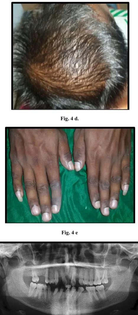
Fig. 4 d.
Fig. 4 e
Fig. 4d. OPG reveals congenitally missing maxillary lateral incisors, mandibular right canine 43, Left central incisor 31,
lateral incisor 32 and 2nd premolar 35
The salivary flow and the lacrimal secretion were found to be adequate. The patient was further subjected to further investigation and was diagnosed with hypohidrotic ectodermal dysplasia.
CASE 3
A patient named Vijayalakshmi aged 22 years came with a chief complaint of missing teeth in the upper and lower arch from birth. She had difficulty in speech, mastication and her esthetics was compromised.Personal history reveals she had severe intolerance to heat and had hyperxemia even in mild exertion.
Fig. 5a.
Fig. 5b.
Fig. 5c.
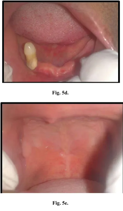
Fig. 5d.
Fig. 5e.
Fig. 5f.
Fig. 5h. Post prosthetic rehabilitation
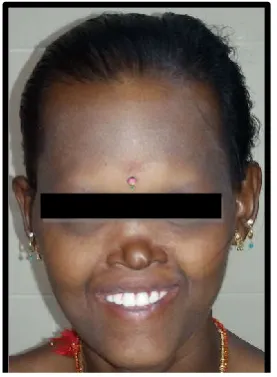
She gave a history of burning sensation all over the body. General examination reveals thinly built mildly elevated temperature with mid face hyperplasia, depressed nasal bridge, linear wrinkles and pigmentation in perioccular and forehead region with decreased eyelashes and loss of eyebrows, soft, dry, scaly skin surface sparse hair ( hypotrichosis), protruding lips, decreased vertical dimension, hypohydrosis, breathy voices noted, underdeveloped breasts. Intra oral examination reveals complete edentulous maxilla with unfavourable arch. Partial edentulous in mandibular arch with grade 2 mobility in right lower C – oligodontia. After being subjected to further investigations she was diagnosed with hypohidrotic ectodermal dysplasia. On radiological examination, the OPG revealed completely resorbed maxillary ridge and thin knife shaped ridges in mandible. Lateral cephalogram revealed loss of absence of frontal sinus, mid face atrophy.
DISCUSSION
Ectodermal dysplasia was first described by Thurnam [Nunnet
al., 2003; Yavuz, 2008] in 1848. Freire-Maia and Pinheiro
proposed the first classification system of the EDs in 1982 and classified it into different subgroups according to the presence or absence of hair anomalies or trichodysplasias, dental
abnormalities, nail abnormalities or onychodysplasias, [Nunn
et al., 2003] and dyshidrosis. In 2003, Lamartine [2003] reclassified it into the following four functional groups based on the underlying pathophysiologic defect:
cell-to-cell communication and signalling,
adhesion,
Development and
Others.
Thurman first reported two male cousins and their maternal grandmother with a hereditarysyndrome associated with sparse hair, missing teeth and dry skin. ED is an X linked recessive disorder affecting males and is inherited through female carriers. Clinical findings in carrier females are same as those in affected males. One third of the carriers appears healthy, another third of them show mild symptoms, and the last third exhibits significant symptoms, but often milder than the affected males. In our cases, both males and females were affected but features were more prominent in female patient.
The presentation of facial deformity, dryskin, and sparse hair in this report is similarto previous reports. These features are dueto anomalies of the skin appendages, whichinclude the hair follicles, sweat glands andsebaceous glands [Berg et al., 1990; Lowry et al., 1996]. Pure ED is characterized by defects in ectodermal structures alone, while ED syndromes are combined with other anomalies like cleft lip and palate. In autosomal recessive condition, there is a total absence of permanent teeth with or without taurodontism of primary molars, which was not observed in our patients. Cases have been reported where both primary and permanent teeth were
congenitally missing, [Vieira, 2007] which was seen in the
female patient vijayalakshmi.
Oral rehabilitation of the ectodermal dysplasiapatient is necessary to improve both the sagittal and vertical skeletal relationship during craniofacial growth and development as well as to provide improvements in esthetics, speech, and masticatory efficiency.2 Although removable prostheses are the most common treatment method, dental implants are also considered to be a treatment option.
Dental implants combined with implantsupported dentures for adolescents over 12 years of age are recommended as a treatment choice in literature. In situations where implant therapy is indicated, the main problem is insufficient bone; if bone atrophy progresses in these already alveolar- deficient patients, implant placement may not be possible without bone grafting.8Conversely, implantation reconstruction surgery is subject to a greater risk of failure compared to more conservative prosthetic treatment, besides its psychological aspects particularly in young children.8,12 Early implant placement in a growing child may cause cosmetic problems because the implants act like ankylosed teeth. With the vertical development of the jaws, implant and are to be treated with proper treatment plan and a continuous follow up. These case reports highlight the importance of accurate treatment planning as well as the influence of anodontia, hypodontia and oligodontia on the diagnosis of ectodermal dysplasia. Since the oral rehabilitation of these cases is often difficult, particularly in pediatric patients, treatment should be administered by a multidisciplinary team involving pediatric dentistry, orthodontics, prosthodontics, and oral-maxillofacial surgery.
Conclusion
Oral physicians are often the first who diagnose these patients. Therefore, they should be aware of the clinical manifestations of this syndrome. This will be helpful in proper diagnosis, early interventions and appropriate therapies for these patients and to provide a better platform for a confident smile in their future journeys.
REFERENCES
Abadi, B., Herren, C. 2001. Clinical treatment of ectodermal dysplasia: a case report. Quintessence Int., 32:743-745. Berg, D., Weingold, D.H., Abson, K.G., Olsen, E.A. 1990.
Sweating in ectodermal dysplasia syndromes: A review. Arch Dermatol. 126:1075–9.
Lamartine, J. 2003. Towards a new classification ofectodermal dysplasias. Clin Exp Dermatol, 28:351-5
Lowry, R.B., Robinson, G.C., Miller, J.R. 1966. Hereditary ectodermal dysplasia. Symptoms, inheritance patterns, differential diagnosis, management. Clin Paediatric 5:395-402.
Nunn, J.H., Carter, N.E., Gillgrass, T.J., Hobson, R.S., Jepson, N.J., Meechan, J.G., et al. 2003. The interdisciplinary management of hypodontia: background and role of paediatric dentistry. BrDent J., 194:245-251.
Paschos, E., Huth, K.C., Hickel, R. 2002. Clinical management of hypohidroticectodermal dysplasia with anodontia: case report. J Clin Pediatr Dent., 27:5-8.
Rad, A.S., Siadat, H., Monzavi, A., Mangoli, A.A. 2007. Full mouth rehabilitation of a hypohidrotic ectodermal dysplasia patient with dental implants: a clinical report. J Prosthodont 16:209-213.
Tarjan, I., Gabris, K., Rozsa, N. 2005. Early prosthetic treatment of patients with ectodermal dysplasia: a clinical report. J Prosthet Dent., 93:419-424.
Tarjan, I., Gabris, K., Rozsa, N. 2005. Early prosthetictreatment of patients with ectodermal dysplasia: A clinical report. J Prosthet Dent., 93:419-24.
Vieira, K.A., Teixeira, M.S., Guirado, C.G., Gaviao, M.B. 2007. Prosthodontic treatment of hypohidrotic ectodermal dysplasia with complete anodontia: case report. Quintessence Int., 38:75-80.
Yavuz, İ., Ülkü, S.Z., Ünlü, G., Kama, J.D., Kaya, S., Adıgüzel, O., Kaya, F.A., Tümen, E.C., Zortuk, M., Bahsi, E. 2008. Arslanoğlu Z. Ectodermal dysplasia: clinical diagnosis. Int Dent Med Disorders., 1:1-10.