R E S E A R C H
Open Access
Recommendations for the management of
MPS IVA: systematic evidence- and
consensus-based guidance
Mehmet Umut Akyol
1, Tord D. Alden
2, Hernan Amartino
3, Jane Ashworth
4, Kumar Belani
5, Kenneth I. Berger
6,
Andrea Borgo
7, Elizabeth Braunlin
8, Yoshikatsu Eto
9, Jeffrey I. Gold
10, Andrea Jester
11, Simon A. Jones
12,
Cengiz Karsli
13, William Mackenzie
14, Diane Ruschel Marinho
15, Andrew McFadyen
16, Jim McGill
17,
John J. Mitchell
18, Joseph Muenzer
19, Torayuki Okuyama
20, Paul J. Orchard
21, Bob Stevens
22, Sophie Thomas
22,
Robert Walker
23, Robert Wynn
24, Roberto Giugliani
25, Paul Harmatz
26, Christian Hendriksz
27*, Maurizio Scarpa
28,
MPS Consensus Programme Steering Committee and MPS Consensus Programme Co-Chairs
Abstract
Introduction:Mucopolysaccharidosis (MPS) IVA or Morquio A syndrome is an autosomal recessive lysosomal storage disorder (LSD) caused by deficiency of theN-acetylgalactosamine-6-sulfatase (GALNS) enzyme, which impairs lysosomal degradation of keratan sulphate and chondroitin-6-sulphate. The multiple clinical manifestations of MPS IVA present numerous challenges for management and necessitate the need for individualised treatment. Although treatment guidelines are available, the methodology used to develop this guidance has come under increased scrutiny. This programme was conducted to provide evidence-based, expert-agreed recommendations to optimise management of MPS IVA.
Methods:Twenty six international healthcare professionals across multiple disciplines, with expertise in managing MPS IVA, and three patient advocates formed the Steering Committee (SC) and contributed to the development of this guidance. Representatives from six Patient Advocacy Groups (PAGs) were interviewed to gain insights on patient perspectives. A modified-Delphi methodology was used to demonstrate consensus among a wider group of healthcare professionals with experience managing patients with MPS IVA and the manuscript was evaluated against the validated Appraisal of Guidelines for Research and Evaluation (AGREE II) instrument by three independent reviewers.
Results:A total of 87 guidance statements were developed covering five domains: (1) general management principles; (2) recommended routine monitoring and assessments; (3) disease-modifying interventions (enzyme replacement therapy [ERT] and haematopoietic stem cell transplantation [HSCT]); (4) interventions to support respiratory and sleep disorders; (5) anaesthetics and surgical interventions (including spinal, limb, ophthalmic, cardio-thoracic and ear-nose-throat [ENT] surgeries). Consensus was reached on all statements after two rounds of voting. The overall guideline AGREE II assessment score obtained for the development of the guidance was 5.3/7 (where 1 represents the lowest quality and 7 represents the highest quality of guidance).
(Continued on next page)
© The Author(s). 2019Open AccessThis article is distributed under the terms of the Creative Commons Attribution 4.0 International License (http://creativecommons.org/licenses/by/4.0/), which permits unrestricted use, distribution, and reproduction in any medium, provided you give appropriate credit to the original author(s) and the source, provide a link to the Creative Commons license, and indicate if changes were made. The Creative Commons Public Domain Dedication waiver (http://creativecommons.org/publicdomain/zero/1.0/) applies to the data made available in this article, unless otherwise stated.
* Correspondence:[email protected]
27Steve Biko Academic Hospital, University of Pretoria, Pretoria, South Africa
(Continued from previous page)
Conclusion:This manuscript provides evidence- and consensus-based recommendations for the management of patients with MPS IVA and is for use by healthcare professionals that manage the holistic care of patients with the intention to improve clinical- and patient-reported outcomes and enhance patient quality of life. It is recognised that the guidance provided represents a point in time and further research is required to address current knowledge and evidence gaps.
Keywords:Morquio a syndrome, Mucopolysaccharidosis, MPS IVA, Management guidelines, Elosulfase alfa, VIMIZIM, Enzyme replacement therapy, ERT, Haematopoietic stem cell transplantation, HSCT, Surgery, Anaesthetics
Background
Mucopolysaccharidoses (MPS) are a clinically hetero-geneous group of rare metabolic disorders referred to as lysosomal storage disorders (LSDs). MPS disorders arise due to deficiencies in enzymes that break down glyco-saminoglycans (GAGs) [1–3]. The build-up of GAGs, either directly or indirectly, cause progressive damage to cells, tissues and various organ systems and result in severe morbidity and reduced life expectancy [4, 5]. Clinical features that are common to all MPS disorders include skeletal and joint abnormalities, dysfunction in vision and hearing, cardiorespiratory problems, hepato-splenomegaly and coarse features [1]. Changes in the central nervous system are characteristic of some of these disorders, with typical imaging findings including white matter lesions, hydrocephalus, cervical spinal canal stenosis and bone abnormalities of the skull and spine [6]. MPS IVA or Morquio A syndrome (253000) is an auto-somal recessive MPS disorder caused by deficiency of the N-acetylgalactosamine-6-sulfatase (GALNS) enzyme (EC 3.1.6.4), which impairs lysosomal degradation of keratan sulphate and chondroitin-6-sulphate [7–11]. Results of a systematic review indicate that the point prevalence of MPS IVA was 1 per 926,000 in Australia, 1 per 1,872,000 in Malaysia and 1 per 599,000 in the UK [12]. Many genetic mutations have been identified and are responsible for the heterogeneous clinical presentation, which make diagnosis challenging. Although elevated urinary GAGs or reduced GALNS activity in dried blood spot results may be indicative of MPS IVA, a definite diagnosis is often only achieved through demonstration of reduced GALNS activity in leukocytes or fibroblasts or with mo-lecular confirmation of two disease-causing mutations in theGALNSgene [13].
MPS IVA is a heterogeneous and progressive disorder. Skeletal and joint abnormalities including genu valgum, joint hypermobility, hip subluxation and dysplasia, spinal cord compression, spinal instability and thoracolumbar kyphoscoliosis are the most prevalent manifestations [8, 14–16], and patients require regular assessment of multi-organ involvement [17]. Respiratory impairment and spinal cord instability are the main cause of morbidity and
mortality [14, 18, 19] and cardiac dysfunction has also been reported [20]. Other manifestations include corneal clouding and hearing loss.
Objectives
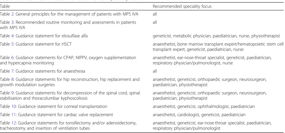
This manuscript provides robust guidance for the man-agement of adult and paediatric patients with MPS IVA using a validated modified-Delphi methodology to gain consensus. The guidance is comprised of a holistic set of recommendations for the timely and appropriate use of medical and surgical interventions and management of the natural history of MPS, with the intention to main-tain and enhance patient quality of life with the intention to maintain and enhance patient quality of life (QoL) and improve and improve clinical- and patient re-ported- outcomes. The guidance is intended for use by healthcare professionals that manage the care of patients with MPS IVA, in particular paediatricians and geneti-cists, and aims to enhance multidisciplinary practice across specialities. It also provides specific guidance for other specialists (Table 1) and stakeholders in the health services who are in contact with patients with MPS and is a useful reference for patient advocates, patients and their families. Table 1 describes the areas of clinical focus covered within this guidance and the correspond-ing recommended speciality focus.
This guidance was developed as part of a broader con-sensus programme that also covered the management of MPS VI, the results of which are published in a compan-ion article (Recommendations for the management of MPS VI: systematic evidence- and consensus-based guidance).
Methods and process
The methods described below cover both MPS IVA and MPS VI; however, the results section focuses on MPS IVA only.
Convening of the steering committee
Co-Chairs made recommendations for 22 SC members from across the world, including experts in: anaesthesia, ear nose and throat (ENT) surgery, cardiology, genetics, endocrinology, hand surgery, HSCT, neurosurgery, oph-thalmology, orthopaedic surgery, paediatrics, pain man-agement and pulmonology. To ensure the patient view was represented, three members from Patient Advocacy Groups (PAGs) formed part of the SC group. The SC de-fined the scope of the programme, including the medical and surgical interventions to be covered in the guidance and provided search terms for the literature review. Fur-ther details, including the competing interests, institu-tions and contribuinstitu-tions of each SC member are listed within the declarations section of this manuscript.
Setting the clinical questions to be addressed by the guidance The clinical questions to be addressed by the guidance were developed by the SC group according to the pa-tient, interventions, comparator and outcome (P.I.C.O.) methodology (Additional file 1: Appendix 1a) and are shown below.
1. What are the general principles for the management of adult and paediatric patients with MPS IVA/VI? 2. What are the recommended routine monitoring
and assessments that should be used to track the natural history of adult and paediatric patients with MPS IVA/VI and indicated interventions to be used during the care of the common symptoms of MPS? 3. For adult and paediatric patients with MPS IVA/VI, what is the impact on clinical outcomes and safety/ tolerability of:
Interventions that address the underlying enzyme deficiency including:
ERT
HSCT
Interventions used to manage the symptoms of MPS IVA/VI including:
Respiratory and sleep disorders
Anaesthesia
Limb and spinal surgeries
Ophthalmic surgeries
Cardio-thoracic surgeries
ENT surgeries
A secondary focus of the programme was to highlight current evidence gaps and provide recommendations for future treatment directions. Topics that were deemed out of scope of this programme included: comprehensive recommendations for diagnosis (e.g. screening, testing and identification of differential disease phenotypes), validation of new clinical outcome assessment tools (e.g. to assess patient-reported outcomes) and defining mi-nimal clinically important differences for MPS IVA/VI.
Patient Advocacy Groups insights
Consultations were conducted with representatives from six global PAGs (including Brazil, Canada, Germany, Turkey, the United Kingdom and the United States) [listed in the acknowledgements section of the manu-script]. To gain an understanding of the experience and views of patients with MPS IVA/VI on the holistic ma-nagement of MPS across several geographies and health-care systems, the consultations addressed: the biggest challenges faced by patients with MPS IVA/VI; the
Table 1Clinical areas of focus and recommended speciality focus
Table Recommended speciality focus
Table2: General principles for the management of patients with MPS IVA all
Table3: Recommended routine monitoring and assessments in patients with MPS IVA
all
Table4: Guidance statement for elosulfase alfa geneticist, metabolic physician, paediatrician, nurse, physiotherapist
Table5: Guidance statement for HSCT anaesthetist, bone marrow transplant expert/hematopoietic stem cell transplant expert, geneticist, paediatrician, nurse
Table6: Guidance statements for CPAP, NIPPV, oxygen supplementation and hypercapnia monitoring
anaesthetist, ear-nose-throat specialist, geneticist, paediatrician, respiratory physician/pulmonologist, nurse
Table7: Guidance statements for anaesthesia all
Table8: Guidance statements for hip reconstruction, hip replacement and growth modulation surgeries
anaesthetist, geneticist, orthopaedic surgeon, neurosurgeon, paediatrician, physiotherapist
Table9: Guidance statements for decompression of the spinal cord, spinal stabilisation and thoracolumbar kyphoscoliosis
anaesthetist, geneticist, orthopaedic surgeon, neurosurgeon, paediatrician, physiotherapist
Table10: Guidance statement for corneal transplantation anaesthetist, geneticist, ophthalmologist, paediatrician
Table11: Guidance statement for cardiac valve replacement anaesthetist, cardiologist, geneticist, paediatrician
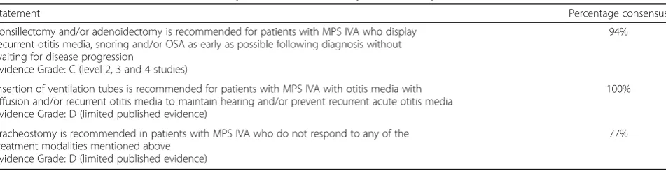
Table12: Guidance statements for tonsillectomy and/or adenoidectomy, tracheostomy and insertion of ventilation tubes
impact of medical/surgical interventions on patient outcomes, including: physical function/performance, mental/emotional outcomes and socioeconomic out-comes/cost implications; the overall benefit/risk profile associated with medical/surgical interventions and how this influences patient choice; barriers to the use of medical/surgical interventions across each region (e.g. reimbursement challenges, cost issues, access issues etc) and facilitators and barriers for application of the guidance.
Systematic literature review methodology
A systematic literature review was independently con-ducted by three Biographical Fellows in accordance with the Preferred Reporting Items for Systematic Reviews and Meta-Analyses (PRISMA) statement [21]. The focus of the literature review was to collate evidence for the clinical questions. Owing to the nature of the re-commendations for general principles and routine moni-toring and assessments, the associated guidance is predominantly based on clinical opinion; however, where available, published evidence has been applied to sup-port these statements.
Literature searches were performed in July 2017 using PubMed, Web of Science, SCOPUS and the Cochrane Database of Systematic Reviews; no early date limit was set. Supplementary searches of Google Scholar were undertaken to identify recent publications. Search strings incorporated Medical Subject Headings and free text key words, and were defined based on the P.I.C.O. methodology to answer each of the clinical questions (Additional file 1: Appendix 1a) [22]. Reproducible search strategies were reviewed by the SC (Additional file 1: Appendix 1b).As part of the screening, duplicates were removed. Exclusion criteria included: in-vitro/animal studies, non-English language studies, studies that did not report an outcome, diagnosis studies, non-systematic re-view articles and studies that did not relate to a specific intervention. Only studies conducted in humans were included and evidence was extrapolated from other MPS types. Single case studies were captured and referred to where evidence was scarce. The bibliographies of identi-fied pre-existing guidelines and review articles were checked for additional relevant studies. The following information was extracted from included studies: refer-ence details, patient population, intervention, comparator (where applicable), outcomes (physical function/perfor-mance, mental/emotional, socioeconomic), safety and tolerability and reported study limitations. Data were extracted by one Bibliographic Fellow and checked by a second. Any discrepancies in screening and data extrac-tion were resolved through discussion or the intervenextrac-tion of a third reviewer. Results were reported according to PRISMA (Additional file1: Appendices 1b and 1c).
The quality of evidence level for each paper was assessed using the Oxford Centre for Evidence-based Medicine (OCEM) criteria. In accordance with the criteria (Additional file 1: Appendix 2) each reference obtained from the systematic literature review was critically appraised and designated an evidence level based on the study design, rigour of methodology and out-comes (Additional files 2 and 3). The recommenda-tion statements were then graded based on the average evidence level for each supporting reference (Additional files 2 and 3). The evidence levels and grades were cross-checked by the SC group and used to assess the strength of each recommendation during the development of the guidance statements.
Development and validation of guidance statements For each clinical area of focus and intervention, the SC developed draft guidance statements accompanied by supporting text, which were based on the results of the literature search, insights from the PAG interviews and expert clinical opinion. This process was facilitated by an independent secretariat via a series of face-to-face and online meetings and email correspondence across a 14-month period.
scale from 1 (strongly disagree) to 9 (strongly agree). Scores of 6 or less were defined as disagreement with the statement and it was mandatory for respondents to provide rationale for their disagreement and suggest amendments to the statement. Scores of 7 or more were defined as agreement with the statement and respondents were given the option to add comments.
Consensus was reached when ≥75% of respondents agreed with a given statement. This consensus threshold was determined by review of literature and applied to the rare disease field by the Steering Committee group. The most commonly reported definition of consensus for Delphi studies is percent agreement, with 75% being the median threshold to define consensus [24].
Statements for which consensus was not achieved in the first round of voting were amended by the SC based on respondent feedback and expert clinical opinion and sent for re-voting. The percentage consensus and assigned evidence grade for each guidance statement is shown within the results section below.
The quality of study reporting in this programme was independently assessed by three reviewers using the A-ppraisal of Guidelines for Research and Evaluation (AGREE) II Instrument (declared in the acknowledge-ments section of the manuscript) [25].
Measures to ensure independence
The programme was funded by BioMarin; the manufac-turer of the recombinant enzymes used to treat MPS IVA and MPS VI, however, they were not involved in any stages of the process and did not influence the scope or content of the programme. BioMarin were absent from all SC meetings, were blinded to the guidance statements and were not involved in the publication process. The programme was managed by an independ-ent secretariat (Lucid Partners Ltd), and the scope of the programme and content, including the development of guidance statements, was led by the SC with editorial support provided by the secretariat. The SC was iden-tified through a systematic expert mapping process, conducted independently of the funder. Conflicts of in-terests for all SC members (shown in the declarations section) were recorded at the start of the consensus programme and updated throughout the programme. Following the systematic expert mapping exercise, it was noted that some of the SC had previously worked on a consultancy basis with the programme sponsor, who hold the marketing authorisation for approved phar-maceutical therapy in MPS IVA. Efforts were therefore taken to ensure representation from leading experts across other treatment modalities, including HSCT/ BMT on the Steering Committee panel and during the modified-Delphi voting process, where a large number
of physicians across multiple specialities and geographies were engaged. At several stages during the process, the SC were also required to provide updated conflict of interest disclosures.
Results
Here we report the results relevant to the management of patients with MPS IVA. The results for MPS VI are published in a companion article (Recommendations for the management of MPS VI: systematic evidence- and consensus-based guidance).
Patient Advocacy Groups insights
The results of the PAG consultations highlighted that the biggest current unmet needs and challenges faced for patients with MPS IVA/VI include: treatment expert-ise, timing and access to treatment, appropriate infra-structure, maintenance of independence and social prejudice. The results also summarised the factors that affect patient prognosis and QoL, key considerations for all surgical interventions with an emphasis of anaesthe-sia, barriers to use of medical/surgical interventions and patient and caregiver considerations.
Detailed insights from all PAG consultations were pre-sented to the SC group by the patient advocates who formed part of the SC and were used to inform the development of the guidance statements and ensure re-presentation of the patient voice. These insights assisted with the development of the general principles for management of patients with MPS IVA/VI, and ensured holistic care was considered across the whole guidance development process.
Modified-Delphi results
In round two of the modified-Delphi voting, 13 statements were sent for re-voting to 145 MPS physi-cians (experts of specialties not relevant to these state-ments were removed from the mailing list). A total of 71 survey submissions were received from 53 institu-tions across 18 countries and two respondents did not meet the minimum experience threshold and their submissions were excluded (further information in-cluding the number of respondents/statements, re-spondent specialities and geographies, and rere-spondent feedback to guidance statements are including within Additional file 5). Consensus was reached on all 13 statements (Additional file 5). In total, 87 guidance statements reached consensus for MPS IVA.
Appraisal of Guidelines for Research and Evaluation (AGREE) II assessment
The methodological rigour and transparency employed within the development of this guidance was evaluated through the review of the manuscripts against the validated AGREE II instrument. Three independent reviewers [listed
in the acknowledgements section of the manuscript] assessed the manuscript and suggested amendments were addressed where possible; a subsequent second round of re-view was conducted by all rere-viewers. Across each AGREE II criteria, the average domain scores obtained were: scope and purpose (85%), stakeholder involvement (85%), rigour of development (73%), clarity of presentation (78%), applic-ability (34%) and editorial independence (64%) (full infor-mation including the scores from the two rounds of AGREE II evaluation can be found in Additional file1: Ap-pendix 3). The guidance documents were given an overall guideline assessment score of 5.3/7 (where 1 represents the lowest quality, and 7 represents the highest quality).
Guidance statements
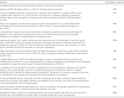
General principles (Table2)
The SC noted that newborn screening in MPS IVA would allow earlier diagnosis if/when available thus leading to earlier intervention, which would likely change the course of disease. As comprehensive re-commendations for diagnosis of MPS IVA is out of
Table 2General principles for the management of patients with MPS IVA
Statement Percentage consensus
All guidance statements are evidence Grade D (level 5 expert clinical opinion)
Diagnosis of MPS IVA during infancy is critical to optimise patient outcomes 98%
The first consultation should be conducted by a physician with experience of treating MPS as soon as possible after diagnosis. This should include a full discussion of disease pathology, progression, treatment options and management. Ongoing information should be provided to optimise patient outcomes
97%
Patients and caregivers should receive ongoing psychosocial support from a social worker and/or psychologist, and should be directed towards an MPS society or relevant patient organisation in their country
94%
A comprehensive medical history and multi-system evaluation should be conducted within days of diagnosis to set a baseline for ongoing assessments and evaluate the physical and neurological manifestations of disease, functional ability and disease burden
88%
Ongoing and regular multi-system monitoring, and assessments are recommended to track the natural history of MPS IVA, monitor the impact of treatment and assess the need for treatment interventions to manage the symptoms of MPS IVA. These should be conducted at every clinic visit, annually or in some cases as clinically indicated (for example pre- and post-operatively)
100%
Timely interventions are recommended where clinically indicated by monitoring, to help avoid irreversible damage caused by the natural history of MPS IVA, and to manage the disease manifestations and maintain long-term QoL
99%
A multidisciplinary team (MDT) of metabolic specialists, surgeons and allied healthcare professionals (including, but not limited to: nurses, physiotherapists, occupational therapists, psychologists and audiologists) is required to manage the diverse range of disease manifestations of MPS IVA
99%
Co-ordination of the entire MDT care team is required prior to any procedure to determine the need for surgery, to discuss the benefits and risks of combining surgeries to minimise the need for multiple anaesthetics and to decide the optimal order of procedures. The decision to combine surgeries should take into consideration the surgical and intubation time, and complexity of procedures
93%
The risks and benefits of any intervention and the competing risks of other medical problems should be assessed and discussed with patients, families and caregivers such that they can make an informed decision on the appropriateness of the therapy/surgery
100%
Surgical procedures should be performed by (or under the guidance of) specialist surgeons and anaesthetists with experience of MPS, in medical centres with intensive care units
99%
Management of pain should be a fundamental part of the care of patients with MPS IVA, with the aim of improving QoL and maintaining mobility. Refer to general guidelines for pain management
scope of this guidance, more detailed discussions of newborn screening can be found elsewhere [17]. General guidelines for pain management in patients with MPS have also been published elsewhere [26,27].
Recommended routine monitoring and assessments (Table3)
Disease-modifying interventions
ERT (elosulfase alfa) in patients with MPS IVA (Table4)
Rationale and evidence baseElosulfase alfa [28] is a recombinant form of the human lysosomal enzyme GALNS, which is deficient in patients with MPS IVA [29]. ERT with elosulfase alfa aims to transiently restore GALNS activity, thereby preventing the accumulation of KS and chondroitin-6-sulphate in lysosomal compart-ments of cells, which is responsible for the clinical mani-festations of MPS IVA. Elosulfase alfa is currently the only disease-specific treatment that is licensed for pa-tients with MPS IVA, having been validated in clinical trials [30–32]. In a Phase III clinical trial, patients who received intravenous elosulfase alfa at a dose of 2 mg/kg/ week experienced reductions in urinary levels of keratan sulphate (a pharmacodynamic biomarker for the disease) [30]. Elosulfase alfa has been shown to improve endur-ance and exercise capacity (as measured by the 6MWT), which may in part relate to improved respiratory func-tion and oxygen utilizafunc-tion [30,33–36]. A trend for im-provement in performance of ADL was also observed in a long-term extension study. The findings suggest that long-term elosulfase alpha ERT is associated with partial recovery of functional abilities, improving Morquio A patients’abilities to perform ADL [36]. The evidence for the effect of elosulfase alfa on bone is currently limited and further research is required.
Elosulfase alfa is well-tolerated [37] and results of a randomized, double-blind, pilot study has shown that treatment reduces pain in some patients with MPS IVA [38]. The majority of adverse events in clinical trials were infusion associated reactions (IARs), which are defined as reactions occurring after initiation of infusion until the end of the day following the infusion. Serious IARs were observed in clinical trials and included ana-phylaxis, hypersensitivity and vomiting [28]. As stated on the US Prescribing Information for elosulfase alfa, the most common symptoms of IARs (occurring in ≥10% of patients treated with elosulfase alfa and≥5% more when compared with placebo) were headache, nausea, vomiting, pyrexia, chills and abdominal pain. IARs were generally mild or moderate and the frequency was higher during the first 12 weeks of treatment and tended to occur less frequently with time [28,39].
Early intervention with elosulfase alfa is associated with a trend towards improvement in growth however
the data is currently limited [40]. Most data related to the use of elosulfase alfa are from patients who initiated ERT relatively later in their disease. The early initiation of ERT will likely change the course of disease in patients with MPS IVA; therefore, additional studies are warranted to determine the long-term outcomes of patients in whom elosulfase alfa treatment is adminis-tered from an early age.
Considerations prior to starting ERTIt is important to evaluate the life-long impact of elosulfase alfa on an individual basis, as the benefits of treatment may not be consistent across all patients and there may be subpopu-lations of patients where the risk-benefit and efficacy versus cost effectiveness is less certain (e.g. less severe phenotypes) [41]. The status of the patient, disease bur-den, comorbidities and prognosis should be fully consid-ered prior to initiation. The first dose of elosulfase alfa should (where possible) be administered by a clinician with experience of metabolic disorders and take place within an infusion centre/hospital with facilities for ef-fective management of allergic/anaphylactic reactions. Home infusion may be considered where possible; this decision should be made by the physician and patient. Careful patient selection, good vascular access and a de-tailed management plan for IARs and anaphylaxis are es-sential for the success of this approach [42]. Consideration should be given to the need for a totally implantable vascular access device (TIVAD) to facilitate long-term venous access for frequent or continuous ad-ministration of ERT; the patient and their families should be made aware of the benefits and risks of using such a device [43, 44]. Patients with acute febrile or re-spiratory illness may be at increased risk of life-threatening complications from hypersensitivity re-actions; therefore, the clinical status of each patient should be evaluated prior to the administration of elosulfase alfa and if necessary, delay of treatment should be considered.
Table 3Recommended routine monitoring and assessments in patients with MPS IVA
Statement Percentage consensus
All guidance statements are evidence Grade D (level 5 expert clinical opinion), unless otherwise stated
Physical examination
A physical examination should be performed during every visit to assess general health, growth, vital signs, abdominal organ size, presence of hernia, neurologic function (including gait), ligamentous laxity, and functions of the eyes, ears, heart and lungs
90%
Routine physical examination can also identify signs of potential respiratory problems, such as an enlarged tongue or sniffing position
90%
Radiology
While X-rays are essential to identify the natural history of disease and response to treatment, efforts should be made to minimise radiation exposure, and images should be requested only when clinically useful
85%
Hips: an anteroposterior (AP) pelvis radiograph should be performed at diagnosis and as clinically indicated (based on physical examination or reports of pain) to quantify hip dysplasia or identify early signs of hip migration
88%
Lower limbs: in patients with clinical evidence of valgus deformity of the lower limbs, standing AP radiographs of lower extremities should be performed prior to guided growth surgery
100%
Spine: standing or sitting plain radiography of the cervical and thoracolumbar spine to examine for spinal deformities is recommended in patients with MPS IVA at diagnosis and every 2–3 years thereafter, or sooner if clinically indicated
85%
Magnetic resonance imaging (MRI) of the whole spine (in neutral position) should be performed annually in children with MPS IVA to assess for spinal cord injury. The frequency may be reduced for adult patients with stable imaging who do not display symptomsa
84%
Flexion/extension MRI of cervical spine may be needed to identify changes in spinal canal and spinal cord 86%
MRI of the brain is recommended at diagnosis in patients with MPS IVA, and should be repeated as needed in individuals with clinical suspicion of hydrocephalus
80%
MRI of the brain and spinal cord in patients with MPS IVA may require sedation or general anaesthesia, depending on patient age and cooperation. General anaesthesia carries substantial risk for patients with MPS
95%
Flexion/extension computerised tomography (CT) of the craniocervical junction may be considered in patients with MPS IVA if MRI is not available or if sedation is not possible
92%
The presence of specific radiological signs may indicate the need for surgical intervention to correct skeletal deformities; however, there is insufficient evidence to support preventative surgery based on radiological findings
88%
Endurance
Choice of assessment depends on the patient’s physical and developmental ability 97%
Baseline assessment is the most important and ideally two values should be obtained as a minimum. Consistent protocols should be used when performing repeat measurements to minimise variability
95%
Annual endurance testing using the 6-min walk test (6MWT) is recommended, as per the American Thoracic Society guidelines [1,45]
Evidence Grade: C (level 4 study and extrapolation from level 1 study) [8,46]
87%
In patients with limited ambulation who are unable to perform the 6MWT, endurance should be assessed via alternative methods such as an adapted timed 25-ft walk test (T25FW)
76%
Endurance testing is also recommended prior to initiation of ERT and annually thereafter as a measure of treatment efficacy and to provide early evidence of possible neurologic or skeletal issues
87%
Growth
Assessment of growth should be performed at each clinic visit (ideally every 6 months) as part of a regular physical examination and should include: standing height (sitting height if the patient is unable to stand), length (supine position), weight, head circumference (≤3 years), Tanner pubertal stage (until maturity) [47]
95%
Height and weight should also be measured before initiation of ERT and at every clinic visit thereafter (ideally every 6 months) to evaluate the impact of treatment [47]
95%
Urinary keratan sulphate (KS)/urinary glycosaminoglycan (uGAG) levels
Where available, tandem mass spectrometry may be used to assess levels of urinary keratan sulphate prior to starting elosulfase alfa and every 6 months thereafter to determine the pharmacodynamic effects of ERT [48] Evidence Grade: D (level 3/4 studies support the statement; [8,49–54] however, one level 3 study [55] does not support use of urinary keratan sulphate for monitoring the therapeutic effect of ERT)
94%
Total uGAG levels are often elevated in neonates and infants with MPS IVA and may overlap with normal values in adults and some teenagers. However, if a specific keratan sulphate assay is not available, measurement of uGAG levels using standard dye-binding methods may be useful. Preferably, measurements should be performed in the same laboratory and assessed against age-related reference values
Table 3Recommended routine monitoring and assessments in patients with MPS IVA(Continued)
Statement Percentage consensus
Cardiac function
Initial cardiac evaluation should be performed at the time of diagnosis and include assessment of vital signs with measurement of oxygen saturation, right arm and leg blood pressure measurements, careful auscultation, full transthoracic two-dimensional and Doppler echocardiogram, and 12-lead electrocardiogram (ECG)
100%
Longer ECG monitoring (prolonged Holter/event monitoring) may be considered in older patients, especially if they have symptoms of black outs, unexpected falls or dizziness
96%
Follow-up in expert centres should be annually initially, but may be extended to every 2–3 years if there is no evidence of cardiac abnormality
92%
Additional cardiac assessment, including a standard ECGb, should be performed prior to any surgical procedure requiring general anaesthesia
92%
Neurological exam
A detailed neurological examination should be performed at every clinic visit (minimally every 6 months) and, where possible, these should correlate with imaging studies of the spine to detect early spinal stenosis or instability compromising the cervical cord. For patients without clinical or radiographic concern, annual neurological examination may be sufficient [56]
87%
Standard MRI of the cervical spine should be performed to assess for presence of spinal cord compression. In the absence of significant spinal cord compression, proceed with flexion/extension MRI to confirm the presence of worsening spinal cord compression with motionc
78%
Respiratory function and sleep disorder
Evaluation of respiratory function by spirometry, including forced vital capacity (FVC) and maximum voluntary ventilation (MVV), should be performed to assess changes in lung volume and obstruction in children over 5 years of age
97%
Respiratory function should be assessed annually until children stop growing, and every 2–3 years thereafter, provided that respiratory symptoms remain unchanged. Additional testing should be performed if respiratory symptoms change or if intercurrent illnesses occur
91%
Normative values are not available, therefore change in absolute volume from patient’s own baseline will be the best indicator of deterioration or improvement
97%
Measurement of respiratory rate and arterial oxygen saturation before and after annual endurance testing is recommended
86%
Evaluation of gas exchange and respiratory function is also recommended before any planned air travel, to ensure safety during the flight
86%
To identify symptoms of sleep apnoead, patients should be asked to report presence of snoring and morning headaches at every clinic visit
100%
An overnight sleep study (polysomnography) is recommended at diagnosis (if possible, and no later than 2 years of age), and every 3 years thereafter or when signs and symptoms of obstructive sleep apnoea (OSA) are noted
94%
Ear-nose-throat (ENT)
ENT examination, including tympanometrye, should be conducted every 3–6 months during childhood and every 6–12 months thereafter
91%
ENT examination in patients with MPS IVA should include visualisation of the upper respiratory tract to determine diagnosis, management and assist in pre-operative planning. Endoscopic examinations should be recorded and kept, to monitor disease progression
92%
Fibreoptic examination in patients with MPS IVA should be performed at diagnosis and at least annually thereafter, or as clinically indicated. For those individuals who require general anaesthesia, ENT examination should be performed during the pre-operative evaluation for other surgical procedures
83%
Upper airway CT, focused on airway anatomy preferably with reconstruction, may be useful to identify the area of the abnormality and possible cause of obstruction in patients with MPS IVA with suspected obstruction or malaciaf
92%
Age-adjusted audiometric assessment as a baseline objective hearing evaluation should be conducted at first clinic visit and repeated annually to assess conductive and sensory-neural hearing loss
Evidence Grade: C (Grade 4 studies) [57,58]
100%
If speech problems are determined during ENT examination, an assessment by a speech pathologist should be conducted [59]
100%
Balance tests should be conducted if the patient has a history of balance problem 95%
Ophthalmological function
Age-appropriate evaluations by an ophthalmologist is recommended every 6 months if possible, or at least annually [60]
evidence reporting the effects of discontinuation of ERT in MPS IVA, and additional research is required.
Considerations for managing specific adverse events of interest Due to the potential for hypersensitivity reac-tions with elosulfase alfa, patients treated in the clinical trial programme received antihistamine premedication, with or without antipyretics, 30 to 60 min prior to the start of the infusion. This approach is broadly followed in clinical practice but there is limited evidence for or against the necessity of premedication. Patients should be closely observed for signs of anaphylaxis during and after admin-istration of elosulfase alfa and if severe hypersensitivity
reaction occurs, hospital admission is advised. Patients with a with previous history of IARs, can be given add-itional premedication, such as H2 receptor blockers or montelukast sodium. Other risk factors may also include history of severe allergic diathesis, status asthmaticus, reactions to other infusion or biologic products, compro-mised airway or pulmonary function and previous his-tory of a significant pause between ERT treatment.
IARs are generally managed by reducing the rate of administration or by temporarily interrupting the infu-sion plus administration of additional antihistamines, an-tipyretics, or for more severe reactions, corticosteroids Table 3Recommended routine monitoring and assessments in patients with MPS IVA(Continued)
Statement Percentage consensus
Ophthalmic assessment may include visual acuity, refraction, slit-lamp examination of cornea, funduscopic evaluation including optic nerve, and measurement of intraocular pressure
100%
Scotopic and photopic electroretinogram may be performed in patients with clinical suspicion of retinopathy or when considering corneal transplantation [60]
100%
Intraocular pressure monitoring and pachymetry may be considered prior to corneal transplant [60] 100%
Evaluation of oral health by dentist
Close monitoring of dental development (at least annually) is recommended to prevent caries and attrition, as is monitoring of occlusion and chewing functions
100%
The need for subacute bacterial endocarditis (SBE) prophylaxis prior to dental procedures should be assessed by a cardiologist
100%
Disease burden
Annual assessment of patient-reported outcomes is recommended for: pain severity, QoL (as assessed by reproducible and age-appropriate questionnaires [e.g. EQ-5D-5 L]), fatigue), and activities of daily living (ADL; as assessed by functional tests [6MWT/T25FW]), age-appropriate ADL questionnaires (e.g. MPS Health Assessment Questionnaire [MPS HAQ]), and assessment of wheelchair/walking aid use [61]
97%
These assessments may have to be adapted both for language, culture, and individual physical limitations, as they have not been validated in specific disorders
97%
Physical therapy
Regular assessments by a physical therapist (lower limb), occupational therapist (upper limb) and rehabilitation medicine specialist should be conducted to assess limb function and provide support as needed
93%
The physical therapist could also assist in suggesting walking aids and other adaptations that may improve QoL 98%
Post-consensus comments by the SC to be taken into consideration:
a
Magnetic resonance imaging (MRI) can also be used to assess for spinal cord compression. The frequency may be reduced for older patients with stable imaging who do not display symptoms
bEchocardiogram (ECHO) should also be performed prior to any surgical procedure requiring general anaesthesia c
This topic was discussed in detail with the neurosurgical and orthopaedic colleagues in the SC group. It was their expert clinical opinion that flexion/ extension MRI is not dangerous to perform within the hands of an experienced team. It is important that the range of motion (ROM), flexion and extension of the patient is evaluated while they are awake immediately before anaesthesia. The ROM during anaesthesia should not exceed the ROM as noted in the awake state, and should only be carried out after it is confirmed that there is no spinal cord compression. See Table9for guidance statements on spinal surgeries including spinal cord decompression
d
Signs and symptoms for sleep apnoea (a type of sleep disordered breathing (SDB)) can be divided into nocturnal and daytime symptoms. Nocturnal symptoms include loud snoring, observed episodes of breathing cessation during sleep, abrupt awakenings accompanied by gasping or choking, and awakening with a dry mouth or sore throat. Daytime symptoms include excessive daytime sleepiness, morning headaches, difficulty concentrating during the day, personality and mood changes including depression or irritability, and high blood pressure. To identify presence of SDB, patients should be asked to report snoring and other signs and symptoms of SDB at every clinic visit
eTympanometry is used to measure the volume of the ear canal/tympanic membrane movement and indirectly assess for fluid accumulation and
opening of pressure equalising tubes
f
Upper airway CT may also be useful to identify the area of the abnormality and possible cause of obstruction in patients with MPS IVA with suspected tracheo bronchomalacia
Table 4Guidance statement for elosulfase alfa
Statement Percentage consensus
Initiation of long-term ERT with elosulfase alfa at a dose of 2.0 mg/kg/week through intravenous infusion is recommended in all patients with MPS IVA as soon as possible after a confirmed diagnosis Evidence Grade: B (level 2 or 3 studies)
(for a 12–18-h period prior to infusion). Due to the risk of sleep apnoea in patients with MPS IVA, use of a non-sedating antihistamine is recommended.
HSCT in patients with MPS IVA (Table5)
Rationale and evidence baseEvidence supporting the use of HSCT in patients with MPS IVA is limited and comprises a small number of case studies and a single institution evaluation of ADL following surgical inter-vention [40,62–66]. Evidence of GALNS expression was confirmed following transplantation, which showed the potential for cross-correction of other tissues [40, 62– 66]. Improvements in height and skeletal dysplasia have not been observed but this may be due to the transplant-ation being performed in older patients [40,62–66].
Due to increasing availability of well-matched donors, improved supportive care and modification to transplant regimens, the risks associated with transplantation have declined in recent years; however, mortality rates still vary between centres depending on experience, and ser-ious risks, including death, remain. A recent study sug-gests that peri-transplantation mortality has improved; however, this data was from two of the most experienced centres with MPS transplantation and involved other subtypes of MPS [67].
Due to the lack of evidence related specifically to MPS IVA, and the recognised risks of transplantation, HSCT cannot be considered as a recommended therapy in patients with MPS IVA. The strongest data for the use of HSCT are for other types of MPS, namely MPS IH (Hurler syndrome) [68]. Patients with MPS IH are treated with HSCT due to the effectiveness of this approach in treating the central nervous system (CNS) manifestations of the disease, which are not improved by ERT [69–73]. In MPS IH, the increased risk of HSCT compared with ERT is considered justified. Given that CNS disease is not prominent in patients with MPS IVA, the risk–benefit profile for the use of HSCT in these patients is less clear. There are reports that the incidence of hydrocephalus and cervical stenosis is decreased in MPS IH patients treated with HSCT as compared to treatment with ERT [74]. As there are no inherent cognitive manifestations in patients with MPS IVA similar to those seen in patients with MPS IH, it is unclear whether HSCT may be advantageous in patients with MPS IVA and caution should be taken when
extrapolating evidence from other MPS types [11,17,75]. There is a need for more research to better understand the long-term efficacy and safety of HSCT in patients with MPS IVA, and for a well-designed comparative study of HSCT and ERT in patients of similar age and disease severity.
Interventions to support respiratory and sleep disorders
Continuous positive airway pressure (CPAP),
non-invasive positive pressure ventilation (NIPPV),
oxygen supplementation and hypercapnia monitoring (Table6)
Rationale and evidence base Respiratory complica-tions are a major cause of morbidity and mortality in pa-tients with MPS IVA and are often among the first symptoms to appear [18,19,76]. Typical features of MPS IVA include upper and lower airway obstruction and re-strictive pulmonary disease which result from a variety of anatomical and functional abnormalities. Upper airway obstruction is attributable to cranial abnormalities, a short neck and progressive deposition of GAGs in the tissues surrounding the supraglottic upper respiratory tract, while lower airway obstruction reflects GAG deposition in the airway walls with resultant tracheal and bronchomalacia. Lung volume and chest expansion are further limited by short stature, chest wall deformities and abdominal organomegaly. Clinical manifestations include recurrent upper and lower respiratory tract infections, obstructive sleep apnoea (OSA) and impaired exercise tolerance [18]. If respiratory complications are not diagnosed and treated appropriately, respiratory failure can ensue, leading to early death [14,77]. There is evidence that long-term ERT is associated with sustained improvements in respiratory function in patients with MPS IVA [34]. In contrast, spe-cific interventions beyond ERT are required to address OSA and other forms of sleep disordered breathing (SDB). CPAP prevents upper airway collapse during inspir-ation and is the mainstay of treatment for OSA in the general population with benefical effects on blood pres-sure, cardiac events, mortality and QoL. [78, 79] A comprehensive review of the evaluation and treatment op-tions for SDB in MPS concludes that applying CPAP can prevent functional airway collapse during inspiration when asleep and may improve obstruction-related pulmonary hypertension, heart failure and/or respiratory failure [80,81].
Table 5Guidance statement for HSCT
Statement Percentage consensus
Due to the lack of evidence, HSCT cannot be recommended for patients with MPS IVA and at this time is considered an investigational procedure
Evidence Grade: D (level 3/4 studies with inconsistent risk/benefit results)
An alternate form of therapy is required for patients that demonstrate either persistent OSA despite CPAP or hypoventilation during sleep. NIPPV provides an in-creased pressure during the inspiratory phase of breathing to augment ventilation. Although outcome data in MPS are scarce, the effectiveness of this therapy has been dem-onstrated in a broad range of diseases including neuro-muscular and chest wall disorders; which are features that are frequently noted in patients with MPS. Evidence sug-gests that this approach results in an improvement in QoL, functional capacity and respiratory failure [81–83].
Supplemental oxygen can be prescribed for individuals that demonstrate persistent nocturnal oxygen desaturation and for patients who do not tolerate therapy with CPAP or NIPPV. Caution is required when prescribing oxygen because of the known suppression of respiratory drive and arousal from sleep with the potential for either worsening pre-existing hypercapnia or inducing onset of hypercapnia in susceptible patients.
Considerations for interventionSDB can be managed by application of CPAP, which delivers air at an elevated pressure via a mask that fits around the nose and/or mouth; however, consideration should be given to facial abnormalities that can make mask-fitting difficult. Patients should be monitored to ensure they do not develop sus-tained hypoventilation. To prevent pneumonia, vaccina-tions against respiratory pathogens causing influenza and pneumococcus infections should be recommended.
Anaesthesia and surgical interventions
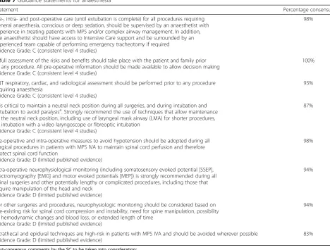
Use of anaesthesia in patients with MPS IVA (Table7)
Rationale and evidence base Patients with MPS IVA will likely require anaesthesia for multiple surgical inter-ventions and investigations during the management of their disease [84,85] but are considered high-risk due to potential difficulties with mask ventilation and endo-tracheal intubation. Other risk factors include: the pres-ence of narrow airways from adeno-tonsillar hypertrophy and deformity of the lower airways, odontoid peg hypopla-sia causing potential instability of the cervical spine, and
other skeletal abnormalities causing thoracic deformity and pulmonary disposition. In addition, cardiovascular and neurological impairment may also be present [86–88]. Adverse events (including fatalities and paralysis) oc-curring during anaesthesia have been reported in the literature [89–91]. Intubation and extubation can be challenging in patients with MPS IVA due to several factors including: restricted mouth opening; short neck length; limited range of motion (ROM) and upper airway obstruction, which is often caused by hypertrophied ton-sillar/adenoidal tissue, a large tongue with micrognathia, subglottic narrowing, and atlanto-axial instability due to odontoid hypoplasia and ligamentous laxity [77, 86, 89, 92–97]. Some patients also experience tracheal obstruc-tion, which in the presence of coexisting upper airway obstruction commonly remains unrecognised and can increase the risk of death during anaesthesia [89, 96]. Retrospective evaluation from 83 intubations of 108 anaesthetics (in 28 patients) demonstrated difficulties in intubation following cervical fusion [89]. Airway abnor-malities including tortuous appearance of the trachea and bronchi as a result of the abnormalities in the hya-line cartilage and deposits of glycosaminoglycans were observed, suggesting that MPS IVA results in abnorma-lities of both upper and large airways [89].
Management of the airway will require care to maintain neutrality of the cervical spine and may necessitate the use of videolaryngoscopy or fibreoptic techniques. Although hypothetical, poor perfusion related to arterial narrowing and reduced foramina di-ameters secondary to dysostosis should be anticipated by an anaesthetist, appropriately monitored with arterial lines, and supported in near-normal range during procedures.
Although epidural anaesthesia has been performed suc-cessfully in patients with MPS IVA [89], it is not currently recommended due to observations of spinal cord infarc-tion following lower extremity surgeries in which patients received an epidural for post-operative pain management [91,98,99]. Although data on peripheral nerve block are lacking, this approach may be considered during the care
Table 6Guidance statements for CPAP, NIPPV, oxygen supplementation and hypercapnia monitoring
Statement Percentage consensus
CPAP therapy is recommended for patients with MPS IVA who display the presence of obstructive sleep apnoea (OSA) that persists after tonsillectomy and/or adenoidectomy
Evidence Grade: D (limited published evidence)
97%
NIPPV therapy is recommended for patients with MPS IVA who display nocturnal hypoventilation and are unresponsive to CPAP, or display daytime hypoventilation with increased PaCO2and/or
serum HCO3levels
Evidence Grade: D (level 5 expert clinical opinion)
91%
Oxygen supplementation during sleep is recommended for patients with MPS IVA who exhibit sleep apnoea with nocturnal hypoxemia, and who do not tolerate CPAP or NIPPV masks Evidence Grade: D (level 5 expert clinical opinion)
77%
Patients with MPS IVA should be monitored for development of hypercapnia after starting oxygen therapy using measurement of PaCO2and/or serum HCO3
Evidence Grade: D (level 5 expert clinical opinion)
of patients with MPS IVA. The use of ultrasound technol-ogy can also aid successful nerve block. Intraoperative neurophysiological monitoring is recommended to pre-vent significant complications in this high-risk population; however, availability is extremely variable.
Considerations for use of anaesthesia Due to the risk of upper airway obstruction, pre-operative seda-tive premedication may be used with caution and only with appropriate monitoring. Assessment of upper and lower airway anatomy (for example, a pre-operative flexible nasopharyngolaryngoscopy and three-dimensional CT scan where feasible), cardiac function, and potential cervical spine instability should be performed prior to any pro-cedure that requires sedation or anaesthesia. MRI scans of the spine in a neutral position or patient-initiated flexion/extension X-ray of the spine can be performed to assess the risk of spinal cord compression and in-stability (flexion extension X-ray measures inin-stability only). Flexion/extension imaging is important to evalu-ate for cervical spine instability prior to anaesthesia. The frequency of imaging depends on both the patient’s age and clinical condition. Owing to potential unstable
cervical and thoracic spinal regions, it is critical to maintain a neutral neck position during all surgeries (including intubation and extubation) to avoid spinal cord injury (which can lead to paralysis), sensory injury with dysesthesia pain and/or loss of proprioception. The anaesthetist should use techniques that allow a neutral neck position to be maintained, including use of a laryngeal mask airway for shorter procedures, or intubation with a video laryngoscope or fibreoptic scope. Use of a seated position [99] can be considered and there should be a range of options available to se-cure the airway and support ventilation. If possible, in a very difficult airway scenario, intubation may be com-pleted while patients are awake, and if the patient is anaesthetised, the use of paralytic agents should be avoided such that spontaneous breathing is maintained until intubation is completed successfully. Displacing the tongue anteriorly prior to intubation by manual re-traction using a ring forceps or a piece of gauze may help to access the larynx in children with MPS IVA [89]. A smaller endotracheal tube should be available and is usually necessary to avoid intraoperative swelling
Table 7Guidance statements for anaesthesia
Statement Percentage consensus
Pre-, intra- and post-operative care (until extubation is complete) for all procedures requiring general anaesthesia, conscious or deep sedation, should be supervised by an anaesthetist with experience in treating patients with MPS and/or complex airway management. In addition, the anaesthetist should have access to Intensive Care support and be surrounded by an experienced team capable of performing emergency tracheotomy if required
Evidence Grade: C (consistent level 4 studies)
98%
A full assessment of the risks and benefits should take place with the patient and family prior to any procedure. All pre-operative information should be made available to allow decision making Evidence Grade: C (consistent level 4 studies)
100%
ENT respiratory, cardiac, and radiological assessment should be performed prior to any procedure requiring anaesthesia
Evidence Grade: C (consistent level 4 studies)
93%
It is critical to maintain a neutral neck position during all surgeries, and during intubation and extubation to avoid paralysisa. Strongly recommend the use of techniques that allow maintenance of the neutral neck position, including use of laryngeal mask airway (LMA) for shorter procedures, or intubation with a video laryngoscope or fibreoptic intubation
Evidence Grade: C (consistent level 4 studies)
87%
Pre-operative and intra-operative measures to avoid hypotension should be adopted during all surgical procedures in patients with MPS IVA to maintain spinal cord perfusion and therefore protect spinal cord function
Evidence Grade: D (limited published evidence)
98%
Intra-operative neurophysiological monitoring (including somatosensory evoked potential [SSEP], electromyography [EMG] and motor evoked potentials [MEP]) is strongly recommended during all spinal surgeries and other potentially lengthy or complicated procedures, including those that require manipulation of the head and neck
Evidence Grade: D (limited published evidence)
94%
For other surgeries and procedures, neurophysiologic monitoring should be considered based on pre-existing risk for spinal cord compression and instability, need for spine manipulation, possibility of hemodynamic changes and blood loss, or extended length of time
Evidence Grade: D (limited published evidence)
94%
Intrathecal and epidural techniques are high-risk in patients with MPS IVA and should be avoided wherever possible Evidence Grade: D (limited published evidence)
83%
Post-consensus comments by the SC to be taken into consideration:
a
of the airway and enable successful extubation. Where possible, patients should be extubated in the operating room and asked to demonstrate movement of the lower extremities. If safe intubation cannot be achieved, tracheostomy may be considered electively prior to pro-longed surgery, or to facilitate post-operative care. Mean arterial pressure should be maintained to maxi-mise perfusion of the spinal cord and reduce the risk of spinal cord injury. Intensive care management is often not required but may be necessary for complicated or prolonged procedures requiring post-operative ventila-tion or peri-operative tracheostomy. If ventilaventila-tion is performed through an endotracheal tube, it is best to aim for early extubation to minimise swelling of the air-ways. When clinically indicated, maintenance of intub-ation overnight following the procedure can be considered to allow resolution of airway swelling. Extu-bation should be performed by an experienced anaesthe-tist capable of inspecting the airway before extubation and, if necessary, performing reintubation. Wherever pos-sible, alternative techniques (e.g. peripheral nerve block under light sedation) should be considered to avoid gen-eral anaesthesia and associated risks. However, the sur-gical team should always be prepared to perform general anaesthesia if required.
Considerations after surgery To reduce airway oedema, intraoperative prophylaxis with steroids is the standard treatment. The use of post-operative treatment steroid prophylaxis may be required in some patients for 24 h following surgery. Standard treatment for patients with upper airway obstruction should be available includ-ing NIPPV, CPAP and continuous monitorinclud-ing of respira-tory and cardiac function. Intensive care management is not mandatory for all patients, but when necessary, should be maintained for 24–48 h post-surgery because of the po-tential complications of oral secretions, thoracic cage stiff-ness, heart and lung failure, apnoea, laryngospasm, bronchospasm, cyanosis and respiratory failure.
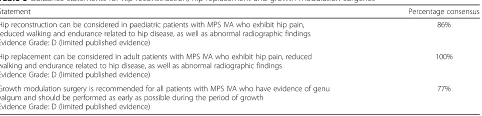
Limb surgeries in patients with MPS IVA (Table8)
Rationale and evidence basePatients with MPS IVA have progressive musculoskeletal involvement; therefore, multiple orthopaedic interventions are usually required
to prevent deformity, improve physical function and re-duce pain [100, 101]. Typical features of MPS IVA that do not occur in other types of MPS include joint hyper-mobility and deformity in the wrists, which lead to floppy wrists with weak grip and loss of fine motor skills. Patients with MPS IVA have subluxation of the hip joints and joint instability in the knees which can exacer-bate genu valgum, patella dislocation and gait abnormal-ities [14–16, 102]. Almost all patients with MPS IVA develop genu valgum to an extent which is serious enough to require surgery [101,103]. A review of the literature for outcomes of orthopaedic surgery suggest that correction of genu valgum by hemi-epiphysiodesis may improve QoL and function including improved walking dis-tance (as measured by 6MWT); however, patients re-main significantly impaired when compared to healthy individuals [104,105]. A case report of knee replacements in two patients with MPS IVA also suggests that correc-tion of genu valgum improves mechanical axis, mobility, ADL and QoL [106]. While there is more evidence avail-able for correction of genu valgum, data for other limb surgeries are limited [104]. Evidence from case reports supports the use of hip reconstruction as early as possible (before the age of 10 years) to minimise progression of subluxation and hip dysplasia and to improve out-comes, reduce pain and facilitate hip replacement fol-lowing surgical intervention. The expert SC commented that improved function has been observed following hip surgery; however, the majority of pub-lished literature suggest that the outcomes of hip surgery are judged largely on radiographic appearance with little correlation with function. Assessment of pelvic radiographs showed no correlation between 6MWT distance and degree of hip migration, and pa-tients with hip migration greater than 40% had no in-creased probability of being wheelchair-bound [105]. Overall, the expert opinion of the SC group is that pa-tients are more mobile following hip surgery; however, the literature is sparse and further data are required to sup-port these observations.
Patient selection for intervention Before orthopaedic intervention in patients with MPS IVA, morbidity and mor-tality risks, pain level, optimal timing, and patient
Table 8Guidance statements for hip reconstruction, hip replacement and growth modulation surgeries
Statement Percentage consensus
Hip reconstruction can be considered in paediatric patients with MPS IVA who exhibit hip pain, reduced walking and endurance related to hip disease, as well as abnormal radiographic findings Evidence Grade: D (limited published evidence)
86%
Hip replacement can be considered in adult patients with MPS IVA who exhibit hip pain, reduced walking and endurance related to hip disease, as well as abnormal radiographic findings Evidence Grade: D (limited published evidence)
100%
Growth modulation surgery is recommended for all patients with MPS IVA who have evidence of genu valgum and should be performed as early as possible during the period of growth
Evidence Grade: D (limited published evidence)
preference should be considered on a case-by-case basis. The need for hip surgery can be determined by presence of hip pain, reduced walking endurance and abnormal radio-graphic findings indicating hip dysplasia or lower limb alignment. Growth modulation surgery should be initiated as soon as the deformity is observed, or if the tibial-femoral angle is greater than 15 degrees. For optimal results, it should be performed early during the period of growth due to the deceleration in growth that occurs as the skel-eton matures, [100] however expert clinical opinion varies regarding the ideal age to perform the surgery. The period following the commencements of ERT may also be a good time to perform growth modulation surgery. Hemi-epiphysiodesis is indicated within the first decade of life, after this point osteotomy should be considered.
Currently there is no hand surgical intervention that can be recommended to improve the weakness of the grip but maintain vital flexibility for transfer and adequate ADL. External custom-made splints can be worn to help with certain tasks e.g. heavy lifting. Occupational thera-pists are vital to help with ADL including providing gadgets to perform necessary tasks. Patients with weak grip can learn to adapt to master necessary ADL.
Considerations for pre- and post-surgical monitor-ing and assessment The primary goal of limb surgery is not to improve or restore joint ROM, but to reduce pain or improve mobility. Goniometer measurement per-formed by a physiotherapist/occupational therapist/ rheumatologist may be useful, but this may not be avail-able in all centres. Post-surgery physical assessments should be performed regularly, as patients with MPS IVA may require repeated surgeries/interventions. Patients who have undergone hemi-epiphysiodesis around the knee at one level (tibial or femoral only) and who show evidence of progression of genu valgum during follow-up should be considered for a second growth modulation procedure on the unoperated level (tibial or femoral).
Considerations for surgeryAll surgeries should be su-pervised by an anaesthetist with experience in treating MPS and/or complex airway management (refer to the
anaesthetics recommendations). Limb surgeries should be performed by an orthopaedic surgeon with a basic understanding of MPS including: clinical presentation, musculoskeletal abnormalities and radiographic findings. An overnight hospital stay is recommended following hip surgery to allow access to intensive care, should this be needed, although this may not be necessary for more minor surgeries, such as hemi-epiphysiodesis. Long-term intensive physical therapy is recommended post-surgery to enhance recovery.
Spinal surgeries in patients with MPS IVA (Table9)
Rationale and evidence base Spinal involvement is a major cause of morbidity and mortality in patients with MPS IVA. Early diagnosis and timely treatment of spinal stenosis/instability is critical in preventing or arresting neurological deterioration and loss of function. Spinal involvement in patients with MPS IVA occurs at three locations. Cervical involvement, particularly instability and compression at C1–C2, is very common and predis-poses patients to myelopathy, paralysis and sudden death [11]. Upper cervical and craniocervical pathology is commonly seen in patients with MPS IVA; dens hypo-plasia in combination with ligamentous laxity can lead to atlantoaxial instability, and subsequently, to spinal canal stenosis and spinal cord compression [107]. Al-though craniocervical junction instability plays a major role in cervical cord pathology in patients with MPS IVA, a retrospective analysis of 28 patients suggests that decompression surgery without occipito-cervical stabilisation may yield good postoperative results [108]. Spinal cord compression can also occur at the cervicothoracic level, and is often missed [56]. Spinal cord compression due to kyphotic deformity at the thora-columbar level is not as common but can lead to paraple-gia [102, 109, 110]. Evidence from small case studies in patients with MPS indicates that thoracolumbar spine fusion was associated with good outcomes [111,112].
ERT and HSCT are of a limited use in preventing the development of skeletal deformities in patients with MPS
Table 9Guidance statements for decompression of the spinal cord, spinal stabilisation and thoracolumbar kyphoscoliosis
Statement Percentage consensus
Decompression of the spinal cord is recommended in patients with MPS IVA who have evidence of spinal cord compression based on clinical and radiographic findingsa
Evidence Grade: C (level 3/4 studies)
97%
Spinal stabilisation of the craniocervical junction with either cervical fusion or occipital-cervical fusion is recommended in patients with MPS IVA who have evidence of significant instability Evidence Grade: D (limited published evidence)
97%
Correction of thoracolumbar kyphoscoliosis is recommended in patients with MPS IVA who present with progressive radiographic deformity, intractable pain and neurological deterioration
Evidence Grade: C (level 3/4 studies)
100%
Post-consensus comments by the SC to be taken into consideration:
a