R E S E A R C H
Open Access
Evaluation of shear bond strength of orthodontic
brackets using trans-illumination technique with
different curing profiles of LED light-curing unit in
posterior teeth
Farzin Heravi
1, Saied Mostafa Moazzami
2, Negin Ghaffari
3*, Javad Jalayer
3and Yasaman Bozorgnia
4Abstract
Background:Although using light-cured composites for bonding orthodontic brackets has become increasingly popular, curing light cannot penetrate the metallic bulk of brackets and polymerization of composites is limited to the edges. Limited access and poor direct sight may be a problem in the posterior teeth. Meanwhile, effectiveness of the trans-illumination technique is questionable due to increased bucco-lingual thickness of the posterior teeth. Light-emitting diode (LED) light-curing units cause less temperature rise and lower risk to the pulpal tissue. The purpose of this study was to evaluate the clinical effectiveness of trans-illumination technique in bonding metallic brackets to premolars, using different light intensities and curing times of an LED light-curing unit.
Methods:Sixty premolars were randomly divided into six groups. Bonding of brackets was done with 40- and 80-s light curing from the buccal or lingual aspect with different intensities. Shear bond strengths of brackets were measured using a universal testing machine. Data were analyzed by one-way analysis of variance test and Duncan's post hoctest.
Results:The highest shear bond belonged to group 2 (high intensity, 40 s, buccal) and the lowest belonged to group 3 (low intensity, 40 s, lingual). Bond strength means in control groups were significantly higher than those in experimental groups.
Conclusions:In all experimental groups except group 6 (80 s, high intensity, lingual), shear bond strength was below the clinically accepted values. In clinical limitations where light curing from the same side of the bracket is not possible, doubling the curing time and increasing the light intensity during trans-illumination are recommended for achieving acceptable bond strengths.
Keywords:Bond strength; Orthodontic brackets; Light cure adhesive; Pulp chamber
Background
Acid etch technique was first introduced by Buonocore in 1955 [1], and since then, it has become increasingly popular among dentists due to its numerous advantages. It has been introduced by Newman [2] in the field of orthodontics and used for direct bonding of brackets for many years.
Using self-polymerizing composites for bonding brac-kets needs significant time for mixing, so it is a time-consuming procedure when multiple mixes are required.
The polymerizing process begins immediately after mix-ing; therefore, limited time is available for positioning the brackets. Another disadvantage of this method is the possibility of air bubble lockup into the adhesive during mixing that would reduce the bond strength of ortho-dontic brackets. However, using light-polymerizing com-posites provides extended working time for prompt bracket positioning and easier residue removal [3]. Moreover, it has been demonstrated that the initial bond strength of brackets would be higher by using light-polymerizing composites rather than self-light-polymerizing materials [4].
* Correspondence:negin.ghaffari@gmail.com
3Mashhad University of Medical Sciences, Mashhad 91735-984, Iran
Full list of author information is available at the end of the article
Since curing light cannot penetrate the metallic bulk, polymerization of composites under metallic brackets is limited to the edges of the bracket base. So it may result in incomplete polymerization and diminished bond strength [3]. Some researchers have suggested that cur-ing the composite layer under metallic brackets can be performed from mesial and distal aspects for 20 s each [5]. However, limited access to and poor direct sight of posterior segments may cause some difficulties especially when curing the composite under the bracket from the distal aspect. Therefore, trans-illumination technique has been proposed to cure the composite under metallic brackets by Tavas and Watts in 1979 [6]. In this tech-nique, light is emitted from the opposite side of the tooth and passes through bucco-lingual thickness toward the composite under the metallic bracket. Although little information can be found in orthodontic literature to confirm its usefulness, there is a general agreement on increasing the curing time while using this technique [7]. King et al. [8] tripled the trans-illumination curing time and found proper shear bond strength values re-gardless of the bucco-lingual thickness of the teeth ran-ging from 3.4 to 7 mm. Oesterle and Shellhart [7] used human maxillary incisors and found out that the bond strength of 50-s curing from the lingual aspect was so close to the control group and there was no significant difference in adhesive remaining index between control and experimental groups.
There are various types of light-curing units with dif-ferent spectral profiles and light intensities. While low-intensity light may lead to inadequate depth of cure and insufficient bond strength, high-intensity light might cause excessive heat during irradiation [9-11]. When using trans-illumination technique, the concerns over pulp chamber temperature rise become more important because the light passes through the bucco-lingual thick-ness of the tooth including the pulp chamber. Many studies have been done to compare pulp chamber temperature rise during polymerization while using dif-ferent types of light-curing units. Yazici et al. [12] and Haiduc et al. [13] reported that using light-emitting diode (LED) light-curing unit results in significantly lower temperature rise in comparison with conventional halogen units.
As in some cases, it is proposed to have a trans-illumination for bonding of posterior brackets due to limited access from the buccal side to their distal aspect and also for being assured from complete polymerization of bonding materials under the metallic brackets. The purpose of this study was to evaluate the effectiveness of trans-illumination technique in bonding metallic brackets to premolars while using different curing profiles of LED light-curing unit.
Methods
Based on the results of Oesterle and Shellhart's study [7] and the following formula, the sample size was found to be 10:
n¼ Nδ2 N−1
ð ÞDþδ2;D¼ B2
4
wherenis the sample size,Nis the population size,δ2is the variance, and B is the error bound. To estimate δ2,
we used an elementary sample with α= 0.05. The power
of the study was measured to be 80%. Sixty human pre-molars that were recently extracted for orthodontic pur-poses were collected. The criteria for tooth selection were intact buccal enamel, no pretreatment of chemical agents (such as derivatives of peroxide, acid, or alcohol), no cracks from forceps, no caries, and no restorations.
The teeth were sterilized in buffered formalin as Lee et al. suggested in 2007 [14]. One week prior to use, the teeth were placed in isotonic normal saline (0.90%w/v of NaCl, 300 mosM/L) in order to avoid any possible effect of the remaining solution on the bonding process. The storage media was changed every day to avoid bac-terial growth. The teeth were embedded in auto-polymerizing polymethyl methacrylate. A mounting jig was used to align the facial surface of the teeth to be perpendicular to the bottom of the mold and its labial surface parallel to the force during the shear bond strength (SBS) test.
Before bonding, the teeth were randomly divided into six groups each containing ten teeth. Specimen prepar-ation was done exactly as was instructed by the manu-facturer. The labial surfaces of the teeth were polished using non-fluoride pumice and then rinsed with water and subsequently dried with moisture-free air. The buc-cal enamel was etched with a 37% phosphoric acid (UltraEtch, Ultradent Products Inc., South Jordan, UT, USA) for 30 s. Afterwards, the etched surface was rinsed for at least 15 s until the etchant was completely re-moved and then dried with oil and moisture-free air source. A thin uniform coat of Transbond XT primer (3 M Unitek, Monrovia, CA, USA) was applied to the etched surfaces. A small amount of adhesive paste of Transbond XT was applied to the bracket bases. Stain-less steel brackets used in this study were Ultratrim Standard Edgewise (Dentaurum, Ispringen, Germany) in all groups. Immediately after placing the adhesive, the brackets were lightly placed on the tooth surface, ad-justed to the final position and then pressed firmly. Ex-cess adhesive material was gently removed from around the bracket base without disturbing it.
The adhesive was cured with High Power (800 mW/cm2)
(Ivoclar, Vivadent, Schaan, Liechtenstein) LED-curing unit in all groups:
Group 1 (control): 40 s light curing with the Low Power program from the buccal aspect (10 s for each mesial, distal, occlusal, and gingival aspect).
Group 2 (control): 40 s light curing with the High Power program from the buccal aspect (10 s for each mesial, distal, occlusal, and gingival aspect).
Group 3 (experimental): 40 s light curing with the Low Power program from the lingual aspect (light cure tip was placed as close as possible to the lingual surface perpendicular to occluso-gingival axis of the tooth).
Group 4 (experimental): 80 s light curing with the Low Power program from the lingual aspect (light cure tip was placed as close as possible to the lingual surface perpendicular to occluso-gingival axis of the tooth).
Group 5 (experimental): 40 s light curing with the High Power program from the lingual aspect (light cure tip was placed as close as possible to the lingual surface perpendicular to occluso-gingival axis of the tooth).
Group 6 (experimental): 80 s light curing with the High Power program from the lingual aspect (light cure tip was placed as close as possible to the lingual surface perpendicular to occluso-gingival axis of the tooth).
After bonding, specimens were stored individually in a normal saline solution at 37°C in a dark environment 24 h prior to testing. The shear bond strength of speci-mens was measured using a Zwick testing machine (Zwick GmbH & Co, Ulm, Germany) at a crosshead speed of 0.5 mm/min. In order to avoid bias, the bond-ing and debondbond-ing procedures were done by two differ-ent operators and the teeth were given codes unrelated to their group numbers. The results of the SBS test were recorded in megapascal.
Statistical analysis
The data was confirmed to be normally distributed using the Kolmogorov-Smirnov test. One-way analysis of variance
(ANOVA) and Duncan'spost hoctests were used to
com-pare SBS values among groups using SPSS 11.5 software.
Results
The one-way ANOVA test showed that there was a sig-nificant difference among shear bond strength amounts in groups (F(5,10) = 719.394, p< 0.05). Also, as it is
shown in Table 1, Duncan'spost hoc tests revealed that
there was a significant difference between every two groups except for groups 1 and 2 (control group).
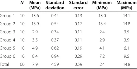
Mean values of SBS are shown in Table 2. The highest mean bond strength was seen in group 2 (40-s curing from the buccal aspect with the intensity of 800 mW/cm2)
and the lowest value was in group 3 (40-s curing from the lingual aspect with the intensity of 650 mW/cm2).
SBS was not significantly different between two con-trol groups; in both groups, the SBS values were above the clinically accepted values according to Reynolds' study [15], and values were significantly higher than ex-perimental groups.
Among four experimental groups, the only group with sufficient SBS values for orthodontic bonding was group 6 (80-s curing from the lingual aspect with the intensity of 800 mW/cm2).
Discussion
Tavas and Watts [6] first introduced trans-illumination technique in 1979, which suggested curing composite adhesive under metallic brackets from the opposite side of the tooth. Many studies have been done on this tech-nique mostly on the anterior teeth, and the effectiveness of this technique has been proved in the anterior region. King et al. [8] studied trans-illumination technique using bovine teeth and lingual brackets. They tripled the Table 1 Duncan'spost hoctest
Group N Subset forα= 0.05
1 2 3 4 5
Group 1 10 13.6120
Group 2 10 13.9920
Group 3 10 2.9190
Group 4 10 3.5210
Group 5 10 4.9590
Group 6 10 8.4420
Significance 1.000 1.000 1.000 1.000 0.151
Group 1 (control): 40 s, 650 mW/cm2
, buccal; group 2 (control): 40 s, 800 mW/ cm2
, buccal; group 3: 40 s, 650 mW/cm2
, lingual; group 4: 80 s, 650 mW/cm2 , lingual; group 5: 40 s, 800 mW/cm2
, lingual; group 6: 80 s, 800 mW/cm2 , lingual. The test showed that there was a significant difference between every two groups except for groups 1 and 2 (control groups).
Table 2 Shear bond strength values in all six groups
N Mean
(MPa)
Standard deviation
Standard error
Minimum (MPa)
Maximum (MPa)
Group 1 10 13.6 0.44 0.13 13.0 14.1
Group 2 10 13.9 0.54 0.17 13.4 14.8
Group 3 10 2.9 0.34 0.11 2.4 3.5
Group 4 10 3.5 0.37 0.11 2.9 3.9
Group 5 10 4.9 0.62 0.19 4.1 6.1
Group 6 10 8.4 0.94 0.29 7.2 9.5
Total 60 7.9 4.59 0.59 2.4 14.8
Group 1 (control): 40 s, 650 mW/cm2
, buccal; group 2 (control): 40 s, 800 mW/ cm2
, buccal; group 3: 40 s, 650 mW/cm2
, lingual; group 4: 80 s, 650 mW/cm2 , lingual; group 5: 40 s, 800 mW/cm2
curing time and found proper shear bond strength values when using trans-illumination regardless of the bucco-lingual thickness of the teeth ranging from 3.4 to 7 mm. Oesterle and Shellhart [7] used human maxillary incisors and compared shear bond strength when curing from the buccal (as control groups) or lingual (trans-illu-mination technique) aspect. They found bond strength values very close to control groups with 50-s curing from the lingual aspect and no statistical difference in adhesive remaining index between control and experi-mental groups.
Although in posterior segments, limited access and poor direct sight cause some difficulties in curing the adhesive from the same side of the bracket (especially when it is needed to be cured from the distal or gingival aspect), there is little in orthodontic literature about the effective-ness of trans-illumination technique in posterior seg-ments. Regardless of the conclusion of each study, there is a general agreement on using higher light energy (light in-tensity or curing time) while directing the light through the bucco-lingual thickness of the tooth [7]. However, pos-sible damaging effects of excessive heat generated during polymerization have been warned [9-11]. Previous studies have shown increase in temperature ranging from 1.5°C to more than 4°C in the pulp chamber of extracted teeth [9,10] which were assumed to be caused by both exother-mic reaction and energy absorbed during irradiation [16-20]. The critical temperature for making irreversible damage to the pulpal tissue is 42°C to 42.5°C [21,22]. As Yazici et al. [12] and Haiduc et al. [13] demonstrated in their studies, using LED units can cause significantly lower temperature rise in the pulp chamber in comparison with halogen units. Consequently, in our study, a polywave LED unit, Bluephase C8 (Ivoclar, Vivadent, Schaan, Liechtenstein), was used in bonding procedures in order to make minimum temperature changes in the pulp chamber. As it is shown in Table 2, the highest shear bond strength was in group 2 (40-s curing from the buccal
as-pect with the intensity of 800 mW/cm2) and the lowest
value was in group 3 (40-s curing from the lingual aspect with the intensity of 650 mW/cm2).
Increase in light intensity from 650 to 800 mW/cm2 be-tween the control groups did not result in any significant increase in SBS values. Among the four experimental groups, the only group with sufficient SBS values for orthodontic bonding was group 6 (80-s curing from the
lingual aspect with the intensity of 800 mW/cm2). In
group 4, increasing the curing time in comparison with group 3 resulted in a significant increase in bond strength, but according to Reynolds' study [15], the values were not acceptable for orthodontic treatment. Also, in group 5, in-creasing the intensity (without inin-creasing curing time) in comparison with group 3 resulted in higher bond strengths, but still the values were not acceptable. This
leads to the deduction that doubling the light curing time and increasing the light intensity to 800 mW/cm2[2] have to be done simultaneously in order to achieve accep-table bond strength values when using trans-illumination technique.
In another similar study on human premolars using metallic and ceramic orthodontic brackets with different curing times of a halogen unit [23], SBS in control groups (cured from the buccal aspect) was significantly higher than that in experimental groups (cured from the lingual aspect), and in one of their experimental groups with 40-s curing from the lingual aspect, the SBS values were lower than the values accepted for clinical ortho-dontic treatment as they were in our study. On the other hand, in the other experimental groups of their study, SBS values were clinically acceptable. This difference be-tween the results can be the consequence of the differ-ence in light-curing units used in each study.
Conclusions
According to the results of this study, using trans-illumination technique in bonding metallic brackets to the premolar teeth (in comparison with light curing from the same side of the bracket) resulted in signifi-cantly lower bond strengths. In clinical limitations where light curing from the same side of the bracket is not possible, doubling the curing time and increasing the light intensity to 800 mW/cm2during trans-illumination with the LED light-curing unit can be done for attaining acceptable bond strength with minimum risk of overheat to the pulpal tissue.
Competing interests
The authors declare that they have no competing interests.
Authors' contributions
The work presented here was carried out in collaboration among all authors. FH designed the methods and experiments and edited the manuscript text. SMM contributed to the design of the study and selection of bonding materials and devices. NG collected and prepared the samples, interpreted the results, and drafted the manuscript. JJ carried out the SBS test and helped in drafting the manuscript. YB bonded the brackets and worked on the data interpretation. All authors read and approved the final manuscript.
Author details
1Dental Materials Research Center, Mashhad University of Medical Sciences,
Mashhad 91735-984, Iran.2Department of Operative Dentistry, Mashhad
University of Medical Sciences, Mashhad 91735-984, Iran.3Mashhad University of Medical Sciences, Mashhad 91735-984, Iran.4Department of
Orthodontics, North Khorasan University of Medical Sciences, Bojnurd 9414974877, Iran.
Received: 6 July 2013 Accepted: 5 November 2013 Published: 21 November 2013
References
1. Buonocore MG.A simple method of increasing the adhesion of acrylic filling material to enamel surfaces.J Dent Res.1955;34:849–53. 2. Newman GV.Epoxy adhesives for orthodontic attachments: progress
3. Sunna S, Rock WP.An ex vivo investigation into the bond strength of orthodontic brackets and adhesive systems.Br J Orthod.1999;26:47–50. 4. Wendl B, Droschl H.A comparative in vitro study of the strength of
directly bonded brackets using different curing techniques.Eur J Orthod. 2004;26:535–44.
5. Oesterle LJ, Messersmith ML, Devine SM, Ness CF.Light and setting times of visible-light-cured orthodontic adhesives.J Clin Orthod.1995;29:31–6. 6. Tavas A, Watts DC.Bonding of orthodontic brackets by trans-illumination of
a light-activated composite: an in vitro study.Br J Orthod.1979;6:207–8. 7. Oesterle LJ, Shellhart WC.Bracket bond strength with trans-illumination
of a light-activated orthodontic adhesive.Angle Orthod.2001;71:307–11. 8. King L, Smith RT, Wendt SL, Behrents RG.Bond strengths of lingual
orthodontic brackets bonded with light-cured composite resins cured by trans illumination.Am J Orthod Dentofacial Orthop.1987;91:312–5. 9. Thompson HF, Gomez HF, Puckett AD.Pulpal temperature changes after
exposure to a light curing source.J Dent Res.1997;76:524.
10. Bennett GE, Blank FJ.Thermal effects of visible-light and chemically cured resins.J Dent Res.1984;63:259.
11. Hannig M, Bott B.In-vitro pulp chamber temperature rise during composite resin polymerization with various light-curing sources.Dent
Mater.1999;15:275–81.
12. Yazici AR, Muftu A, Kugel G, Perry RD.Comparison of temperature changes in the pulp chamber induced by various light curing units, in vitro.Oper Dent.2006;31:261–5.
13. Haiduc C, Dodenciu D, Sinescu C, Negrutiu M, Draganescu G, Mara V.Pulp chamber temperature variations using three types of light curing units.
Eur Cell Mater.2007;13:19–23.
14. Lee JJ, Nettey Marbell A, Cook A Jr, Pimenta LA, Leonard R, Ritter AV.Using extracted teeth for research: the effect of storage medium and sterilization on dentin bond strengths.J Am Dent Assoc.2007;138:1599–603.
15. Reynolds JR.A review of direct orthodontic bonding.Br J Orthod.1975; 2:171–80.
16. McCabe JF.Cure performance of light activated composites by differential thermal analysis (DTA).Dent Mater J.1985;1:231–4. 17. LIoyd CH, Joshi A, McGlynn E.Temperature rises produced by light
sources and composites during curing.Dent Mater J.1986;2:170–4. 18. Masutani S, Setcos JC, Schnell RJ, Phillips RW.Temperature rise during
polymerization of visible light activated resins.Dent Mater J.1988;4:174–8. 19. Smail SRJ, Petterson CJW, McLaundie AC, Strang R.In vitro temperature
rises during visible-light curing of a lining material and a posterior composite.J Oral Rehabil.1988;15:361–6.
20. Hanson EK, Asmussen E.Correlation between depth of cure and temperature rise of a light-activated resin.Scand J Dent Res.1993; 101:176–9.
21. Zach L, Cohen G.Pulp response to externally applied heat.Oral Surg Oral
Med Oral Pathol.1965;19:515–30.
22. Pohto M, Scheinin A.Microscopic observations on living dental pulp II. The effect of thermal irritants on the circulation of the pulp in the lower rat incisor.Acta Odontol Scand.1958;16:303–27.
23. Heravi F, Bayani S.Changes in shear bond strength of ceramic and stainless steel brackets with different visible light curing times and directions.Aust Orthod J.2009;25:153–7.
doi:10.1186/2196-1042-14-49
Cite this article as:Heraviet al.:Evaluation of shear bond strength of orthodontic brackets using trans-illumination technique with different curing profiles of LED light-curing unit in posterior teeth.Progress in
Orthodontics201314:49.
Submit your manuscript to a
journal and benefi t from:
7Convenient online submission 7Rigorous peer review
7Immediate publication on acceptance 7Open access: articles freely available online 7High visibility within the fi eld
7Retaining the copyright to your article