Background:
Atrial deptal defect device closure has become the preferred method in the treatment of atrial septal
defect. We aim to study the in-hospital complications of atrial septal defect device closure procedure.
Methods:
It was a single center, retrospective study conducted from Febuary 2016 to January 2019. Cardiac
catheterization laboratory records of all consecutive patients who underwent atrial septal defect device closure was
included and the in-hospital complications were been retrospectively reviewed.
Results:
During the study period, a total of 566 patients were attempted for device closure. In 557 (98.4%) of cases
device was implanted. Among the 557 patient in which device was implanted 401(71.9%) were female. Age ranged
from 5 years to 72 years with the mean of 30.9 years. Transient ST segment elevation 15 (2.6 %)was the commonest
complication followed by pericardial tamponade 4 (0.7%), and cardiac arrhythmias 3 (0.5%).
Conclusions: Atrial deptal defect device closure can be done safely with a high success rate and a low complication
rate.
Keywords:
Amplatzer duct occluder; atrial septal defects; in hospital complications; transcatheter device closure.
In Hospital Complications of Atrial Septal Defect
Device Closure at Shahid Gangalal National Heart
Centre, Kathmandu, Nepal
Chandra Mani Adhikari,1 Kiran Prasad Acharya,1 Amrit Bogati,1 Sachin Dhungel,1 Manish Shrestha2
1Department of Cardiology, Shahid Gangalal National Heart Centre, Kathmandu, Nepal, 2Department of Pediatric Cardiology, Shahid Gangalal National Heart Centre, Kathmandu, Nepal.
Correspondence:
Dr Chandra Mani Adhikari, Department of Cardiology, Shahid Gangalal National Heart Centre Kathmandu, Nepal. Email: topjhap@gmail. com, Phone: +9779851212111.ABSTRACT
J Nepal Health Res Counc 2019 Oct-Dec;17(45): 474-8
INTRODUCTION
Atrial septal defect (ASD) is one of the common congenital heart disease and accounts for 8-10% of the congenital heart diseases. Secundum ASD is the most common type and accounts for 70% of all ASDs.1 Surgical closure of ASD remained a gold standard treatment for secundum ASD.2 Trans-catheter device closure of ASD was first attempted by King and Mills in 1976 and soon became an effective and safe alternative to surgery. It was accepted as the gold standard of therapy for closure of all suitable secundum ASD at the beginning of the millennium.3 Device closure for secundum ASD has increasingly become the preferred strategy because of its relatively high efficacy, lower morbidity and low complication compared to surgery.4, 5 Device closure can also avoid the thoracotomy and Sternotomy scar. However complication in any interventional procedure cannot be avoided. We aimed to study in-hospital complications of ASD device closure in Shahid Gangalal National Heart Centre, Kathmandu, Nepal.
METHODS
This study was a cross sectional, single center, retrospective study conducted at Shahid Gangalal National Heart Centre, Kathmandu, Nepal. Cardiac catheterization laboratory records and Medical records of all patients who were attempted for ASD device closure from February 2016 to January 2019 were retrospectively reviewed. In the mean time, Performa was designed to collect the patient information which includes age, gender, ASD size, device closure procedural approach, and device type and size and in-hospital complications. This study was approved by institute review committee of Shahid Gangalal National heart center, Kathmandu, Nepal. All the variables were entered into the Statistical Package for Social Sciences software, version 14 (SPSS Inc) for data analysis. Descriptive statistics were computed and presented as means for continuous variables categorical variables were reported in percentages.
ASD device closure procedure was done under general
DOI
https://doi.org/10.33314/jnhrc.v17i4.1957
anesthesia in five patients and under the transesophageal echocardiogram guidance in the other ten patients. In all other cases procedure was done under local anesthesia assisted with transthoracic echocardiogram. In children local anesthesia along with intravenous anesthesia was done. After performing the femoral vein puncture, heparin (100 IU/kg) and antibiotic prophylaxis were given routinely in all the patients. Implantation was performed under fluoroscopic and echocardiographic control. Conventional device delivery technique was the preferred technique. When necessary, left upper pulmonary vein technique, right upper pulmonary vein technique, Balloon assisted technique, catheter assisted technique were used. The proper position of the device in addition to the interference of the device with the caval or pulmonary veins or with the atrioventricular valves were checked and confirmed with echocardiography.
If all the findings were satisfactory, the device was released. All patients underwent clinical examination, electrocardiography, chest radiography in two projections and transthoracic echocardiography before discharge.
RESULTS
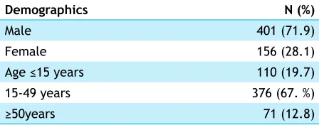
The ASD device was successfully implanted in 557 (98.4%) patients out of the 566 patients who were scheduled for the ASD device closure. Unfortunately, the device could not be implanted in nine cases, due to pericardial effusion in three case and due to unstable device in other six patients. Furthermore, age of the patients ranged from 5 to 72 years with the mean of 30.9 years. Undoubtedly, most of the patients were female as shown in the Table 1. The ASD size ranged from 7 mm to 37mm with the mean of 20.8 mm and the device size ranged from 8 to 42 mm with the mean of 26.5 mm. In addition to that, most of the patients were in the age group of 15 to 45 years as elaborated in the Table 1.
Table 1. Demographics of successful implanted cases (n=557).
Demographics N (%)
Male 401 (71.9)
Female 156 (28.1)
Age ≤15 years 110 (19.7)
15-49 years 376 (67. %)
≥50years 71 (12.8)
Four different types of device were used with the Amplatzer septal occluder in 527 (94.6%) patients, hyperion (Comed) device in 10 (1.7%) patients, Memopart (Lepu) device in 19 (3.4%) patients and Cera (Life tech)
device in 1(0.1%) patients as enlisted in the Table 2.
Table 2. Type of ASD device among successful procedure (n=557).
Types of devices N (%)
Amplatzer septal occlude 527 (94.6)
Hyperion (Comed) device 10 (1.7)
Memopart (Lepu) 19 (3.4)
Cera (Life tech) 1 (0.1)
Table 3. Complications among attempted cases (n=566).
Complications N (%)
Transient ST segment elevation 15 (2.6)
Pericardial tamponade 4(0.7)
Cardiac arrhythmias 3 (0.5)
Residual leak 2(0.3)
Air embolization to right heart 2(0.3)
Complete heart block 1 (0.1)
Infection (pneumonia) 1(0.1)
Groin hematoma 1(0.1)
Cardiac arrest 1 (0.1)
Device embolism 1(0.1)
Among the 566 attempted cases, a total of 31 patients developed some form of complications. The most common one encountered in 15 patients (2.6%) was Transient ST segment elevation in the electrocardiogram. It was followed by pericardial effusion leading to cardiac tamponade encountered in 4 patients (table 3). Three of them had the cardiac tamponade during the procedure, in which the device was not been implanted. Two patients underwent pericardiocentesis in the catheterization laboratory. Unfortunately, one patient underwent surgical drainage for the large pericardial effusion due to a tear in the left upper pulmonary vein and left atrial junction. Regrettably, the patient died next day of the surgery. In addition, one patient developed cardiac tamponade three hours after the device closure in which pericardiocentesis was done in the ward but the patient developed hypoxic cerebral encephalopathy.
Although, the arrhythmic complications were infrequently encountered, one of the patients developed atrial fibrillation and the other developed supraventricular tachycardia during the procedure. However, in one patient supraventricular tachycardia developed after the procedure. One patient developed groin hematoma which required surgical intervention. Furthermore, two patients had air embolism to the right heart after device implantation but with no apparent sequelae. One of the patients developed pneumonia after the procedure and was treated with antibiotics. Unfortunately, one of the patient required cardiopulmonary resuscitation (CPR) during the procedure but luckily the cardiac activity was revived in less than a minute of CPR.
DISCUSSION
ASD device closure is associated with the risks inherent in any interventional cardiac catheterization procedure. Our study highlights the common complications of the ASD device closure in the real world.
Transient ST elevation during device closure is thought to be a rare complication.6 It was the most common complication in our cases as in the study done by Jose M de la Torre Hernandez et al.7 This complication was experienced in only in 1.2% cases in a study done in Bangladesh.8 The same rate was observed 1 case in a series of 100 cases in a study done by Chan KC, et al.6
This complication has been attributed to the possible embolization of small air bubbles. In any case, whatever is the etiological mechanism, the only avoidable factor is to prevent the embolization of air, so that it is important to be very careful with the external manipulation of the device, checking several times that there are no air bubbles.7 Careful attention should be given during the process of device introduction into the sheath.
Pericardial effusion which occurred in only four cases in our study is thought to be one of the rare complications of device closure of an ASD, with a reported incidence of 0.5-1.5%. The reported etiology ranges from wire perforation to catastrophic device erosion. 9
After 24 hour of device closure, residual leak occurred in 0.3% of the cases in this study. A study from Bangladesh reported a residual leak in 0.7% cases.8 Trivial/smoky residual shunt was detected in 15 (10.0%) patients, and a persistent residual shunt was observed in 1 (0.6%) patient.10 Among the different device available residual shunts is very low with Amplatzer septal occluder.4,11,12 Kazmi et al reported that only 3 of 202 patients had small residual leaks immediately after procedures.13
In our study, one of the patients had had device embolized to right pulmonary artery which was successfully retrieved and successful device closure was performed with the another device. In case of device embolization, it can be retrieved with a gooseneck and attempt a device closure or patient can be referred to the surgeon for device retrieval and surgical closure of ASD at the same time.4 Chessa et al reported on a large series of 417 patients who had catheter closure of secundum ASDs.4 The most common complication was device embolization/malposition occurred in 3.5% of cases. Of the 15 patients in whom devices embolized or were malposition, 10 required surgical retrieval while in the remainder the devices were retrieved by catheter techniques. In a multicenter UK experience, Chan et al reported on 101 Amplatzer septal occluder procedures performed in 100 patients, device embolization in one patient.6 The rate of embolization reported in 2004 by AGA was 21 out of 3824 patients (0.55%).14 According to the US FDA Manufacturer and User Facility Device Experience (MAUDE) database,there were 114 (0.62%) device embolization’s out of 18,333 Amplatzer septal occluder implants.15 Other meta-analyses of transcatheter closure of ASD/PFO showed device embolization rates were 0.2-0.43%.16,17
In this study very few patients had supraventricular arrhythmias. Atrial fibrillation or supraventricular tachycardia after ASD device implantation, is possibly due to stretching of the interatrial septum by the central waist of the device.4
Complete heart block after the procedure was very rare in our study. The reported prevalence of advanced heart block was less than 1%.16, 17Advanced heart block could occur as early as device deployment. Most of the atrioventricular (AV) block is transient and recovered within a short period. Corticosteroid treatment has been used in different centers. 18
Groin hematoma and femoral arteriovenous fistula are not infrequent and are probably under-reported.19 They result from inadvertent arterial puncture and/or the introduction of relatively large venous sheaths and probably in elderly or obese patients. 12 Hematomas may rarely require blood transfusions and surgical repair. The post catheterization groin should be treated with meticulous care. In our study only one patients developed groin hematoma which required surgical treatment.
device embolization (3.5%), and pericardial effusion (0.5%).20 In our study the complication rate is less compared to studies worldwide.
Spence and Qureshi suggest that, avoidance of complications is dependent on careful case selection, sizing of the defect, selection of the correct size of device, aseptic technique, proper heparinization during the procedure, avoidance of air embolism and testing of device stability during the procedure, and antibiotic prophylaxis.12 Vigilance is also required to detect complications early so that remedial action can be taken promptly to minimize the risk of serious adverse outcomes.
The study only give glimpse of procedures performed at one center with lack of long term follow up. A nationwide survey of the procedures might give a more accurate pictures of the complications.
CONCLUSIONS
Transient ST segment elevation is commonest complication of atrial septal defect closure followed by pericardial tamponade. Vigilance regarding potential complication can guide for early management of patients and minimise adverse events.
ACKNOWLEDGEMENTS
To all the doctors, Nurses and Catheterization laboratory staff of Shahid Gangalal National Heart center who are involved in Atrial Septal Defects device closure.
![HECS and the farmer's son [Commentary]](data:image/gif;base64,R0lGODlhAQABAIAAAP///wAAACH5BAEAAAAALAAAAAABAAEAAAICRAEAOw==)