Fine Needle Aspiration Cytology of
Kikuchi-Fujimoto Disease
Adhikari RC
11Department of Pathology, Tribhuvan University Teaching Hospital, Maharajgunj, Kathmandu, Nepal.
ABSTRACT
Correspondence:
Ram Chandra Adhikari, Department of Pathology, Om Hospital
& Research Centre, Chabhil, Kathmandu, Nepal. E-mail: [email protected],
INTRODUCTION
Kikuchi-Fujimoto disease (KFD) was fi rst described
in 1972 simultaneously by Kikuchi1 and Fujimoto
and colleagues.2 It is characterized by acute onset
febrile illness and lymphadenopathy, predominantly affecting young women with predilection for cervical lymphadenopathy. Despite many studies, the etiology of this disease remains unclear. The diagnosis of KFD is made on histological examination of excised lymph node.
The experience with Fine Needle Aspiration cytology (FNAC) technique to diagnose KFD is limited to either
case reports or small series.3-6 Fine needle aspiration
cytology fi ndings in 13 histopathologically proven cases of KFD is presented here.
METHODS
Thirteen cases of KFD with both FNAC and excisional biopsy of lymph node were retrieved from the department of Pathology, Tribhuvan University Teaching Hospital and Om Hospital & Research Centre Pvt. Ltd., Kathmandu, Nepal from August 2009 to July 2013. The cytological smears were reviewed retrospectively from these 13 cases with histological diagnosis of KFD.
All aspirates were performed prior to biopsy using 21
gauzed needle and air dried & wet fi xed smears were
prepared for Giemsa stain and H&E stain respectively. All excised lymph nodes were routinely processed and H&E staining was performed.
Background:
Kikuchi-Fujimoto disease is an acute onset febrile illness of unknown etiology, predominantly
affecting young women with predilection for cervical lymphadenopathy.
Methods:
The study included 13 cases of Kikuchi-Fujimoto disease with both fine needle aspiration cytology
and excisional biopsy of lymph node available and data & slides were retrieved from the department of Pathology,
Tribhuvan University Teaching Hospital and Om Hospital & Research Centre Pvt. Ltd., Kathmandu, Nepal from
August 2009 to July 2013.
Results:
The mean age of the patients was 27.6 years with a range of 17 to 38 years. Twelve of 13 patients
had cervical lymphadenopathy. Cytomorphological features included cellularity, karyorrhectic debris,
crescentichistiocytes, necrosis and cellular polymorphism. Histologically, Lymph nodes showed partially effaced
architecture by paracortical pale foci with karyorrhectic debris. These foci were composed of phagocytic &
non-phagocytic histiocytes, plasmacytoid monocytes, immunoblasts and lymphocytes.
Conclusions:
Kikuchi-Fujimoto disease, in most cases, can be diagnosed cytologically on the basis of identification
of karyorrhectic debris and crescentic macrophages with reactive background.
Keywords:
fine needle aspiration cytology, Kikuchi-Fujimoto disease, lymph node.
RESULTS
The clinical details of all 13 cases are summarized in Table 1.
Table 1. Clinical features of Patients with KFD.
Case no.
Age (Yr)
Sex Clinical feature Cytological diagnosis Lymph node aspirated
1 25 M Fever & cervical lymphadenopathy
for last 3 weeks
Tuberculosis Right cervical lymph
node, III
2 28 F Cervical lymphadenopathy for last
3 months
Tuberculosis Right cervical lymph
node, V
3 27 F Axillary lymphadenopathy Reactive lymphadenitis Right axillary lymph
node
4 28 F Low grade fever & enlarged left
cervical lymphnode
Kikuchi-Fujimoto disease Left cervical lymph node, supraclavicular
5 24 F Left sided neck swelling Kikuchi-Fujimoto disease Left cervical lymph
node, II
6 30 F Bilateral cervical
lympha-denopathy for 2 months
Non-Hodgkin lymphoma Left cervical lymph
node, IV
7 28 F Matted tender cervical lymph node
for last 15 days; On & off fever
Kikuchi-Fujimoto disease Left cervical lymph node, II & III
8 28 F Enlarged right cervical lymph
noode, 2x1.5 cm sized
Kikuchi-Fujimoto disease Right cervical lymph node, supraclavicular
9 38 F Fever with cervical
lymphadenopathy for 15 days
Kikuchi-Fujimoto disease Right cervical lymph node, III
10 17 F Enlarged right cervical lymph node
for 3 months
Kikuchi-Fujimoto disease Right cervical lymph node, II
11 23 F Multiple nodular swelling, neck for
2 months
Kikuchi-Fujimoto disease Left cervical lymph node, II,III
12 35 F Tender cervical lymph node for 10
days
Kikuchi-Fujimoto disease Right cervical lymph node, II & III
13 28 F Cervical lymphadenopathy Kikuchi-Fujimoto disease Right cervical lymph
node, III & IV
Twelve of 13 patients were female. The mean age of the patients was 27.6 years with a range of 17 to 38 years. Twelve of 13 patients had cervical lymphadenopathy, while one had axillary lymphadenopathy. Most of the cases were clinically suspected as tuberculosis. In all cases, FNAC was followed by excisional biopsy.
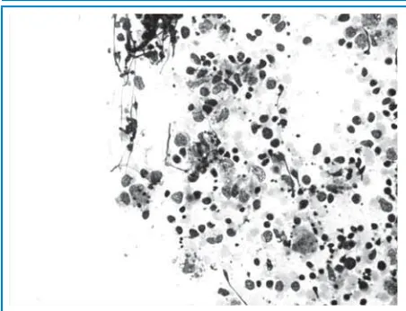
Cytomorphological features are tabulated in Table 2. Special attentions were given on: cellularity, karyorrhectic debris, crescentic histiocytes, necrosis and cellular polymorphism. Hypercellularity and karyorrhectic debris were seen in all cases and karyorrhectic debris (Figure 1) were intracellular as well as extracellular. Crescentic histiocytes (Figure 2) were
prominent fi ndings in 10 cases only. These histiocytes
have nuclear debris in their cytoplasm and eccentric crescent shaped nucleus. Necrosis (Figure 3) was seen only in three cases. In 12 of 13 cases, a polymorphous lymphoid population (Figure 4) of small lymphocytes, plasmacytoid lymphocytes and large immunoblastic cells
were observed. No granulomas or epithelioid cells and neutrophils were observed.
Figure 2. FNAC lymphnode,
Scatteredcrescentichistiocytes. Giemsa stain, X400.
Figure 3. FNAC, Necrosis. Giemsa stain, X400.
Figure 4. FNAC, Polymorphous lymphoid population, Giemsa stain, X40.
Table 2. Cytomorphological features.
Morphological features No. of cases (%)
Hypercellularity 13 (100)
Cellular polymorphism 12 (92)
Karyorrhectic debris 13 (100)
Crescentic histiocytes 10 (77)
Absence of neutrophils 13 (100)
Necrosis 3 (23)
Granulomas
-Figure 5. Lymphnode biopsy, Paracortical pale focus (below), H&E stain, X100.
Figure 6. Lymphnode biopsy, Apoptosis with karyorrhectic debris, H&E stain, X400.
apoptosis was seen in three cases (Figure 6). Focally preserved lymphoid follicles were seen in seven cases. None of the lymph nodes showed granulomas.
DISCUSSION
The role of FNAC has been established in the diagnosis of reactive as well as neoplastic disorders of the lymph node. KFD is one of the lymph node disorders,
which have a characteristic histological fi nding and
cytomorphological picture. This study is one of the series of KFD diagnosed on cytological material in most of the cases with subsequent histological confi rmation.
The diagnosis of KFD is possible given an adequately sampled, well-prepared specimen in which characteristic karyorrhectic debris with admixed crescentic
macrophages on a reactive background are evident.3 In
this study, karyorrhectic debris, crescentic macrophages and reactive background are found in most of the cases on retrospective examination of the smears.
In one case (case no. 3), because of paucity of necrosis, crescentic macrophages & karyorrhectic debris, the diagnosis of reactive lymphadenitis was made cytologically. However, review of smears showed little karyorrhectic debris. In addition, all cases lack neutrophils, granulomas or epithelioid cells.
Karyorrhectic debris are common fi ndings and they were
both intracellular and extracellular. These debris with reactive background may be seen in lupus lymphadenitis and these two diseases are indistinguishable
cytologically.5 Some authors claim that KFD may be a
self liming form of systemic lupus erythematosus.7-10
Crescentic macrophages have eccentrically placed crescent shaped nucleus and abundant cytoplasm with debris in some of them.
The background was reactive with polymorphism in 12 out of 13 cases in this study, while one case showed monotonous population of lymphoid cells mimicking Non-Hodgkin lymphoma, so the original cytological diagnosis was Non-Hodgkin lymphoma. However, the review of cytological smears revealed little karyorrhectic debris. These debris sometimes may be considered secondary to probable tumor and may be a cause of false positive
diagnosis on cytology4 as well as a diagnostic problem
histologically.11,12
The another differential diagnosis based on clinical fi nding is tuberculosis in regions where it is more prevalent.11,13
However, tuberculous lymphadenitis and KFD can easily be distinguished on cytological preparations as well as histologically. The epithelioid cell granulomas seen in tuberculosis are not a feature of KFD. However, the diagnosis of tuberculosis was suspected cytologically in
two cases in this study because of abundant necrosis that had caseous appearance. The retrospective review of cytological smears in these cases showed few crescentic macrophages and even little debris.
Kuo TT proposed three histological phases of KFD:
proliferative, necrotizing and xanthomatous.14 The
proliferative phase is characterized by presence of histiocytes, plasmacytoid monocytes, transformed monocytes, transformed lymphocytes, karyorrhectic nuclear fragments and apoptotic debris. The necrotic
phase is defi ned by the presence of non-suppurative
coagulative necrosis. If foamy histiocytes predominate in the lesions, the case is categorized as being in the xanthomatous phase despite the presence or absence of
necrosis. The cytological fi nding also depends on these
phases. When FNAC reveals mixed background with karyorrhectic debris, this probably denotes proliferative phase. The presence of abundant necrosis on cytological smears indicate necrotic phase and the presence of numerous crescentic macrophages favors xanthomatous phase.
CONCLUSIONS
Kikuchi-Fujimoto disease, in most cases, can be diagnosed cytologically on the basis of identifi cation of karyorrhectic debris and crescentic macrophages with reactive background in the adequate clinical context (young female with cervical lymphadenopathy).
REFERENCES
1. Kikuchi M. Lymphadenitis showing focal reticulum cell hyperplasia with nuclear debris and phagocytosis. Nihon KetsuekiGakkaiZasshi. 1972;35:379-80.
2. Fujimoto Y, Kozima Y, Yamaguchi K. Cervical subacute necrotizing lymphadenitis: A new clinicopathological entity. Naika. 1972;30:920-7.
3. Osborn M, Agel N, Levine TS. The fine needle aspiration appearances of Kikuchi’s lymphadenitis. Cytopathology. 2009;20:36-43.
4. Viguer JM, Jimenez-Heffernan JA, Perez P, Lopez-Ferrer P, Gonzalez-Peramato P, Vicendi B. Fine needle aspiration cytology of Kikuchi’s lymphadenitis: A report of ten cases. DiagnCytopathol. 2001;25:220-4.
5 Greenberg ML, Cartwright L, McDonald DA. Histiocytic necrotizing lymphadenitis (Kikuchi’s disease): Cytologic diagnosis by fine-needle biopsy. DiagnCytopathol. 1993;9:444-7.
6. Hsueh EJ, Ko WS, Hwang WS, Yam LT. Fine-needle aspiration of histiocytic necrotizing lymphadenitis (Kikuchi’s disease). DiagnCytopathol. 1993;9:448:52.
8. Tumiati B, Belleli A,Portioli I, Prandi S. Kikuchi’s disease in systemic lupus erythematosus; an independent or dependent event? ClinRheumatol. 1991;10:90-3.
9. Litwin MD, Kikham B, Henderson DRF, Milazzo SC. Histiocytic necrotizing lymphadenitis in systemic lupus erythematosus. Ann Rhuem Dis. 1992;51:805-7.
10. el-Ramahi KM, Karrar A, Ali MA. Kikuchi’s disease and its association with systemic lupus erythematosus. Lupus. 1994;3:409-11.
11. Chamulak GA, Brynes RK, Nathwani BN. Kikuchi-Fujimoto disease mimicking malignant lymphoma. Am J SurgPathol. 1990;14:514-23.
12. Menasce LP, Banerjee SS, Edmondson D, Harris M. Histiocytic necrotizing lymphadenitis (Kikuchi-Fujimoto disease): continuing diagnostic difficulties. Histopathol. 1998;33:248-54.
13. Adhikari RC, Sayami G, Lee MC, Basnet RB, Shrestha PK, Shrestha HG. Kikuchi-Fujimoto disease in Nepal: A study of 6 cases. Arch Pathol Lab Med. 2003;127:1345-8.