R E S E A R C H
Open Access
Automatic seizure detection using
three-dimensional CNN based on multi-channel
EEG
Xiaoyan Wei
1, Lin Zhou
2, Ziyi Chen
3, Liangjun Zhang
1and Yi Zhou
1*From2018 Sino-US Conference on Health Informatics Guangzhou, China. 28 June - 01 July 2018
Abstract
Background:Automated seizure detection from clinical EEG data can reduce the diagnosis time and facilitate targeting treatment for epileptic patients. However, current detection approaches mainly rely on limited features manually designed by domain experts, which are inflexible for the detection of a variety of patterns in a large amount of patients’EEG data. Moreover, conventional machine learning algorithms for seizure detection cannot accommodate multi-channel Electroencephalogram (EEG) data effectively, which contains both temporal and spatial information. Recently, deep learning technology has been widely applied to perform image processing tasks, which could learns useful features from data and process multi-channel data automatically. To provide an effective system for automatic seizure detection, we proposed a new three-dimensional (3D) convolutional neural network (CNN) structure, whose inputs are multi-channel EEG signals.
Methods:EEG data of 13 patients were collected from one center hospital, which has already been inspected by experts. To represent EEG data in CNN, firstly time series of each channel of EEG data was converted into the two-dimensional image. Then all channel images were combined into 3D images according to the mutual correlation intensity between different electrodes. Finally, a CNN was constructed using 3D kernels to predict different stages of EEG data, including inter-ictal, pre-ictal, and ictal stages. The system performance was evaluated and compared with the traditional feature-based classifier and the two-dimensional (2D) deep learning method.
Results:It demonstrated that multi-channel EEG data could provide more information for increasing the specificity and sensitivity in cpmparison result between the single and channel. And the 3D CNN based on multi-channel outperformed the 2D CNN and traditional signal processing methods with an accuracy of more than 90%, an sensitivity of 88.90% and an specificity of 93.78%.
Conclusions:This is the first effort to apply 3D CNN in detecting seizures from EEG. It provides a new way of learning patterns simultaneously from multi-channel EEG signals, and demonstrates that deep neural networks in combination with 3D kernels can establish an effective system for seizure detection.
Keywords:Seizure detection, Epilepsy, Three-dimensional, Convolutional neural network, Multi-channel
* Correspondence:[email protected]
1Department of Biomedical Engineering, Zhongshan School of Medicine, Sun
Yat-sen University, Guangzhou 510080, Guangdong Province, China Full list of author information is available at the end of the article
Background
An epileptic seizure is a critical clinical problem [1] and Electroencephalogram (EEG) is one of the most prominent ways to study epilepsy and capture changes in electrical brain activities that could indicate an imminent seizure [2]. The diagnosis of epilepsy relies on manual inspection of EEG, which is time-consum-ing and error-prone. Research from Elger and Hoppe found that only less than half of epileptic seizures which patients document were able to record accur-ately, and more than half of the seizures captured in long-term video EEG monitoring were not reported [3]. It is of great significance to develop practical and reliable intelligent diagnosis algorithm for automatic seizure detection. Although many efforts have been taken to push the field, we must conclude that seizure detection analysis has not made its way into the clin-ical practice yet [4].
The task of seizure detection includes distinguishing different stages of seizures, which are generally divided into inter-ictal, pre-ictal and ictal periods [5]. In gen-eral, the seizure detection procedure is separated into two parts: feature extraction and classification. There are numerous technological researches based on artifi-cial features and machine learning classifiers [6]. On the one hand, the time-frequency analysis [7], nonlin-ear dynamics [8], complexity, synchronization [9] and
increments of accumulated energy [10] methods were
used as feature extraction method. On the other hand, the machine learning classifier includes a Bayes net-work, traditional neural network and support vector machine (SVM) etc. In fact, feature-classifier engineer-ing techniques have been used successfully in seizure
detection tasks [11]. However, the features were
extracted based on a limited and pre-fined set of hand-engineer operations. Most importantly, given that seizure characteristics vary among different pa-tients and may change over time, automatically extracting and learning informative features from EEG data is necessary.
Recent advances in deep learning in the past decade have attracted more attention in detective and predict-ive data analytics, especially in health care and medical practice [12, 13]. It is a powerful computational tool that enables features to be automatically learned from data. Previous studies have proven the deep multi-layer perceptron neural network performs better than the traditional methods such as logistic regression [14] and
support vector machine [15]. Related research has
shown a 13-layer deep Convolutional neural net-work(CNN) algorithm achieved an accuracy, specifi-city, and sensitivity of 88.67, 90.00, and 95.00% respectively in the small Bonn University public data
[16]. The ensemble of pyramidal one-dimensional
CNN models [17] was proposed to reduce memory
space and detection time. Recurrent convolutional neural network learned the general spatially invariant representation of a seizure, exceeding significantly pre-vious results obtained on cross-patient classifiers [18]. The deep unsupervised neural network such as denois-ing sparse auto-encoder (DSAE) was used in automat-ically detecting the seizures timely, but may miss important information because of sparse strategy [19]. Other technologies such as deep belief network, trans-fer learning and so on are also applied to seizure detec-tion [20,21]. These algorithms based on deep learning lay the foundation of seizure detection research [22].
Nevertheless, the deep neural network was well suited for time series classification [23, 24], it is diffi-cult to learn the corresponding information of multiple electrodes simultaneously. One of the multi-channel analysis is to study different electrodes respectively
and finally integrate them [25]. Another method is
used by two-dimensional (2D) CNN to learn multi-electrodes, neglecting the relationship between the electrodes [26]. Therefore, we present CNN for seizure detection with a three-dimensional (3D) kernel that is accurate and fully automated to an individual’s need. This method was originally designed to solve the prob-lem of ignoring the inter-frame information recogni-tion of image sequences in the 2D CNN.
In this study, the time series of each channel of EEG data are transformed into images. All channel images consequently were combined as 3D images. In addition, the CNN based on 3D kernels was constructed to per-form the classification of different epileptic EEG stages of image datasets. The main contributions of this work are as follows:
1) An efficient method was proposed to preprocess raw EEG data into a 3D image form suitable for a CNN, which integrate multi-channel information; 2) This is the first time that the deep CNN with 3D
kernels was applied into the epileptic datasets. In addition, we proposed instructive settings to help the CNN perform well in the seizure detection task. 3) The performance of the 3D CNN methodology was validated by test data, compared to both 2D CNN and traditional machine learning techniques that have been previously evaluated in the literature.
Methods
Data resource and data preparation
Data resource
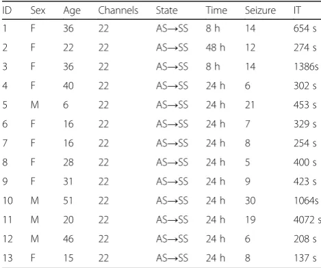
frequency was 500 Hz and the electrodes were located the international 10–20 system. Clinical experts have labeled every seizure. The specific information of epi-leptic patients was shown in Table1.
Different seizures have different signal characteristics, and the performance of seizure detection is related to the type of epileptic seizure [27]. So in this paper, the patients’ data with complex partial seizures’were selected. The ex-perimental data included 13 patients, the age ranged from six to 51 years old. One hundred fifty-nine times of sei-zures were recorded. The average number of seisei-zures per patient was 12.2. The observation time of each patient was 24 h and the total seizure time was 9956 s.
Data preparation
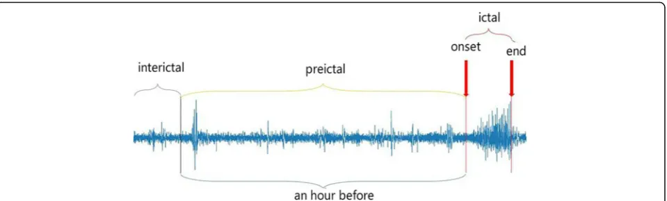
Numerous investigations have demonstrated a gradual transition between the inter-ictal state and ictal state, which is defined as the pre-ictal stage [28]. Thus, the seizure detection could be considered as the classifica-tion of three states. In this study, the EEG data col-lected from clinical patients were divided into three stages: inter-ictal, pre-ictal and ictal stage, as depicted in Fig.1. The details are as follows respectively:
Pre-ictal state: Segment with an hour duration before each seizure was defined as the pre-ictal state [29].
Ictal state: Neurophysiology experts labeled the clinical seizures.
Inter-ictal state: The EEG signal data of each patient which was neither pre-ictal nor ictal state were de-fined as the inter-ictal state.
System design
The overall study design consists of typical blocks (see Fig. 2). Firstly, due to the multiple electrodes, the
multi-channel EEG time series were constructed as 3D images by means of the position of electrodes on the brain. 3D convolutional kernels were tunable to suit the 3D images input. Moreover, deep CNN automatic-ally learned the patterns of different stages from the EEG signal, and then the training model was used to test in the held-out data. Training and inference phase for 13 patients were calculated using a high-perform-ance computer.
Preprocessing
Time window selection A sliding window analysis usually split the raw EEG data into segments for fea-ture extraction, including overlapping sliding window
and non-overlapping sliding window [30]. Since EEG
signals are non-stationary data, time window should ensure the stability of data. The overlapping sliding window can guarantee the continuity of data, but it is easy to cause information redundancy. Depending on the pre-experiment, the sliding time window for the ictal data is 2500 points (5 s), while for the non-onset period, the sliding time window size is 10 s, and no overlap occurs.
3D image reconstruction
Since a 3D CNN is built in this work, it is inevitable to convert the multi-channel EEG signal into a 3D array (just like the multi-channel image). The conversion must enable to keep most information from the ori-ginal data. In total, the procedure was divided into two major steps. Firstly, the time series were formed into 2D images. In order to suit the CNN kernels, the image was designed as a square, which resolution is equal to the number of points (like 5000*5000). And the image compression was used to reduce the image down to 256*256 for reducing the complexity of com-putation. Then the successive relationship of the differ-ent electrodes was selected according to the adjacdiffer-ent degree of the electrodes [31], and the corresponding 2D EEG images were fused to form a 3D multi-channel image. Its structure is [256,256, 22], which is presented in the Fig.3.
The proposed 3D CNN structure
The 3D convolution method was proposed in the ac-tion recogniac-tion in video tasks, which is most widely
used as C3D model [32]. Since the CNN based on the
3D kernels has not been used for epileptic classifica-tion, there is no optimal network architecture for re-ferring in the literature. Thus, we construct a new CNN structure with the 3D kernel in this experiment,
as described in Table 2, which is different from the
C3D model and suitable for seizure detection. Table 1The details of collected data
ID Sex Age Channels State Time Seizure IT
1 F 36 22 AS→SS 8 h 14 654 s
2 F 22 22 AS→SS 48 h 12 274 s
3 F 36 22 AS→SS 8 h 14 1386s
4 F 40 22 AS→SS 24 h 6 302 s
5 M 6 22 AS→SS 24 h 21 453 s
6 F 16 22 AS→SS 24 h 7 329 s
7 F 16 22 AS→SS 24 h 8 254 s
8 F 28 22 AS→SS 24 h 5 400 s
9 F 31 22 AS→SS 24 h 9 423 s
10 M 51 22 AS→SS 24 h 30 1064s
11 M 20 22 AS→SS 24 h 19 4072 s
12 M 46 22 AS→SS 24 h 6 208 s
13 F 15 22 AS→SS 24 h 8 137 s
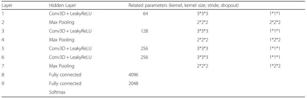
Feature extraction Convolution neural network is a type of neural network with spatial invariance charac-teristics. In addition, the 3D convolution layer has the ability to collect spatial-temporal information, which preserves the input signal after every convolution op-eration. We empirically find that 3 × 3 × 3 convolution kernel for all layers to work best among the limited set of explored architectures. The architecture is shown in Fig. 4. As stated in the experiment, the size of the 3D convolution kernel is 3*3*3 and the step length is 1*1*1, with the Leaky Rectified Linear Unit (ReLU) ac-tive function whose coefficient is set to 0.01. The pool layer uses the maximum pool and the size of 2*2*2. The step length of the first layer is 2*2*2, and the rest of the layer is 1*2*2, reducing the attenuation of the feature. The third layer is directly connected to the fourth layer to retain the channel characteristic infor-mation as far as possible, the full connection layer of 4096 units and 2048 units followed. Finally, the soft-max classifier was used for epileptic classification tasks. Our model was implemented in Python 2.7 with Tensor flow 1.6.0.
Reduce overfitting stage For the sake of limited avail-able datasets, it is important to prevent the CNN from overfitting and improve the performance of the model. Firstly, the equal three stage datasets were adopted. Then the dropout strategy was applied in the both of the fully connected layers. Dropout strategy makes re-sults in the dysfunction of the weight of some hidden layer nodes. Thirdly, considering the size of the epoch,
group normalization proposed by He [33] have
re-placed batch normalization algorithm [34] in 3D CNN. Group normalization can divide the data into groups, then calculate the mean and variance in each group. It improves network generalization ability and accelerates the model convergence. The comparison results are shown in Table3.
Classification stage In this stage, each CNN branch can learn features from different stages. The input of the several branches is the data processed in 3D image re-construction stage. After the feature extraction stage and reduce overfitting stage, the features obtained by each
Fig. 1The single-channel EEG recordings illustrating typical brain states. The typical brain states of epilepsy patients include pre-ictal, ictal and inter-ictal three states. An hour segment before each seizure was defined as a pre-inter-ictal state. Neurophysiology experts annotated inter-ictal state. EEG data of the signal that were neither pre-ictal nor ictal defined as inter-ictal states. The figure represents the whole process of brain electrical signal seizure
a b
branch are merged. The outputs of the model are the predicted category labels.
Training and inference phase A total of 36,000 images dataset was split into a training dataset (30,000 images), a validation dataset (3000 images) and a test dataset (3000 images). The training dataset was used to train the parameters of the model. The validation samples used to validate the model. The test dataset was used to evaluate the trained model.
The classification procedure includes the training phase and inference (test) phase. In the training
phase, we trained our model using a 10-fold
cross-validation strategy. The dataset is randomly scrambled and divided into 10 equal parts. One is selected as the validation dataset to validate the model, and the rest is the training set to complete the training process, each fold data was verified. The
aim of this method is to prevent overfitting of the CNN model during training. In the inference phase, the independent test data was used to evaluate the performance of the model.
According to the pre-experiments, we proposed in-structive settings to help the CNN perform well with the seizure detection task. The batch size is set as 10, an epoch iteration is 6000 times, and a total of 200 epochs are trained. The cross-entropy loss function is selected as the cost function, using the Adaptive Mo-ment Estimation (Adam) optimizer (initial learning rate = 0.01, β1 = 0.9, β2 = 0.999, decay = 0). For the learning rate strategy: if 10 consecutive Epochs, when the error on the verification set remains unchanged, the current learning rate will be reduced by 10 times. Otherwise, the learning rate is divided by 10 after each 40 Epoch. Repeat the above three operations until training all epochs.
a
b
Fig. 32D and 3D image reconstruction for multi-channel EEG.a2D image reconstruction on a multi-channel time series results in an image in 2D (multiple frames as multiple channels).b3D image reconstruction on multi-channel time series results in 3D image volume, preserving temporal information of the input signal. The z-axis is the channel number, x is the size of the time window, y is the value of the signal
Table 2The parameters of the 3D CNN
Layer Hidden Layer Related parameters (kernel, kernel size, stride, dropout)
1 Conv3D + LeakyReLU 64 3*3*3 1*1*1
2 Max Pooling 2*2*2 2*2*2
3 Conv3D + LeakyReLU 128 3*3*3 1*1*1
4 Max Pooling 2*2*2 1*2*2
5 Conv3D + LeakyReLU 256 3*3*3 1*1*1
6 Conv3D + LeakyReLU 256 3*3*3 1*1*1
7 Max Pooling 2*2*2 1*2*2
8 Fully connected 4096
9 Fully connected 2048
Compared 2D CNN structure
The 2D CNN developed rapidly with the help of com-puter vision, the representative convolution neural net-work mainly includes LeNet, AlexNet, Inception, ResNet, DenseNet, Xception, MobileNet, ShuffleNet,
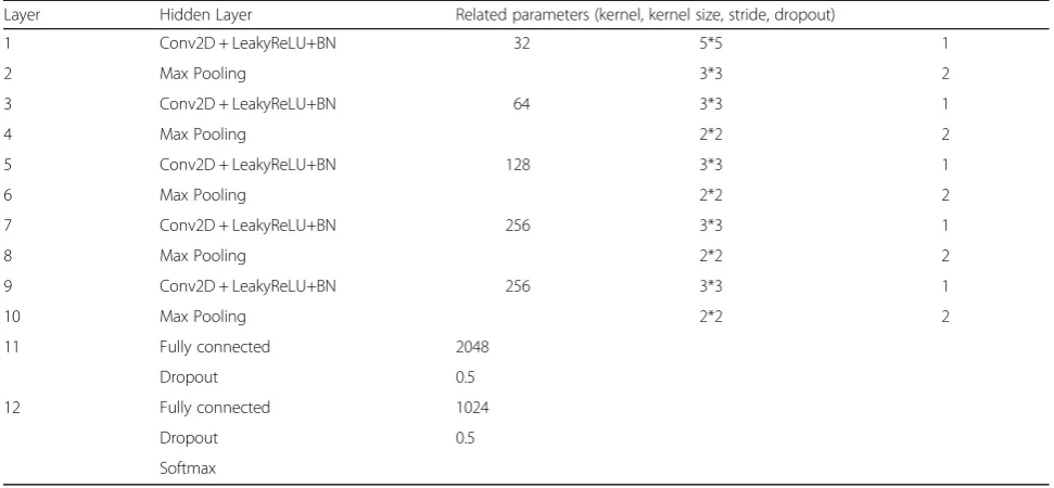
Capsule network etc. [35]. We constructed the 12
layers 2D CNN structure shown in Table4 and Fig. 5.
For the feature extraction stage, the most difference was 2D convolution layer which was applied to collect EEG image information, and every convolution layer adopted the batch Normalization to reduce the changes in the distribution of internal neurons. For the reduction of overfitting stage, the fully connected layers applied the dropout strategy with a dropout rate of 0.5. For the training phase, the setting including earning rate, epochs and cost function etc. is the same as the 3D CNN.
System evaluation
To evaluate the seizure detection performance, we used the metrics in Table5[36].
Standard measurements including sensitivity, specifi-city, and accuracy were adopted to evaluate the model. According to the above performance parameters, the evaluation indexes are defined as:
Accuracy¼TPþFN=TPþTNþFPþFN ð1Þ
Specificity¼TN=TNþFP ð2Þ
Sensitivity recall rate or true positiveð Þ
¼TP=TPþFN ð3Þ
False negative rate FNRð Þ ¼FN=TPþFN
¼1−sensitivity ð4Þ
False positive rate FPRð Þ ¼FP=FPþTN
¼1−specificity ð5Þ
Results
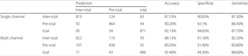
In this paper, the 2D CNN model was used to test the single- electrode EEG data and multi-electrode EEG data respectively, and the 3D CNN model was tested for demonstrating the 3D kernels’effeteness compared to other methods. The results are shown in Tables 6,7
and8.
According to Table 6, the accuracy rate of the
net-work based on the single electrode data test was 89.95%, the FNR was 15.07%, and the FPR was 7.53%. While the accuracy of the multi-channel was 89.91%, the FNR was 15.13% and the FPR was 7.57%. It demon-strated that more channels from EEG data carried more information and could increase the specificity and sensitivity in medical analysis.
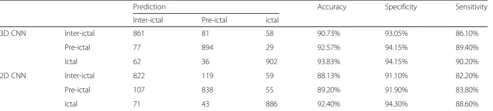
In Table 7, the accuracy of 3D CNN based on
multi-channel was 92.37%, the FNR is 11.43%, and the FPR is 6.22%. While the accuracy of the 2D CNN was 89.91%, the FNR was 15.13% and the FPR was 7.57%. The overall recognition rate of the 3DCNN model was higher than that of the 2D CNN, and the recognition rate for the ictal time segment was the highest, followed by the recognition rate of the pre-ictal EEG data.
Table8 lists the comparison of the 3D CNN based
al-gorithm with traditional machine learning alal-gorithms as well as the 2D CNN, all of the above methods were trained and tested with the data used in this study. Ac-cording to the results, the method proposed in this paper not only achieved the best performance but also reduce the hand–engineered time.
Discussion
People with uncontrolled epilepsy suffer uncertainty when a seizure occurs, the diagnosis of seizure was a lack in remote areas because of limited medical ser-vices [37]. For examining epilepsy patients efficiently, we hope to develop an automatic seizure detection sys-tem to guide doctors.
Fig. 4The architecture of 3D CNN. 3D CNN network has 4 convolution layers, 3 max-pooling layers, and 2 fully connected layers, followed by a softmax output layer. All conv3D kernels are 3*3*3 with stride 1 in both three dimensions; all pooling layer kernels are 2*2*2. The first fully connected layer has 4096 output units and the second fully connected layer has 2048 output units
Table 3Comparison between batch normalization and group normalization
Method Batch size = 10
Epoch = 1 Epoch = 50 Epoch = 200
BN 74% 84% 89%
GN 79% 87% 90%
Deep learning opens the new gate of intelligent diag-nosis in medical healthcare, especially in EEG signal processing. The LSTM network was able to predict all 185 seizures, providing high rates of seizure prediction sensitivity based on different pre-ictal time window in the public datasets [38]. The proposed deep learning approach combined the time-frequency and CNN achieves a sensitivity of 81.4, 81.2, and 75% in public
dataset [39]. The deep learning applied to the hidden layer makes the expression of data as specific as pos-sible so as to obtain a more efficient representation of EEG signals.
However, most deep learning researches adopt the 2D network, which ignores the fact of multi-channel
signal processing [40], Table 6 shows that the more
channels EEG signal could improve the performance of Table 4The details of 2D CNN structure
Layer Hidden Layer Related parameters (kernel, kernel size, stride, dropout)
1 Conv2D + LeakyReLU+BN 32 5*5 1
2 Max Pooling 3*3 2
3 Conv2D + LeakyReLU+BN 64 3*3 1
4 Max Pooling 2*2 2
5 Conv2D + LeakyReLU+BN 128 3*3 1
6 Max Pooling 2*2 2
7 Conv2D + LeakyReLU+BN 256 3*3 1
8 Max Pooling 2*2 2
9 Conv2D + LeakyReLU+BN 256 3*3 1
10 Max Pooling 2*2 2
11 Fully connected 2048
Dropout 0.5
12 Fully connected 1024
Dropout 0.5
Softmax
the network. We proposed the 3D image reconstruc-tion approach to relate multi-channel informareconstruc-tion, just like in video processing [41]. In addition, the group normalization, as well as the oversampling techniques were applied to overcome the overfitting of the limited
datasets [42]. Compared with the 2D CNN shown in
Table 7, our strategy achieved a mean accuracy of
more than 90%. It demonstrated that there was a reli-able and automatic seizure detection system. This is the first study to introduce 3D kernel CNN’s for seiz-ure detection.
To evaluate our approach, we have measured the proposed algorithm against three studies using the
same data, summarized in Table 8. The first method
[43] extracted pre-defined features from the EEG data and use conventional machine learning techniques to classify epileptic stages. This requires much time and it is possible that some information is fully or partly missed in the selected features. The next two deep learning method including 2D CNN and 3D CNN have introduced before, which could learn data patterns automatically. On average, the proposed 3D CNN method performs better than 2D CNN in terms of the multi-channel information, and it outperforms the hand-engineered method with less time and high accuracy. A recent competition on Kaggle held the seizure detection contest, the top three winner
algo-rithms [44] includes the hand –engineered and deep
learning methods, but they relied on complex fea-tures selected. Therefore, the method presented here can be run on an online platform and tested on
more data, satisfying the power, resource, and
computation that can be implemented in the wear-able device.
However, limitations of this work have to be admit-ted. Firstly, this method, all deep learning technology requires sufficient data to train the model and the de-sign of the network is much harder to guarantee to be optimal. Maybe other research gets better performance just tuning the small parameters. Secondly, few clinical experts in one center labeled the model data. Thirdly, the experiment just involves the EEG data type, which neglects other data types from a multi-scale perspec-tive. In order to have a more generalizable clinical val-idation, the methods should be tested on an extensive and multi-center dataset. Further relevant information sources can be readily incorporated into the deep neural networks, such as video, weather patterns, bio-markers, or clinical notes [45,46]. Detection algorithm which incorporates these additional inputs and the data types is the focus of ongoing work.
Conclusion
This study proposed a new approach for epileptic EEG classification, which constructed the 3D CNN for multi-channel EEG data. The main advantage of the method is fully utilizing the multi-channel signal infor-mation without hand-engineered. The 3D CNN model outperformed the previously heuristic detectors. To our best knowledge, this study is the first try of using 3D CNN algorithm for seizure detection. Therefore, it may serve as a benchmark for new work exploring deep learning enabled seizure detection in terms of multi-channel EEG data. Further studies need to carry Table 5Obfuscation matrix of prediction results and actual results
Prediction Total
Object Non-object
Actual Object True Postive(TP) False Postive(FP) TP + FP
Non-object False Negtive(FN) True Negtive(TN) FN + TN
Total TP + FN FP + TN TP + FP + FN + TN
Each row of the matrix represents the instances in a predicted class while each column represents the instances in an actual class
Table 6Classification result based on 2DCNN model using single and multi-channel
Prediction Accuracy Specificity Sensitivity
Inter-ictal Pre-ictal ictal
Single channel Inter-ictal 813 124 63 87.53% 90.65% 81.30%
Pre-ictal 92 864 44 90.20% 92.1% 86.40%
Ictal 95 34 871 92.13% 94.65% 87.10%
Multi channel Inter-ictal 822 119 59 88.13% 91.10% 82.20%
Pre-ictal 107 838 55 89.20% 91.90% 83.80%
out to validate this algorithm in the multi-center data-set. We expect more advances in signal processing, network design, model validation to shape the future of automatic seizure detection.
Abbreviations
2D:Two-dimensional; 3D: Three-dimensional; ApEn: Approximate entropy; AS: Awake stage; CNN: Convolutional neural network; DWT: Discrete wavelets transform; EEG: Electroencephalogram; IT: Ictal time; ReLU: Rectified Linear Unit; SS: Sleep stage; SVM: Support vector machines
Acknowledgements
The authors would like to thank Zhen Zhang for insightful discussion and language assistance. And the authors thank the anonymous reviewers for their careful review and valuable comments.
Funding
This research/work was supported by the National Natural Science Foundation of China (NSFC) [No. 61876194], The National Key Research and Development Program of China [No. 2018YFC0116902, No.2018YFC0116904, No. 2016YFC0901602], Joint Foundation for the NSFC and Guangdong Science Center for Big Data (No. U1611261], Science and Technology Program of Guangzhou [No. 201604020016], Science and Technology Program of Huizhou [No. 2014B010118003] and Natural Science Foundation of Guangdong Province [No. 2018A030313345]. This article has not received sponsorship for publication.
Availability of data and materials
All data were collected from the patients who had been admitted to the hospital, Because of the hospital’s regulations, the data cannot be made publicly available.
About this supplement
This article has been published as part ofBMC Medical Informatics and Decision Making Volume 18 Supplement 5, 2018: Proceedings from the 2018 Sino-US Conference on Health Informatics.The full contents of the supplement are available online athttps://bmcmedinformdecismak.biomedcentral.com/articles/ supplements/volume-18-supplement-5.
Authors’contributions
Study concept and data provision: YZ, XYW and ZYC. Data preparation and processing: XYW. Formulation of model: LZ, XYW, and YZ. Wrote and revised
the manuscript: XYW and LJ Z. The manuscript review: LZ, ZYC and YZ. All the authors read and approved the final version of the manuscript. The authors declare that they have no competing interests.
Ethics approval and consent to participate
Not applicable.
Consent for publication
Not applicable.
Competing interests
The authors declare that they have no competing interests.
Publisher’s Note
Springer Nature remains neutral with regard to jurisdictional claims in published maps and institutional affiliations.
Author details
1
Department of Biomedical Engineering, Zhongshan School of Medicine, Sun Yat-sen University, Guangzhou 510080, Guangdong Province, China.
2Software Engineering, School of Computer and Data Science, Sun Yat-sen
University, Guangzhou 510006, Guangdong Province, China.3Department of
Neurology, The First Affiliated Hospital, Sun Yat-sen University, Guangzhou 510080, Guangdong Province, China.
Published: 7 December 2018
References
1. Jory C, Shankar R, Coker D, Mclean B, Hanna J, Newman C. Safe, and sound? A systematic literature review of seizure detection methods for personal use. Seizure. 2016;36:4–15.
2. Beniczky S, Ryvlin P. Standards for testing and clinical validation of seizure detection devices. Epilepsia. 2018;59(Suppl 1):9.
3. Elger CE, Hoppe C. Diagnostic challenges in epilepsy: seizure under-reporting and seizure detection. Lancet Neurol. 2018;17(3):279–88. 4. Mormann F, Andrzejak RG. Seizure prediction: making mileage on the long
and winding road. Brain. 2016;139(6):1625–7.
5. Trinka E, Kälviäinen R. 25 years of advances in definition, classification and treatment of status epilepticus. Seizure Eur J Epilepsy. 2017;44:65. 6. Cogan D, Birjandtalab J, Nourani M, Harvey J, Nagaraddi V.
Multi-biosignal analysis for epileptic seizure monitoring. Int J Neural Syst. 2016;27(01):345–55.
7. Fiscon G, Weitschek E, Cialini A, Felici G, Bertolazzi P, Salvo SD, et al. Combining EEG signal processing with supervised methods for Alzheimer’s patient's classification. BMC Med Inform Decis Mak. 2018;18(1):35. 8. Zhou Y, Huang R, Chen Z, Chang X, Chen J, Xie L. Application of
approximate entropy on dynamic characteristics of epileptic absence seizure. Neural Regen Res. 2012;07(8):572–7.
9. Gao ZK, Cai Q, Yang YX, Dong N, Zhang SS. Visibility graph from adaptive optimal kernel time-frequency representation for classification of epileptiform EEG. Int J Neural Syst. 2017;27(4):1750005.
10. Gigola S, Ortiz F, D’Attellis CE, Silva W, Kochen S. Prediction of epileptic seizures using accumulated energy in a multiresolution framework. J Neurosci Methods. 2004;138(1–2):107–11.
Table 7Classification results based on 2D and 3DCNN model using multi-electrode
Prediction Accuracy Specificity Sensitivity
Inter-ictal Pre-ictal ictal
3D CNN Inter-ictal 861 81 58 90.73% 93.05% 86.10%
Pre-ictal 77 894 29 92.57% 94.15% 89.40%
Ictal 62 36 902 93.83% 94.15% 90.20%
2D CNN Inter-ictal 822 119 59 88.13% 91.10% 82.20%
Pre-ictal 107 838 55 89.20% 91.90% 83.80%
Ictal 71 43 886 92.40% 94.30% 88.60%
Table 8Performance comparison of different methods
Method Mean accuracy Running time
ApEn+DWT + SVM [43] 91.25% 85.1 s
2D CNN 89.91% 3.8 s
3D CNN 92.37% 8.6 s
11. Subasi A, Gursoy MI. EEG signal classification using PCA, ICA, LDA and support vector machines. Expert Syst Appl. 2010;37(12):8659–66. 12. Litjens G, Kooi T, Bejnordi BE, Aaa S, Ciompi F, Ghafoorian M, et al. A
survey on deep learning in medical image analysis. Med Image Anal. 2017;42(9):60–88.
13. Xun G, Jia X, Zhang A. Detecting epileptic seizures with
electroencephalogram via a context-learning model. BMC Med Inform Decis Mak. 2016;16(2):70.
14. Subasi A, Erçelebi E. Classification of EEG signals using the neural network and logistic regression. Comput Methods Programs Biomed. 2005;78(2):87–99.
15. Mirowski PW, Lecun Y, Madhavan D, Kuzniecky R, editors. Comparing SVM and convolutional networks for epileptic seizure prediction from intracranial EEG. Machine Learning for Signal Processing, 2008 MLSP 2008 IEEE Workshop on; 2008.
16. Acharya UR, Oh SL, Hagiwara Y, Tan JH, Adeli H. Deep convolutional neural network for the automated detection and diagnosis of seizure using EEG signals. Computers in biology and medicine. 2018; 100:270–8. 17. Ullah I, Hussain M, Qazi EUH, Aboalsamh H. An automated system for
epilepsy detection using EEG brain signals based on deep learning approach. Expert Syst Appl. 2018;107:61–71.
18. Thodoroff P, Pineau J, Lim A. Learning robust features using deep learning for automatic seizure detection; 2016.
19. Lin Q, Ye S-q, Huang X-m, Li S-y, Zhang M-z, Xue Y, Chen W-S. Classification of epileptic EEG signals with stacked sparse autoencoder based on deep learning. InInternational Conference on Intelligent Computing: 2016:802–810.
20. Movahedi F, Coyle JL, SejdićE. Deep belief networks for
electroencephalography: A review of recent contributions and future outlooks. IEEE J Biomed Health Inform. 2017;PP(99):1.
21. Jiang Y, Wu D, Deng Z, Qian P, Wang J, Wang G, et al. Seizure Classification from EEG Signals using Transfer Learning, Semi-Supervised Learning and TSK Fuzzy System. IEEE Trans Neural Syst Rehabil Eng. 2017;PP(99):1. 22. Zhang Q, Yang LT, Chen Z, Li P. A survey on deep learning for big data. Inf
Fusion. 2018;42:146–57.
23. Gupta A, Singh P, Karlekar M. A Novel Signal Modeling Approach for Classification of Seizure and Seizure-free EEG Signals. IEEE Trans Neural Syst Rehabil Eng. 2018;PP(99):1.
24. Makinson CD, Tanaka BS, Sorokin JM, Wong JC, Christian CA, Goldin AL, et al. Regulation of Thalamic and Cortical Network Synchrony by Scn8a. Neuron. 2017;93(5):1165–79.e6.
25. Paesschen WV. The future of seizure detection. Lancet Neurol. 2018;17(3):200–2. 26. Quigg M, Leiner D. Limitations of single-channel EEG on the forehead for
neonatal seizure detection. J Perinatol. 2009;29(3):237–42. 27. Ulate-Campos A, Coughlin F, Gainza-Lein M, Fernández IS, Pearl P,
Loddenkemper T. Automated seizure detection systems and their effectiveness for each type of seizure. Seizure. 2016;40:88–101. 28. Cook MJ, O'Brien TJ, Berkovic SF, Murphy M, Morokoff A, Fabinyi G, et al.
Prediction of seizure likelihood with a long-term, implanted seizure advisory system in patients with drug-resistant epilepsy: a first-in-man study. Lancet Neurol. 2013;12(6):563–71.
29. Bandarabadi M, Rasekhi J, Teixeira CA, Karami MR, Dourado A. On the proper selection of preictal period for seizure prediction. Epilepsy Behav. 2015;46:158.
30. Zhang Z, Chen Z, Zhou Y, Du S, Zhang Y, Mei T, et al. Construction of rules for seizure prediction based on approximate entropy. Clin Neurophysiol. 2014;125(10):1959–66.
31. Weis JA, Miga MI, Yankeelov TE. Three-dimensional image-based mechanical modeling for predicting the response of breast cancer to neoadjuvant therapy. Comput Methods Appl Mech Eng. 2016;314:494–512. 32. Du T, Bourdev L, Fergus R, Torresani L, Paluri M, editors. Learning
Spatiotemporal Features with 3D Convolutional Networks. IEEE International Conference on Computer Vision; 2016.
33. Crytzer TM, Keramati M, Anthony SJ, Cheng Y-T, Robertson RJ, Dicianno BE. Exercise prescription using a group-normalized rating of perceived exertion in adolescents and adults with spina bifida. PM&R. 2018;10(7):738–47. 34. Ioffe S, Szegedy C. Batch normalization: accelerating deep network training
by reducing internal covariate shift; 2015. p. 448–56.
35. Alom MZ, Taha TM, Yakopcic C, Westberg S, Hasan M, Essen BCV, et al. The history began from AlexNet: a comprehensive survey on deep learning approaches. 2018.
36. Assi EB, Dang KN, Rihana S, Sawan M. Towards accurate prediction of epileptic seizures: a review. Biomed Signal Process Control. 2017;34:144–57. 37. Ryvlin P, Beniczky S. Seizure detection and mobile health devices in
epilepsy: Update and future developments. Epilepsia. 2018;59:7-8. 38. TsiourisΚΜ, Pezoulas VC, Zervakis M, Konitsiotis S, Koutsouris DD, Fotiadis
DI. A Long Short-Term Memory deep learning network for the prediction of epileptic seizures using EEG signals. Computers in biology and medicine. 2018;99:24–37.
39. Truong ND, Nguyen AD, Kuhlmann L, Bonyadi MR, Yang J, Ippolito S, Kavehei O. Convolutional neural networks for seizure prediction using intracranial and scalp electroencephalogram. Neural Networks. 2018;105: 104-111.
40. Denmark T, Fridrich J, Comesañaalfaro P. Improving selection-channel-aware steganalysis features. Electron Imaging. 2016;2016(8):1–8.
41. Ji S, Xu W, Yang M, Yu K. 3D convolutional neural networks for human action recognition. IEEE Trans Pattern Anal Mach Intell. 2012;35(1):221–31. 42. Brinkmann BH, Wagenaar J, Abbot D, Adkins P, Bosshard SC, Min C, et al.
Crowdsourcing reproducible seizure forecasting in human and canine epilepsy. Brain. 2016;139(6):1713–22.
43. Kumar Y, Dewar ML, Anand RS. Epileptic seizures detection in EEG using DWT-based ApEn and artificial neural network. SIViP. 2014;8(7):1323–34. 44. Baldassano SN, Brinkmann BH, Ung H, Blevins T, Conrad EC, Leyde K, et al.
Crowdsourcing seizure detection: algorithm development and validation on human implanted device recordings. Brain A J Neurol. 2017;140(6):1680. 45. Kiral-Kornek I, Roy S, Nurse E, Mashford B, Karoly P, Carroll T, et al. Epileptic
seizure prediction using big data and deep learning: toward a Mobile system. EBiomedicine. 2017;27(C):103–11.