R E S E A R C H A R T I C L E
Open Access
Initial presentations and final outcomes of primary
pyogenic liver abscess: a cross-sectional study
Chang-Hua Chen
1,5*, Shung-Sheng Wu
2, Hung-Chi Chang
3and Yu-Jun Chang
4Abstract
Background:Although pyogenic liver abscess (PPLA) fatalities are decreasing owing to early diagnosis and effective treatments, PPLA-associated complications still exist. The purpose of this study was to analyze the characteristic features of initial presentations and final outcomes of PPLA caused by different pathogens. Methods:This retrospective study collected and analyzed information regarding initial presentations and final outcomes in patients diagnosed with PPLA at admitted at Changhua Christian Hospital from January 1 to December 31, 2010.
Results:During the study period, we analyzed the records of a total of 134 patients with documented PPLA. There were no significant causative pathogen-related differences in symptoms at initial presentation. Compared with the survivor group, patients in the mortality group were characterized by male gender (p< 0.001), malignancy (p< 0.001), respiratory distress (p=0.007), low blood pressure (p= 0.024), jaundice (p= < 0.001), rupture of liver abscess (p< 0.001), endophthalmitis (p= 0.003), and multiple organ failure (p< 0.001). No patients received liver transplantation or were diagnosed with HIV during the study period. According to univariate logistic regression analysis, gender (OR = 1.185, 95% CI: 0.284–11.130,p= 0.006), malignancy (OR = 2.067, 95% CI: 1.174–13.130,p= 0.004), respiratory distress (OR = 1.667, 95% CI: 1.164–14.210,p= 0.006), low blood pressure (OR = 2.167, 95% CI: 2.104–13.150,p= 0.003), jaundice (OR = 1.9, 95% CI: 1.246–3.297,p= 0.008), rupture of liver abscess (OR = 5.167, 95% CI: 2.194–23.150,p= 0.003), endophthalmitis (OR = 2.167, 95% CI: 1.234–13.140,p= 0.005), and multiple organ failure (OR = 3.067, 95% CI: 1.184–15.150,p= 0.001) differed
significantly between the mortality and survivor groups.
Conclusion:Although the initial presentations of PPLA caused by different pathogens were similar, there were significant differences in mortality in cases involving: (1) male patients, (2) malignancy, (3) initial respiratory distress, (4) initial low blood pressure, (5) jaundice, (6) rupture of liver abscess, (7) endophthalmitis, , and (8) multiple organ failure. We strongly recommend using a severity score of the disease to determine the risk of mortality for each patient with PPLA. In order to prevent complications and reduce mortality, more attention must be paid to high-risk PPLA patients.
Keywords:Primary pyogenic liver abscess, Initial presentation, Outcome complications, Mortality, Risk factors
Background
Primary pyogenic liver abscess (PPLA) is not a rare disease in Taiwan [1]. With the introduction of percutaneous drain-age, surgical draindrain-age, and effective antibiotics, the treat-ment of PPLA has improved over the last 10 years [2]. However, PPLA still causes significant morbidity and
mortality and if not diagnosed and treated early, is almost always fatal [3].
The clinical presentation of liver abscess in Taiwan has changed over the years owing to antibiotic overuse and as-sociated drug resistance, higher prevalence of chronic or malignant diseases, and population ageing [4]. Despite the increased understanding of this new invasive syndrome [4], only earlier diagnosis and effective treatment can minimize the occurrence of sequelae and improve clinical outcomes. However, the literature describing initial clinical presentations of PPLA and early ultrasonography findings is sparse [5].
* Correspondence:[email protected] 1
Division of Infectious Diseases, Department of Internal Medicine, Changhua Christian Hospital, 135 Nanxiao St., Changhua City, Changhua County 500, Taiwan
5Department of Nursing, College of Medicine & Nursing, Hung Kuang
University, No. 1018, Sec. 6, Taiwan Boulevard, Shalu District, Taichung 43302, Taiwan
Full list of author information is available at the end of the article
Since the clinical symptoms of PPLA are variable, and some of the PPLA causative pathogens, such asKlebsiella pneumoniae (K. pneumoniae), are highly resistant to the most effective antibiotic therapies, it is very important to develop accurate and effective diagnosis and treatment ap-proaches for PPLA. Early diagnosis of patients with liver abscess is one of the most challenging tasks for a phys-ician. Over the past 3 decades, ultrasound has had an im-portant role in the diagnosis of this disease. Even with aggressive treatment with antibiotics and drainage, how-ever, the mortality rate of liver abscess remains high [6-8].
It is often difficult to promptly diagnose the initial stage of PPLA because ultrasound findings are typically negative or non-specific and subclinical symptoms and signs are absent. We performed a cross-sectional study of all of the confirmed cases of PPLA in central Taiwan in order to (1) elucidate the risk factors for PPLA, (2) compare the most common ultrasound findings between PPLA cases caused by different pathogens, and (3) iden-tify the prognostic factors for mortality.
Methods
Setting and population
The population of Changhua is mostly served by the Changhua Christian Hospital System (CCHS), which has maintained comprehensive clinical records since 1867. Changhua County is located in central Taiwan, and it had a population of 140,277 in 2000. Changhua Christian Hos-pital (CCH), a 1,800-beds tertiary referral medical center located in northern Changhua County, is a component of CCHS. CCH has been a Joint Commission International-accredited hospital since 2009. This cross-sectional retro-spective study of PPLA was conducted by reviewing the computerized medical records collected at CCH from January 1 to December 31, 2010. The study was approved by the institutional review board of Changhua Christian Hospital (CCH IRB No. 131116).
Case findings and analysis
Cases of PPLA at CCH were identified using the microbiological databases and medical records in the International Classification of Diseases, Ninth revision, Clinical Modifications (ICD-9-CM). We used ICD-9-CM code number 572.0 to find the cases. Each case had a med-ical chart that contained medmed-ical diagnoses, surgmed-ical inter-ventions, and other key data from the medical records. The medical records of all the PPLA cases were manually reviewed by the primary investigator (C.C.H.) to confirm the diagnosis (using CCHS resources). Cases that were judged ambiguous were reviewed by the secondary investi-gator (S.S.W.). For example, although in some patients the abscess culture revealed Corynebacterium urealyticum, blood cultures grew Staphylococcus epidermidis, possibly because of skin contamination. Discrepancies between the
abscess and blood cultures were also observed in some cases with secondary pyogenic liver abscess, such as after foreign body penetration of the alimentary tract. Only the first episode of PPLA in each person during the study period was included in statistical analyses.
PPLA was diagnosed based on clinical presentation, imaging findings of ultrasonography, abdominal com-puted tomography (CT) scans, and microbiological re-sults. The treatment protocol for PPLA is described in the Additional file 1. The choice of the antimicrobial agent for the treatment was based on the results of the antibiotic susceptibility test. The following patient details were recorded: age, sex, date of admission, severity of underlying disease, comorbidity, diagnosis, clinical signs and symptoms, microbiological results, response to ther-apy, complications, and outcome. Only primary cases were collected, whereas those cases with distant metas-tasis from significant pyogenic infectious sources other than the liver were excluded. The severity of underlying disease was categorized according to the McCabe & Jackson criteria [9]. Microbiological data were collected only at the initial stage before the antibiotic therapy. All bacterial pathogens were collected, including anaerobes. If both blood and pus cultures were positive, we ana-lyzed only the pus culture taken directly from the liver abscess. If there were recurrent PPLA episodes, only the first instance was taken into account.
Microbiological data were collected by reviewing the computerized records of the Microbiological Laboratory of CCH dated from January 1 to December 31, 2010. Blood culture analysis was performed for every case with sus-pected sepsis. We used a BACTEC NR-860 system (Becton Dickinson Diagnostic Instrument Systems, Cockeysville, MD, USA) to detect pathogens, Gram staining to deter-mine morphology, and the API-20 series kit (bioMerieux Vitek, Hazelwood, MO) for biochemical reactions. Anti-biotic susceptibilities were determined using the disk diffusion method (BBL™Sensi-Disc™, Becton Dickinson) ac-cording to the guidelines of the Clinical and Laboratory Standards Institute [10].
Definitions
(including microbiological results), and image findings ob-served within 48 h of admission. Patient delay was defined as the time interval between the onset of symptoms and presentation to the hospital. Delay of antibiotic therapy was defined as the time interval between the presentation to the hospital and the start of antibiotic therapy. Pro-longed delay was defined as a delay greater than the me-dian. We defined final outcome as either a status with complications or patient’s death. PPLA complications were rupture of liver abscess, endophthalmitis, and multiple (more than 3) organ failures attributable to PPLA. The
K. pneumoniae group included patients whose microbio-logical culture revealedK. pneumoniae. The non-Klebsiella
Gram-negative bacilli (GNB) group comprised patients whose microbiological culture revealed GNB other than
K. pneumoniae, including Escherichia coli, K. terrigena,
Morganella morganii, Pseudomonas aeruginosa, and Pro-teus mirabilis. The aerobic Gram-positive cocci (GPC) group included patients whose microbiological culture re-vealed GPC, including Staphylococcus aureusand viridans streptococci. The anaerobes group was composed of pa-tients with anaerobic organisms in the microbiological cul-ture, includingFusobacterium necrogenes,F. varium, andF. nucleatum. The mixed infection group included patients with more than 1 pathogen in the microbiological culture. Patients with polymicrobial infections were assigned to the mixed infection group. The mortality group included indi-viduals who died within 6 weeks during the hospitalization from PPLA-related causes. The survivor group included those who did not die during the hospitalization. Abscesses were considered cryptogenic in origin when no causative le-sions were apparent.
To analyze case characteristics and laboratory parame-ters of each group, descriptive statistics were calculated using SPSS version 10.0 for Windows (SPSS Inc., Chicago, IL, USA). Chi-square and Fisher’s exact tests were used for analysis. Results were considered significant if the p-value was <0.05.
Results
One hundred fifty PPLA cases were diagnosed from January 1 to December 31, 2010. Sixteen cases were ex-cluded because of inadequate data, leaving a total of 134 patients with documented PPLA. The incidence of PPLA in adults served by CCH was 275.4/100,000 patient-years in 2010. The male to female ratio was 0.3 (29.1). The mean age was 59.4 years (mean ± SD: 59.4 ± 16.3; me-dian: 62.9; range: 9–88). The mean length of hospitalization was 14.6 days (mean ± SD: 14.2 ± 7.3; median: 15; range: 1– 58). Clinically, 118 of 134 (88.1%) patients had fever, and 38 (26.7%) had low blood pressure (<90/60 mmHg) upon ad-mission for PPLA. One hundred nine (85.2%) patients had right upper quarter pain at admission. The abscesses were located at the right liver lobe in 92 (68.7%), left lobe in 21,
and both lobes in 7 cases. The liver abscess size ranged from 1 to more than 5 cm in diameter. One hundred twenty-seven (94.8%) patients had a single abscess, and 7 had multiple abscesses. Ninety-five (70.9%) out of 134 pa-tients had abscesses that resulted from infection with
K. pneumoniae. There were no statistically significant dif-ferences in the initial clinical presentations among the 134 patients, and symptoms at initial clinical presentations in-cluded fever, chills, abdominal pain, anorexia, malaise, nausea, emesis, body temperature higher than 38.3°C, right upper quadrant tenderness, Murphy's sign, hepato-megaly, disturbance of consciousness, and ascites. The ini-tial ultrasonographic findings and the results of the iniini-tial ultrasound-guided aspiration biopsy are listed in Table 1.
Every patient received treatment according to the PPLA treatment protocol (see Additional file 1). In every case, an empirical antibiotic treatment was started at the onset of the clinical signs of infection. Antibiotic therap-ies were changed based on the results of antibiotic sus-ceptibility tests. Twenty-one (15.7%) patients developed complications, including endophthalmitis, PPLA rup-ture, and multiple organ failure. Of 134 patients, 6 died, leading to a crude mortality rate of 4.5% (6/134). The risk factors for mortality associated with PPLA included extra-hepatic complications, PPLA rupture, and multiple organ failure. One of 6 patients did not receive drainage because of bleeding tendency and ascites.
We compared the survivor and mortality groups to analyze the risk factors of the final outcome among the 134 patients with PPLA (Table 2). When compared with the survivor group, patients with mortality were male (26.6% vs. 83.3%,p< 0.001), malignancy (28.9% vs. 66. 7%,
p< 0.001), respiratory distress (11.7% vs. 50%, p= 0.007), low blood pressure (29.7% vs. 33.3%, p= 0.001), jaundice (15.6% vs. 83.3%,p< 0.001), rupture of liver abscess (0% vs. 33.3%, p< 0.001), endophthalmitis (5.6% vs. 33.3%, p= 0.003), and multiple organ failure (3.1% vs. 100%,p< 0.001). In our study, no patients received liver transplantation, and none were diagnosed with HIV during the study period.
According to the results of univariate logistic regression analysis, gender (OR = 1.185, 95% CI: 0.284–11.130, p= 0.006), malignancy (OR = 2.067, 95% CI: 1.174–13.130,p= 0.004), respiratory distress (OR = 1.667, 95% CI: 1.164– 14.210,p= 0.006), low blood pressure (OR = 2.167, 95% CI: 2.104–13.150,p= 0.003), jaundice (OR = 1.9, 95% CI: 1.246– 3.297,p= 0.008), rupture of liver abscess (OR = 5.167, 95% CI: 2.194–23.150, p= 0.003), endophthalmitis (OR = 2.167, 95% CI: 1.234–13.140,p= 0.005), and multiple organ failure (OR = 3.067, 95% CI: 1.184–15.150,p= 0.001) differed sig-nificantly between the mortality and survivor groups.
Discussion
ultrasonographic findings in patients with PPLA caused by different pathogens. Importantly, in 6 (7.5%) of 80 pa-tients with bacteremia, infectious foci at the initial stage were not detected during the abdominal ultrasonographic examination (Table 1), but were revealed with abdominal CT. Lin, et al. showed that 38 (14.1%) of 268 PPLA pa-tients had false-negative findings on ultrasonography at initial presentation [5]. Appropriate targeted antimicrobial treatments, combined with drainage of liver abscesses, have already increased PPLA survival rates [1,4]. Earlier diagnosis of PPLA could minimize sequelae and improve clinical outcomes [4]. Therefore, clinicians should pay spe-cial attention to bacteremic patients without obvious in-fectious foci. In particular, abdominal CT may be useful in patients at high risk of PPLA if initial results of abdominal ultrasonography are negative.
Although 9 of the 134 (6.7%) patients from this study did not undergo ultrasound-guided aspiration biopsy be-cause of aspiration difficulties, their final clinical outcomes did not differ from those of the rest of the patients (Table 2). Evaluation of the final outcomes and duration of the treatment with antimicrobial agents revealed no sig-nificant differences between the patients without gas-forming and multiple abscesses (Table 1). In contrast to the results of Chou et al. [11], the current study and some previous studies [12,13] suggest that abscess multilocula-tion is not the single risk factor for PPLA mortality.
We compared the clinical presentations among different causative pathogens, including K. pneumonia, the main cause of liver abscesses in Taiwan with increasing infection prevalence [14]. There were no significant differences among the causative pathogens. In agreement with the previous reports from Taiwan, K. pneumoniae was the most predominant pathogen [15]. Sachdev et al. suggested that serotype K1 is an important factor of complicated en-dophthalmitis inK. pneumonia-caused PPLA [16]. We did not test the serotype ofK. pneumoniaebecause of the lack of adequate equipment. In brief, there was no significant difference inK. pneumonia-related PPLA (97.9% vs. 2.1%,
p= 0.297) between the survival and mortality groups in this study. The different of our results from previous
studies [4,14,15] could be related to multiple factors, in-cluding initial broad-spectrum antibiotics prescription, and fewer K1 serotype of K. pneumoniae in Changhua County, and it’s needed further experiments to elucidate these points.
With the development of appropriate antimicrobial treatments and drainage methods for liver abscesses, PPLA survival rates have increased during this century [1,4]. In our study population, the crude mortality rate was 4.5% (6/134). Overall, the crude PPLA mortality rate has varied in the recent decade, with the average value be-ing around 5% [7,16-18]. The mortality rates varied ac-cording to the differences in the geographic origins, study designs, study arms, and patient populations [4,17]. A lit-erature review indicated that malignancy on presentation is an important risk factor for PPLA mortality [18-20], similar to our results. In our study, the risk factors associ-ated with PPLA mortality included gender, jaundice, rup-ture of liver abscess, and multiple organ failure. Some of these factors have been described previously, including jaundice [21], rupture of liver abscess [21,22], and endoph-thalmitis [22]. Although the differences between genders could be related to testosterone levels [23], further experi-ments will be need to verify this. Multiple organ failure, initial low blood pressure, and initial respiratory distress are poor prognostic factors that result from higher disease severity, contributing to the higher mortality rates in the corresponding groups. In agreement with the results of Lin et al. [24], our study revealed an association between the prolonged delay of effective antibiotics and aspiration/ drainage therapies and the risk of mortality. Although in the present study the time intervals between the presen-tation and initiation of effective antibiotics therapy varied within a wide range because of the non-specific and highly variable nature of PPLA presentations, we strongly recommend that an early, empirical, short-course, broad-spectrum antibiotics therapy is consid-ered when persistent fever or unstable hemodynamics occur during the initial stages of the empirical therapy. The Lin’s report identified 6 independent risk factors predicting severe complications ofK. pneumonia-related
Table 1 Initial ultrasonographic findings and initial aspiration among 134 pyogenic liver abscess patients
Klebsiella pneumonia(n=95)
Non-Klebsiella GNB (n=31)
Aerobic GPC (n=5)
Anaerobes (n=1)
Mixed infection (n=2)
Total (n=134)
P-value
Negative or equivocal findings of ultrasonographic examinations at first examination
8 1 1 0 1 11 1.000
Difficulty at initial aspiration 8 1 0 0 0 9 0.284
Gas formation 21 6 0 0 1 28 0.591
Multiple abscesses 2 4 1 0 0 7 0.022
Complications 5 3 1 0 1 10 0.001
Mortality 6 3 1 0 0 11 0.297
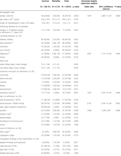
Table 2 Analysis of demographic data, clinical features, concomitant diseases and outcome among 134 patients with primary pyogenic liver abscess
Survivor Mortality Total Univariate logistic regression analysis
N = 128 (%) N = 6 (%) N = 134(%) p value Odds ratio 95% confidence interval
P-value
Demographic data
Gender, male (%) 34 (26.6%) 5 (83.3%) 39 (29.1%) <0.001 1.879 2.284–11.130 0.006
Age, mean ± SD*1(years) 53.6 ± 18.9 67.5 ± 7.4 59.4 ± 16.3 0.705
Length of hospitalization, mean ± SD (days) 10.2 ± 8.3 17.2 ± 6.3 14.2 ± 7.3 0.612
Underlying diseases & co-morbidity
Category C of disease severity
on admission a*2, mean ± SD 15 (11.7%) 2 (33.3%) 17 (10.4%) 0.025
Coexisting diseases, no. (%)
Diabetes mellitus 84 (65.6%) 2 (33.3%) 86 (64.2%) 0.055
Biliary disorders*3 44 (34.4%) 4 (66. 7%) 48 (35.8%) 0.055
Alcoholism 45 (35.2%) 2 (33.3%) 47 (35.1%) 1.000
Liver cirrhosis 46 (35.9%) 3 (50%) 49 (36.6%) 0.530
Malignancy*4 37 (28.9%) 4 (66. 7%) 41 (30.6%) <0.001 2.067 1.174
–13.130 0.004
Uremia 49 (38.3%) 3 (50%) 52 (37.6%) 0.750
Delay issue
Patient delay (days), mean (range) 10 (1–124) 9 (1–91) 0.569
Total delay (days), mean (range) 19 (1–124) 17 (1–91) 0.459
Symptoms and signs on admission, no. (%)
Fever/chills 118 (92.2%) 4 (66.7%) 122 (91.0%) 0.069
Abdominal pain 117 (91.4%) 5 (83.3%) 122 (91.0%) 0.256
Anorexia 12 (9.4%) 2 (33.3%) 14 (10.5%) 0.056
Malaise 115 (89.9%) 5 (83.3%) 120 (89.6%) 0.322
Nausea/emesis 114 (89.1%) 4 (66.7%) 118 (12.7%) 0.057
Respiratory distress*5 15 (11.7%) 3 (50%) 18 (13.4%) 0.007 1.667 1.164
–14.210 0.006 Signs on admission, no. (%)
Body temperature > 38.3°C 111 (86.7%) 6 (100%) 117 (87.3%) 0.358
Blood pressure < 90/60 mmHg 38 (29.7%) 2 (33.3%) 40 (29.8%) 0.001 2.167 2.104–13.150 0.003
Right upper quadrant tenderness 109 (85.2%) 3 (50%) 112 (83.6%) 0.701
Jaundice 20 (15.6%) 5 (83.3%) 25 (18.7%) <0.001 1.900 1.246–3.297 0.008
Murphy's sign*6 21 (16.4%) 1 (16.7%) 22 (16.4%) 0.869
Hepatomegaly 22 (17.2%) 0 (0%) 22 (16.4%) 0.125
Disturbance of consciousness 23 (17.8%) 1 (16.7%) 24 (17.9%) 0.980
Ascites 24 (18.8%) 1 (16.7%) 25 (18.7%) 1.000
Source of infection, no. (%)
Biliary origin*7 32 (25%) 4 (66.7%) 36 (26.9%) 0.069
Cryptogenic origin 33 (25.8%) 1 (16.7%) 34 (25.4%) 0.729
Sonographic findings at first examination, no (%)
Negative findings and equivocal 5 (83.3%) 1 (16.7%) 6 (4.5%) 0.077
Single abscesss (77%) 101 (98.1%) 2 (1.9%) 103 (77%) 0.063
Right lobe (72.3%) 95 (97.9%) 2 (2.1%) 97 (72.3%) 0.071
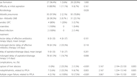
liver abscess: thrombocytopenia (<100,000/mm3), alkaline phosphatase > 300 U/L, gas formation in the abscess, APACHE III score > 40, use of cefazolin (instead of extended-spectrum cephalosporin), and delayed drain-age [24,25].
Our study has several strengths. Most importantly, it represents an reliable reference for evaluating the initial clinical features of PPLA, providing valuable epidemio-logical information regarding the confirmed PPLA cases in central Taiwan. In addition, the prognostic factors, including the initial presentations, were analyzed. The limitations of this study include the retrospective cross-sectional design. The true prevalence of PPLA may be under-estimated because we collected only the cases with definitive diagnosis and positive microbiological findings in order to reduce potential confounding factors. Every case of polymicrobial liver abscess was considered a single case, with the predominant pathogen determined based on the results of microbiological evaluation. Similarly, only the results of liver abscess pus culture were taken into ac-count if both blood and pus cultures were positive. Fur-thermore, since recurrent PPLA was counted as one case,
the true incidence of PPLA is likely to be under-estimated. Finally, we did not calculate the time interval between de-finitive diagnosis and the initiation of effective antibiotic treatment.
Conclusions
Because of early diagnosis and availability of more effective treatments, PPLA mortality is currently decreasing. Never-theless, complications and mortality were still present in the current study. Although the initial presentations of PPLA were not significantly different among the groups with different causative pathogens, the disease was treated successfully in the majority of the patients. The mortality of the patients with PPLA was associated with: (1) male gen-der, (2) presence of malignancy, (3) initial respiratory dis-tress, (4) initial low blood pressure, (5) jaundice, (6) rupture of liver abscess, (7) endophthalmitis, and (8) multiple organ failure. We strongly recommend using a disease severity score to determine the risk of mortality for each patient with PPLA. In order to prevent complications and reduce mortality, more attention must be paid to high-risk PPLA patients.
Table 2 Analysis of demographic data, clinical features, concomitant diseases and outcome among 134 patients with primary pyogenic liver abscess(Continued)
Gas formation 27 (96.4%) 1 (3.6%) 28 (20.9%) 1.000
Difficulty at initial aspiration 8 (88.9%) 1 (11.1%) 9 (6.7%) 0.161
Microbiology 0.028
Klebsiella pneumonia 93 (97.9%) 2 (2.1%) 95 (70.8%)
Non-KlebsiellaGNB 28 (90.3%) 3 (9.7% ) 31 (23.1%)
Aerobic GPC 4 (80%) 1 (20%) 5 (3.7%)
Anaerobes 1 (100%) 0 1 (0.4%)
Mixed infection 2 (100%) 0 2 (1.4%)
Treatment
Doctor delay of effective antibiotics therapy (days), mean (range)
8 (0–33) 4 (0–37) 0.101
Prolonged doctor delay of effective antibiotics therapy (>8 days)
78 (61.5%) 2 (33.3%) 0.118
Delay of aspiration/drainage (days), mean (range) 9 (0–33) 7 (0–27) 0.201
Prolonged delay of aspiration/drainage therapy (>9 days)
78 (61.5%) 1 (16.7%) 0.008
Complications, no. (%)
Rupture of liver abscess 0 (0%) 2 (33.3%) 2 (1.5%) <0.001 5.167 2.194–23.150 0.003
Endophthalmitis, related to PPLA 7 (5.6%) 2 (33.3%) 9 (6.7%) 0.003 2.167 1.234–13.140 0.005
Multiple organ failure, related to PPLA 4 (3.1%) 6 (100%) 10 (7.5%) <0.001 3.067 1.184–15.150 0.001
P-value by Chi-square test or Fisher's exact test when appropriated. Abbreviation:GNBGram negative bacilli,GPCGram positive cocci.
Notes:*1SDstandard deviation,*2Disease severity was categorized according to McCabe & Jackson criteria, and Category C meant the rapid fatal group;*3Biliary disorders including biliary stone diseases (cholelithiasis, choledocholithiasis, or hepatolithiasis) and prior hepatobiliary; surgery;*4
Malignancy including lung cancer, pancreatic cancer, breast cancer, esophagus cancer, stomach cancer, colon cancer, lymphoma, prostate cancer, hematological malignancy, oral cancer, solid tumor and large intestines. Non-digestive system cancer;*5
Respiratory distress including shortness of breath, dyspnea, chest distress, hypoxemia (saturation of oxygen is less than 95%);*6
Murphy’s sign: deep inspiration or cough during subcostal palpation of RUQ producing increased pain and inspiratory arrest;*7
Ethical approval
The study was approved by the institutional review board of Changhua Christian Hospital (CCH IRB No. 131116).
Additional file
Additional file 1:Treatment protocol for pyogenic liver abscesses.
Abbreviations
CCH:Changhua Christian hospital; CCHS: Changhua Christian Hospital System; CT: Computed tomography; GNB: Gram-negative bacilli; GPC: Gram-positive cocci; PPLA: Primary pyogenic liver abscess.
Competing interests
The authors declare that they have no competing interests.
Authors’contributions
C-HC, S-SW, H-CC performed the majority of clinical services. C-HC coordinated this study and wrote the manuscript. Y-JC handled the statistical analysis. All authors read and approved the final manuscript.
Acknowledgements
All authors thank the assistant of the Clinical Microbiology Laboratory of Changhua Christian Hospital. This research project would not have been possible without the support of many people. The author wishes to express her gratitude to staffs of Division of Infectious Diseases and Division of Gastroenterology of Changhua Christian Hospital who were abundantly helpful and offered patient care, invaluable assistance, and support.
Author details
1
Division of Infectious Diseases, Department of Internal Medicine, Changhua Christian Hospital, 135 Nanxiao St., Changhua City, Changhua County 500, Taiwan.2Division of Gastroenterology, Department of Internal Medicine, Changhua Christian Hospital, 135 Nanxiao St., Changhua City, Changhua County 500, Taiwan.3Department of Surgery, Changhua Christian Hospital, 135 Nanxiao St., Changhua City, Changhua County 500, Taiwan. 4
Epidemiology and Biostatistics Center, Changhua Christian Hospital, 135 Nanxiao St., Changhua City, Changhua County 500, Taiwan.5Department of Nursing, College of Medicine & Nursing, Hung Kuang University, No. 1018, Sec. 6, Taiwan Boulevard, Shalu District, Taichung 43302, Taiwan.
Received: 9 July 2014 Accepted: 18 July 2014 Published: 28 July 2014
References
1. Cheng HP, Chang FY, Fung CP, Siu LK:Klebsiella pneumoniae liver abscess in Taiwan is not caused by a clonal spread strain.J Microbiol Immunol Infect2002,35:85–88.
2. Hope WW, Vrochides DV, Newcomb WL, Mayo-Smith WW, Iannitti DA: Optimal treatment of hepatic abscess.Am Surg2008,74:178–182. 3. Cerwenka H, Bacher H, Werkgartner G, El-Shabrawi A, Kornprat P, Bernhardt GA,
Mischinger HJ:Treatment of patients with pyogenic liver abscess.
Chemotherapy2005,51:366–369.
4. Siu LK, Yeh KM, Lin JC, Fung CP, Chang FY:Klebsiella pneumoniae liver abscess: a new invasive syndrome.Lancet Infect Dis2012,12:881–887. 5. Lin AC, Yeh DY, Hsu YH, Wu CC, Chang H, Jang TN, Huang CH:Diagnosis of
pyogenic liver abscess by abdominal ultrasonography in the emergency department.Emerg Med J2009,26:273–275.
6. Seeto RK, Rockey D:Pyogenic liver abscess: changes in etiology, management and outcome.Medicine1996,75:99–113.
7. Yang CC, Chen CY, Lin XZ, Chang TT, Shin JS, Lin CY:Pyogenic liver abscess in Taiwan: emphasis on gas-forming liver abscess in diabetics.
Am J Gastroenterol1993,88:1911–1915.
8. Mischinger HJ, Hauser H, Rabl H, Quehenberger F, Werkgartner G, Rubin R, Deu E:Pyogenic liver abscess: studies of therapy and analysis of risk factors.World J Surg1994,18:852–858.
9. McCabe W, Jackson CG:Gram-negative bacteremia: I. etiology and ecology.Arch Intern Med1962,36:1020–1027.
10. Clinical and Laboratory Standards Institute (CLSI):Methods for dilution antimicrobial susceptibility tests for bacteria that grow aerobically: approved standard—seventeenth edition: document M100-S17.Wayne, PA, USA: CLSI; 2007. 11. Chou FF, Sheen-Chen SM, Chen YS, Chen MC:Single and multiple
pyogenic liver abscesses: clinical course, etiology, and results of treatment.World J Surg1997,21:384–389.
12. Tan YM, Chung AY, Chow PK, Cheow PC, Wong WK, Ooi LL, Soo KC:An appraisal of surgical and percutaneous drainage for pyogenic liver abscesses larger than 5 cm.Ann Surg2005,241:485–490.
13. Pang F, Fung T, Samra J, Hugh T, Smith R:Pyogenic liver abscess: an audit of 10 years’experience.World J Gastroenterol2011,17:1622–1630. 14. Chang FY, Chou MY:Comparison of pyogenic liver abscesses caused by
Klebsiella pneumoniae and non-K pneumoniae pathogens.J Formos Med Assoc1995,94:232–237.
15. Fung CP, Lin YT, Lin JC, Chen TL, Yeh KM, Chang FY, Chuang HC, Wu HS, Tseng CP, Siu LK:Klebsiella pneumoniae in gastrointestinal tract and pyogenic liver abscess.Emerg Infect Dis2012,18:1322–1325. 16. Sachdev DD, Yin MT, Horowitz JD, Mukkamala SK, Lee SE, Ratner AJ:
Klebsiella pneumoniae K1 Liver abscess and septic endophthalmitis in a U.S. resident.J Clin Microbiol2013,51:1049–1051.
17. McDonald MI, Corey GR, Gallis HA, Durack DT:Single and multiple pyogenic liver abscesses. Natural history, diagnosis and treatment, with emphasis on percutaneous drainage.Medicine (Baltimore)1984,63:291–302.
18. Tachopoulou OA, Vogt DP, Henderson JM, Baker M, Keys TF:Hepatic abscess after liver transplantation: 1990–2000.Transplantation2003, 75:79–83.
19. Tsai FC, Huang YT, Chang LY, Wang JT:Pyogenic liver abscess as endemic disease, Taiwan.Emerg Infect Dis2008,14:1592–1600.
20. Chen SC, Yen CH, Lai KC, Tsao SM, Cheng KS, Chen CC, Lee MC, Chou MC: Pyogenic liver abscesses with Escherichia coli: etiology, clinical course, outcome, and prognostic factors.Wien Klin Wochenschr2005,117:809–815. 21. Barakate MS, Stephen MS, Waugh RC, Gallagher PJ, Solomon MJ, Storey DW,
Sheldon DM:Pyogenic liver abscess: a review of 10 years' experience in management.Aust N Z J Surg1999,69:205–209.
22. Karatassas A, Williams JA:Review of pyogenic liver abscess at the Royal Adelaide Hospital 1980–1987.Aust N Z J Surg1990,60:893–897. 23. Lotter H, Helk E, Bernin H, Jacobs T, Prehn C, Adamski J, González-Roldán N,
Holst O, Tannich E:Testosterone increases susceptibility to amebic liver abscess in mice and mediates inhibition of IFNγsecretion in natural killer T cells.PLoS One2013,8:e55694.
24. Lin JC, Chang FY:Pyogenic liver abscess associated with septic pulmonary embolism.J Chin Med Assoc2008,71:603–604.
25. Yeh KM, Kurup A, Siu LK, Koh YL, Fung CP, Lin JC, Chen TL, Chang FY, Koh TH: Capsular serotype K1 or K2, rather than magA and rmpA, is a major virulence determinant for Klebsiella pneumoniae liver abscess in Singapore and Taiwan.J Clin Microbiol2007,45:466–471.
doi:10.1186/1471-230X-14-133
Cite this article as:Chenet al.:Initial presentations and final outcomes of primary pyogenic liver abscess: a cross-sectional study.BMC Gastroenterology 201414:133.
Submit your next manuscript to BioMed Central and take full advantage of:
• Convenient online submission
• Thorough peer review
• No space constraints or color figure charges
• Immediate publication on acceptance
• Inclusion in PubMed, CAS, Scopus and Google Scholar
• Research which is freely available for redistribution