R E S E A R C H
Open Access
The retinoic acid receptor-
α
modulators ATRA
and Ro415253 reciprocally regulate human
IL-5+ Th2 cell proliferation and cytokine
expression
Daniel L Wansley, Yuzhi Yin and Calman Prussin
*Abstract
Background:Th2 cytokine responses are enhanced by all trans retinoic acid (ATRA), the bioavailable form of vitamin A. Retinoic acid receptor alpha (RARα) is the high affinity receptor for ATRA that mediates these pro-Th2 effects. We have previously characterized two major human Th2 subpopulations: IL-5- Th2 (IL-5-, IL-4+, IL-13+) and IL-5+ Th2 cells (IL-5+, IL-4+, IL-13+), which represent less and more highly differentiated Th2 cells, respectively. We hypothesized that the pro-Th2 effects of ATRA may differentially affect these Th2 subpopulations.
Methods:Specific cytokine producing Th2 subpopulations were identified using intracellular cytokine staining. Proliferation was measured using the Cell Trace Violet proliferation tracking dye. Apoptotic cells were identified using either annexin-V or active caspase 3 staining. Th2 gene expression was measured using quantitative polymerase chain reaction.
Results:ATRA increased the output of Th2 cells from house dust mite allergen (HDM) specific short-term cell lines, and this enhancement was limited to the IL-5+ Th2 subpopulation. Conversely, the RARαantagonist Ro415253 decreased Th2 cell output from these cultures, and this effect was again limited to the IL-5+ Th2 subpopulation. ATRA and Ro415253 respectively augmented and inhibited Th2 cell proliferation, and this affect was more pronounced for the IL-5+ vs. IL-5- Th2 subpopulation. ATRA and Ro415253 respectively augmented and inhibited the expression of IL5 in a significant manner, which was not found for IL4 or IL13.
Conclusions:We report that the reciprocal regulation of Th2 cytokine expression and proliferation by RARα
modulators are largely limited to modulation of IL-5 gene expression and to proliferation of the highly differentiated IL-5+ Th2 subpopulation. These results suggest that RARαantagonism is a potential means to therapeutically target allergic inflammation.
Trial registration:Clinicaltrials.gov identifier: NCT01212016
Keywords:Interleukin 5, All-trans retinoic acid, T helper 2, Retinoic acid receptor alpha, House dust mite antigen, Retinoic acid response element
* Correspondence:[email protected]
National Institute of Allergy and Infectious Disease, National Institutes of Health, Laboratory of Allergic Diseases, NIH, 10 Center Drive, MSC-1881, Bethesda, MD 20852-1881, USA
0 103 104 105 0
103 104 105
0 103 104 105 0
103 104 105
0 103 104 105 0
103 104 105
0 103 104 105 0
103 104 105
0 102 103 104 105 0
103 104 105
0 102 103 104 105 0
103 104 105
0 102 103 104 105 0
103 104 105
0 102 103 104 105 0
103 104 105
Vehicle ATRA 1nM ATRA 10nM Ro41 100nM
Vehicle ATRA 1nM ATRA 10nM Ro41 100nM
1.5 73.4
0.05 25.1
2.1 5.4 3.6 4.1 1.7
23.3 9.7 24.7 26.3 22.4 0.27 4.69 3.6 0.04 62.7 33.7 0.24 9.1 0.07 30.5 60.3 0.01 83.116.7
Cell Trace Violet (CTV)
IL-13 IL-5 IL-5
A
E
1000 10000 100000 1000000 100 1000 10000 100000 Vehicle ATRA 1nM ATRA 10nM Ro41 100nM Vehicle ATRA 1nM ATRA 10nM Ro41 100nM sll e c 2 h T + 5-LI )r e b m u n(n.s. n.s.
G
F
ll e c 2 h T - 5-LIs )re b m u n( 0.1 1 10 100 0.1 1 10 100 Vehicle ATRA 1nM ATRA 10nM Ro41 100nM Vehicle ATRA 1nM ATRA 10nM Ro41 100nM*p = 0.04 n.s.
sll ec 2 h T + 5-LI ) %( sll e c 2 h T - 5-LI ) %(
C
1000 10000 100000 1000 10000 100000 1000 10000 100000 sll e c + 5-LI )r e b m u n( Vehicle ATRA 1nM ATRA 10nM Ro41 100nM Vehicle ATRA 1nM ATRA 10nM Ro41 100nM Vehicle ATRA 1nM ATRA 10nM Ro41 100nMn.s. n.s. n.s.
sll e c + 4-LI )r e b m u n( sll e c + 3 1-LI )r e b m u n( 0.1 1 10 100 0.1 1 10 100 0.1 1 10 100 ) %( sll e c + 5-LI Vehicle ATRA 1nM ATRA 10nM Ro41 100nM Vehicle ATRA 1nM ATRA 10nM Ro41 100nM Vehicle ATRA 1nM ATRA 10nM Ro41 100nM
*p = 0.04 *P=0.03 *P=0.03
D
) %( sll e c + 4-LI ) %( sll e c + 3 1-LI 0 20 80 100 P r o li fe r at io n (% C T V lo w)B
40 60 n.s. Vehicle ATRA 1nM ATRA 10nM Ro41 100nMBackground
The association between vitamin A and effective im-mune responses dates back to the early 20th century
when vitamin A was described as an “anti-infective
agent” [1,2]. Large placebo controlled studies in vitamin
deficient populations in developing countries show that vitamin A supplementation is associated with decreased childhood mortality from diarrheal disease and measles, underscoring the critical role for vitamin A in immunity. Studies on vitamin A deficiency have consistently de-monstrated an indispensable role for vitamin A in main-taining host immunity to a variety of pathogens [3-5].
Retinoic acid (RA) is the bioactive form of vitamin A; all-trans-retinoic acid (ATRA) is the most abundant form of RA found in the circulation. RA influences immunity via multiple mechanisms. CD4 T cells are the immune component most prominently affected by RA. RA augments the differentiation of inducible T regulatory cells, which is abrogated both in vitro and in vivo in either vitamin A deficiency states or using ret-inoic acid receptor (RAR) deficient mice [5]. In vitamin A deficient mice, RA restores CD4+ T cell-mediated im-munity, homeostasis, and activation [4,5]. Previous in-vestigations suggest that retinoic acid receptor alpha
(RARα) mediates these RA-induced effects on T cells
[5-8]. ATRA exerts its bioactivity via binding to retinoic acid receptor ligand-activated transcription factors, and ATRA is a high-affinity ligand for retinoic acid receptor
alpha (RARα). ATRA binding to RAR monomers then
induces hetero-dimerization with retinoid X receptors (RXRs). The RAR-RXR complex in turn binds to retinoic acid response elements (RARE) which are present in the promoters of RA responsive genes and result in gene activation [9].
Th2 cells were initially characterized as expressing IL-4, IL-5, and IL-13 [10]. IL-4 is the major factor dri-ving Th2 differentiation, IgE class switching, and alterna-tive macrophage activation, whereas IL-13 functions as an effector molecule that mediates eosinophilic inflam-mation, airway hyper-responsiveness, and mucus secre-tion. IL-5 is the major eosinophil-active cytokine and induces eosinophilopoiesis and eosinophil release from the bone marrow, enhances eosinophil survival, and acts
as a costimulator for eosinophil activation [11,12]. In vivo, vitamin A is associated with eosinophilic tissue inflammation, which is both a protective component of anti-helminth immunity and a major contributor to asthma pathogenesis. Vitamin A deficiency inhibits para-site expulsion due to reduced eosinophilia and IL-5 secretion by antigen-specific lymphocytes in vivo, and vitamin A supplementation restores parasite immunity [11-14]. Vitamin A deficiency diminishes and high-level vitamin A supplementation restores Th2 cytokines and eosinophilia induced by experimental asthma [15].
RARα agonists and RARα antagonists exert opposite
effects on the production of Th2 cytokines by in vitro stimulated T cells [6,16]. We have recently characterized two major subpopulations within the Th2 lineage: IL-5+ Th2, which express IL-5, IL-4, and IL-13, and IL-5- Th2 cells, which only express the latter two cytokines [17]. IL-5+ Th2 cells are more highly differentiated and have greater pro-inflammatory activity than IL-5- Th2 cells [18,19]. IL-5+ Th2 cells are tightly linked to blood eosinophilia [20]. Given the known pro-Th2 activity of RA, we sought to determine if RA drives the diffe-rentiation of IL-5- Th2 to IL-5+ Th2 cells or otherwise enhances the generation of IL-5+ Th2 cells. Using a va-riety of human in vitro Th2 model systems, in this work we demonstrate the reciprocal regulation of cytokine
production and proliferation by RARα modulators are
specific for the IL-5 gene and these effects are limited to the highly differentiated IL-5+ Th2 cell subpopulation.
Materials and methods Subjects
Subjects underwent lymphapheresis (National Institutes of Health Clinical Center Department of Transfusion Medicine), and PBMC were isolated as described [17]. Donors used for lymphapheresis included healthy non-allergic control, eosinophilic gastrointestinal disease and allergic asthmatic subjects. Allergic asthmatic subjects had a minimum one-year history of episodic
broncho-spasm relieved by β-agonist medications and three or
more positive skin test responses (≥3 mm) out of a panel
of 10 aeroallergens. The National Institute of Allergy and Infectious Diseases Institutional Review Board (See figure on previous page.)
approved the clinical protocols used for this study. All subjects signed informed consent.
Cells and culture
For Th2 differentiation, naïve T cells were obtained from PBMC using the naïve CD4+ T cell isolation kit (Miltenyi Biotec, Auburn, CA). Naïve cells were Th2 polarized as published [17]. Th2 polarization cycles were repeated at weekly intervals, where 1×Th2, 2×Th2, and 3×Th2 repre-sent 1, 2, and 3 serial 7d cycles of differentiation. For each
7 d round of stimulation, ATRA/vehicle was added on day 1 and, as replenishment, on day 4 of culture. After dif-ferentiation, Th2-polarized cells were cryopreserved in
liquid N2, and subsequently thawed and recovered in
complete media containing IL-2 (50–100 U/ml) for at
least 24 h prior to RA experiments.
For proliferation assays, PBMC and Th2-polarized
cells were labeled with 5 μM of Cell Trace Violet
prolif-eration tracking dye (CTV; Invitrogen, Carlsbad, CA) ac-cording to instructions. Using PBMC, 10d cultures were
0 20 40 60 80 100 0 20 40 60 80 100 0 20 40 60 80 100
Proliferation (% CTV
)
low
Proliferation (% CTV
)
low
Proliferation (% CTV
)
low
IL2 + V ehicle
IL2 + ATRA
TCR + IL2 + V ehicle
TCR + IL2 + ATRA
TCR + IL2 + Ro41
Unstimulated IL2 + V
ehicle IL2 +
ATRA
TCR + IL2 + V ehicle
TCR + IL2 + ATRA
TCR + IL2 + Ro41
Unstimulated
IL2 + V ehicle
IL2 + ATRA
TCR + IL2 + V ehicle
TCR + IL2 + ATRA
TCR + IL2 + Ro41
Unstimulated
Gate:
Total Th2 cells
0 102 103 104 105
0 200 400 600 800 1000
0 102 103 104 105
0 500 1000 1500 2000
0 102 103 104 105
0 500 1000 1500 2000
0 102 103 104 105
0 200 400 600 800
0 102 103 104 105
0 500 1000 1500
0 102 103 104 105
0 100 200 300 400 500
0 102 103 104 105
0 500 1000 1500
0 102 103 104 105
0 500 1000 1500 2000
0 102 103 104 105
0 500 1000 1500
0 102 103 104 105
0 300 600 900 1200
0 102 103 104 105
0 500 1000 1500
0 102 103 104 105
0 300 600 900 1200
Cell Trace Violet (CTV)
IL-5
Cell number
Un-stimulated Vehicle ATRA 10nM Vehicle ATRA 10nM Ro41 100nM
IL-2 anti-CD3/CD28 + IL-2
0 102
103 104 105 0 103 104 105
0 102
103 104 105 0 103 104 105
0 102
103 104 105 0 103 104 105
0 102
103 104 105 0 103 104 105
0 102
103 104 105 0 103 104 105
0 102
103 104 105 0 103 104 105
***p = 0.01 **p = 0.01 *p = 0.05 **p = 0.01
Total Th2 cells IL-5+ Th2 cells IL-5- Th2 cells
65.9 46.8 36.9 44.4 36.2 46.7
34.1 31.4 33.6 17.3 24.7 11.5
0 7.8 12 17.7 23.5 13.9
0 14.1 17.5 20.5 15.7 27.9
0 30 38.5 56 56.7 60.1
0 34.4 45.3 41.4 40 46.4
A
B
C
D
IL-5+ Th2 cells
IL-5- Th2 cells
used to observe optimal proliferation; in such prolonged proliferation experiments, both the media control (data not shown) as well as the allergen activated cultures
yielded 2 CTV bright “negative” peaks (Figure 1A, lower
right hand quadrant). Using 12d antigen activated cultures,
similar doublet“negative”peaks were described by Givan
and Wallace [21,22] and are thought to be an artifact due to homeostatic proliferation during the prolonged culture.
PBMC proliferation assays were performed using complete media containing 5% heat inactivated human AB serum (Invitrogen). Th2 cell proliferation assays were performed with complete media containing 10% heat inactivated FBS (Invitrogen). Th2 cell cultures used for qRT-PCR were performed using media containing either 10% heat inactivated FBS or 10% charcoal-stripped heat inactivated FBS (Invitrogen). Similar re-sults were obtained with both sources of FBS (data not shown).
All-trans retinoic acid (Sigma, St. Louis, MO) was
reconstituted to 1 mM in DMSO. Ro41-5253 (Ro41, Enzo Life Sciences, Farmingdale, NY) was reconstituted to 10 mM in DMSO. For all experiments ATRA, Ro41, and vehicle (DMSO) were first diluted 1:1000 in 1×PBS and then added to the culture medium at the de-sired concentration. For HDM proliferation assays, D. Pteronyssinus extract (ALK, Round Rock TX) was used at 40 AU/mL.
Intracellular cytokine staining (ICCS) and flow cytometry
ICCS was performed according to published methods [17]. Briefly, after activation, cells were washed once with cold PBS, labeled on ice with Live/Dead Fixable Aqua Dead Cell Stain (Invitrogen), washed, and fixed in 4% paraformaldehyde (Electron Microscopy Sciences, Hatfield, PA). Cells were then suspended in PBS with
10% DMSO (Sigma-Aldrich) and cryopreserved at−80°C.
All antibodies and clones used for flow cytometry ana-lysis have been described [17]. Samples were acquired on an LSR II flow cytometer (BD Biosciences, Franklin Lakes, NJ) and analyzed using FlowJo software (Tree Star, Ashland, OR). Typical forward versus side scatter identified lymphocytes, and cell doublets were excluded using forward scatter area versus height. After gating on viable CD3+, CD4+ cells, data were plotted for various cytokines based on absolute percentages or cell num-bers within the respective gate.
qRT-PCR
RNA was extracted from cell pellets using the RNeasy mini kit (Qiagen, Gaithersburg, MD), treated with DNase (Qiagen), and cDNA was prepared from
100–300 ng RNA using Qscript cDNA Supermix
(Quanta Biosciences, Gaithersburg, MD). qRT-PCR ana-lysis was performed using the delta Ct method of com-parison on the Step-One-Plus Real-Time PCR System Thermal Cycling Block (Applied BioSystems, Carlsbad, CA). All FAM-MGB-labeled TaqMan probe and primer sets for IL-4 (Hs00166237_m1), IL-5 (Hs01548712_g1), IL-13 (Hs00174379_m1), and GAPDH (Hs_03929097_g1) were purchased from Applied Biosystems.
Statistical analysis
Specific statistical methods used are noted in each Figure
Legend. qRT-PCR fold change was calculated as;
Treated ðATRA or Ro41Þ
Vehicle control ,when this quotient was <1, the
nega-tive of the reciprocal value was calculated as described [23].
Results
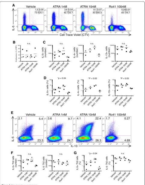
Reciprocal regulation of allergen-specific Th2 cytokine response by RARαmodulators
ATRA enhances human Th2 cell cytokine expression in polyclonally activated T cells [6,16]. To examine the rele-vance of these findings to allergic disease, we analyzed the effects of ATRA and the inhibitor Ro415253 (Ro41) on house dust-mite antigen (HDM) stimulated PBMC from allergic asthmatic subjects. Cell Trace Violet (CTV) was used to track CD4+ HDM-specific memory Th2 cells,
which were identified by gating on CTVlowcells that had
proliferated in response to HDM antigen (Figure 1A).
Nei-ther RARαagonist (ATRA), nor RARα antagonist (Ro41)
significantly affected the total number of CD4 T cells pro-liferating in response to HDM (Figure 1B). However, the output of HDM-specific proliferated IL-5+ Th2 cells was dose-dependently enhanced by ATRA, and reciprocally suppressed by Ro41 (Figure 1C, D).
We have previously characterized two major human Th2 subpopulations: IL-5- Th2 (IL-5-, IL-4+, IL-13+) and IL-5+ Th2 cells (IL-5+, IL-4+, IL-13+), which represent less and more highly differentiated Th2 cells, respectively [17]. We next analyzed HDM-expanded T cells to deter-mine how ATRA and Ro41 affect these Th2 cell
sub-populations (Figure 1E). RARα modulators reciprocally
(See figure on previous page.)
0102 103 104 105 0 102 103 104 105
0102
103 104 105 0 102 103 104 105
0102
103 104 105 0 102 103 104 105
0102
103 104 105 0 102 103 104 105
0102
103 104 105 0 102 103 104 105
0102
103 104 105 0 102 103 104 105
0102
103 104 105 0 102 103 104 105
0102 103 104 105
0 102
103
104
105
0102
103 104 105 0 102 103 104 105
0102
103 104 105 0 102 103 104 105
0102 103 104 105
0 102
103
104
105
0102
103 104 105 0 102 103 104 105
0102
103 104 105 0 102 103 104 105
0102
103 104 105 0 102 103 104 105
0102
103 104 105 0 102 103 104 105
0102
103 104 105 0 102 103 104 105
0102 103 104 105
0 102
103
104
105
0102 103 104 105
0 102
103
104
105
0102 103 104 105
0 102
103
104
105
0102 103 104 105
0 102
103
104
105
0102 103 104 105
0 102
103
104
105
0102
103 104 105 0 102 103 104 105
0102
103 104 105 0 102 103 104 105
0102
103 104 105 0 102 103 104 105
0102
103 104 105 0 102 103 104 105
0102
103 104 105 0 102 103 104 105
0102
103 104 105 0 102 103 104 105
0102
103 104 105 0 102 103 104 105
0102
103 104 105 0 102 103 104 105
0102
103 104 105 0 102 103 104 105
0102
103 104 105 0 102 103 104 105
0102 103 104 105
0 102
103
104
105
0102 103 104 105
0 102
103
104
105
0102 103 104 105
0 102 103 104 105 0 2 4 6 8
Activated Caspase 3
(% positive)
Vehicle ATRA 10nM
Th1
HDM line2xTh2 3xTh2 4xTh2 5xTh2 Activated Caspase 3
Live/Dead
Cell line #1 Cell line #2 Cell line #3 Cell line #1 Cell line #2 Cell line #3
ATRA 10nM Vehicle HDM 5xTh2 4xTh2 3xTh2 2xTh2 Th1
4.6 5.5 4.6 4.1 5.7 4.9
4.4 2.3 5.9 4.1 2.6 6.0
5.3 2.8 3.8 5.7 2.2 3.2
3.5 8.4 4.0 4.2 7.5 5.1
3.2 6.5 4.7 4.0 4.9 4.4
6.8 5.1 6.4 5.8
regulated Th2 cell output in the IL-5+ Th2 but not IL-5-Th2 subpopulation (Figure 1E, F).
In sum, these data demonstrate that ATRA increases the output of IL-5+ Th2 cells from allergen driven cultures.
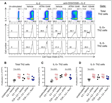
Intrinsic regulation of IL-5+ Th2 cell proliferation by RARα modulators
This greater output of allergen specific IL-5+ Th2 cells ob-served in ATRA treated samples in Figure 1 may be due to several potential mechanisms, including: enhanced Th2 differentiation, enhanced Th2 cytokine expression, pre-ferential Th2 proliferation, or inhibition of Th2 apoptosis. To test if ATRA intrinsically regulates Th2 cell prolifera-tion, we employed in vitro differentiated human Th2 cells and examined their proliferation after modulation with
RARα agonist and antagonist treatment (Figure 2). As
expected, T cell receptor (TCR) plus IL-2 stimulation induced many more generations of cell division than did IL-2 alone (Figure 2A). In the presence of IL-2 alone ATRA significantly increased Th2 cell proliferation (Figure 2B). With maximal activation (anti-CD3, CD28 plus IL-2), ATRA did not further augment Th2 cell proliferation.
We next examined whether RARα modulation
diffe-rentially affected the proliferation of human Th2 sub-populations. Among in vitro differentiated Th2 cells, ATRA augmented IL-2 induced proliferation of both the IL-5+ and IL-5- subpopulations (Figure 2C, D). However, Ro41 significantly inhibited the proliferation of the IL-5+, but not the IL-5- subpopulation. This Ro41 inhibition further demonstrates the differential re-sponsiveness of the IL-5+ vs. IL-5- Th2 subpopulations
to RARαmodulators.
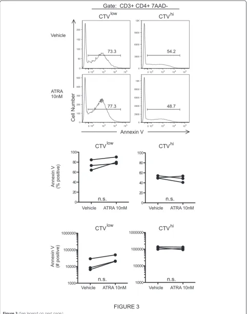
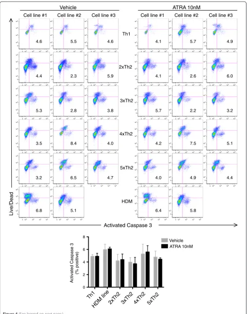
To further address the cellular mechanisms responsible for the ATRA mediated increase in IL-5+ Th2 cell output, we examined ATRA induction of apoptosis. Notably, ATRA did not alter annexin V expression by CD4+ T cells in Th2 dominant HDM proliferation cultures (Figure 3). Additionally, ATRA did not affect caspase-3 activation in CD3 activated Th1 or Th2 cell lines (Figure 4).
In sum, these data demonstrate that ATRA positively regulates Th2 cell proliferation via T cell intrinsic
me-chanisms, and that RARαmodulators have specificity for
the IL-5+ Th2 subpopulation. Additionally, apoptosis is not playing a major role in the preferential outgrowth of IL-5+ Th2 cells induced by ATRA.
ATRA inhibits in vitro Th2 cell differentiation
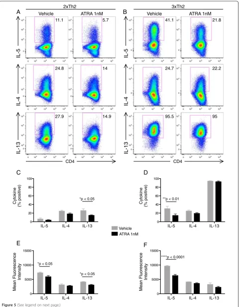
We next examined whether the pro-Th2 effects of ATRA may be due to augmentation of Th2 differen-tiation. We hypothesized that the addition of ATRA to in vitro Th2 cell differentiation cultures would enhance the frequency and/or kinetics of appearance of Th2 cytokines. Counter to our hypothesis, ATRA inhibited Th2 cell differentiation of both 2×Th2 and 3×Th2 cells (Figure 5).
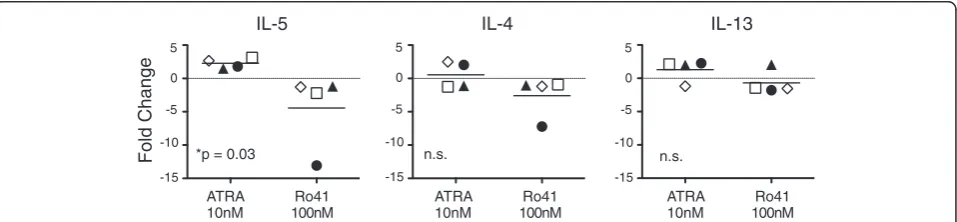
ATRA and Ro41 reciprocally regulate IL5, but not IL4 or IL13, gene expression
We next tested the hypothesis that RARα modulators
directly regulate Th2 cytokine gene expression via Th2 cell intrinsic mechanisms. IL-2 activation (in the absence of TCR signals) increased expression of IL5 message, which was significantly modulated by ATRA and Ro41, in a re-ciprocal manner (Figure 6). No differences were observed in IL4 or IL13 gene expression. These data demonstrate that in addition to their effects on IL-5+ Th2 cell
proli-feration, RARα modulators directly regulate IL5 gene
expression.
Discussion
Here we report that the pro-Th2 effect induced by reti-noic acid is primarily a direct result of Th2 cell-intrinsic
regulation of the cytokine IL-5 by RARα. Additionally,
these data demonstrate that RARα regulates the
pro-liferation of IL-5+ but not IL-5- Th2 cells. These results imply that the highly differentiated IL-5+ effector Th2 cell subpopulation is the primary Th2 cell population
affected by RARα modulators; in contrast, the less
dif-ferentiated IL-5- Th2 cells were significantly less af-fected. Both the proliferation of IL-5+ Th2 cells as well
as IL5 gene expression was suppressed by the RARα
antagonist Ro41, which suggests that RARα antagonism
might provide a therapeutic approach to inhibit the function of pathogenic pro-inflammatory IL-5+ Th2 cells.
The ATRA/RARα pathway is a well-known inducer of
Th2 cytokine responses both in vitro and in vivo, working through both T cell intrinsic and extrinsic mechanisms [6,16,24]. Our results provide evidence, that among the
three major Th2 cytokines, RARα modulators
predo-minantly regulate IL-5 expression. We and other groups have recently characterized IL-5+ Th2 cells as a more (See figure on previous page.)
Vehicle ATRA 1nM IL-5 IL-4 IL-13
0 20 40 60 80 100
e
ni
k
oty
C
)
e
vit
i
s
o
p
%(
*p < 0.05
IL-5 IL-4 IL-13
0 5000 10000 15000
IL-5 IL-4 IL-13
0 5000 10000 15000
IL-5 IL-4 IL-13
0 20 40 60 80 100
IL-5
0 102 103 104 105 0
103 104 105
0 102 103 104 105 0
103 104 105
0 102 103 104 105 0
103 104 105
0 102 103 104 105 0
103 104 105
0 102 103 104 105 0
102 103 104 105
0 102 103 104 105 0
102 103 104 105
0 102 103 104 105 0
103 104 105
0 102 103 104 105 0
103 104 105
0 102 103 104 105 0
104 105
0 102 103 104 105 0
104 105
0 102 103 104 105 0
102 103 104 105
0 102 103 104 105 0
102 103 104 105
CD4 CD4
IL-5
IL-4
IL-13
IL-4
IL-13
E
F
D
C
A
Vehicle ATRA 1nMB
2xTh2
Vehicle ATRA 1nM
3xTh2
F
n
a
e
Me
c
n
e
c
s
er
o
ul
yti
s
n
et
nI
e
c
n
e
c
s
er
o
ul
F
n
a
e
M
yti
s
n
et
nI
11.1 5.7
24.8 14
27.9 14.9 95.5 95
22.2 24.7
41.1 21.8
**p < 0.01
*p < 0.05 *p < 0.05
****p < 0.0001
e
ni
k
ot
y
C
)
e
vit
i
s
o
p
%(
highly-differentiated Th2 subpopulation with greater pro-inflammatory function [17-19]. This current work demon-strates that some of the pro-Th2 activity of ATRA is due to increases in Th2 proliferation, particularly that of the IL-5+ Th2 subpopulation. Notably, despite this overall pro-IL-5 activity, ATRA did not enhance Th2 differen-tiation (Figure 5).
Notably, whereas ATRA promoted IL-5+ Th2
re-sponses, the RARα antagonist Ro41 actually inhibited
IL-5+ Th2 responses. Such inhibition may be due to
Ro41 acting as a neutral antagonist blocking RARα
acti-vation by endogenous ATRA in the cell culture media or by Ro41 acting as an inverse agonist [25]. Notably, the latter activity has not been previously reported for Ro41.
Previous human studies showing ATRA-induced Th2 cytokine production have utilized PBMC or CD4 T cells activated with polyclonal stimuli [6,16]. This study is not-able for using allergen specific Th2 cells from allergic asthmatic subjects as well as highly differentiated Th2 cell lines. The use of such pathogenically relevant Th2 cells lines confirm and extend previous observations using mitogen activated PBMC from healthy donors.
This current work showing ATRA augmentation of pathogenic allergen specific Th2 responses underscores the potential clinical relevance of these findings. RA agonists are available both as prescription and over the counter formulations; these data suggest that RA sup-plementation may potentially augment Th2 responses and thus promote allergic disease, as observed in the mouse model of asthma [15]. Alternatively, other pathways may
augment local ATRA levels by the upregulation of retinal-dehyde dehydrogenase 2, which is required for the bio-synthesis of retinoic acid. To that end, Shreffler and colleagues have characterized a previously undescribed peanut protein that upregulates retinaldehyde dehydro-genase 2 in myeloid dendritic cells [26].
We studied the intrinsic regulation of Th2 cell func-tion (proliferafunc-tion and cytokine producfunc-tion) by reacti-vating highly differentiated Th2 cells. Since this culture system utilizes APC-free in vitro differentiated Th2 cells, the ATRA/Ro41-mediated effects are, by definition, me-diated by Th2 cell intrinsic mechanisms. Using this APC-free system, ATRA did not augment Th2
diffe-rentiation, suggesting that the pro-Th2 effects of RARα
modulation are not through enhanced T cell-intrinsic effects on Th2 differentiation.
Using either Annexin V or activated caspase 3 to
iden-tify apoptotic cells, RARαmodulators did not affect the
frequency of apoptosis (Figures 3 and 4). Taken together, these findings suggest that ATRA augments Th2 re-sponses by promoting Th2 cell proliferation and gene expression and not through differential modulation of cell death or apoptosis.
An inherent limitation of studies using pharma-cological inhibitors is the potential for off-target effects. Indeed, Ro41 has been shown to activate peroxisome
proliferator activated receptor-γ (PPAR-γ) at
concen-trations of 1μM [27], which is 10-fold greater than the
concentrations used in this study. To further address
whether off-target effects of Ro41 on PPAR-γcould have
(See figure on previous page.)
Figure 5ATRA inhibitsin vitroTh2 cell differentiation.Naïve CD4 T cells were isolated by MACS and differentiated under Th2 culture conditions in the presence of 1nM ATRA or DMSO vehicle control.(A, B)After 2 and 3 rounds of differentiation (2xTh2 and 3xTh2, respectively) cells were restimulated with PMA and ionomycin and analyzed by ICCS.(C, D)Percentages and(E, F)mean fluorescence intensity (MFI) of cytokine positive 2xTh2 (C, E) and 3xTh2 (D, F) cells are plotted from 3 experiments. Data is presented as mean +/- SEM. 2-way ANOVA was performed and Bonferroni multiple comparisons p values are displayed.
Fold Change
ATRA 10nM
Ro41 100nM
ATRA 10nM
Ro41 100nM
ATRA 10nM
Ro41 100nM
IL-5 IL-4 IL-13
*p = 0.03 n.s. n.s.
5
0
-5
-10
-15
5
0
-5
-10
-15 5
0
-5
-10
-15
been responsible for its inhibition of IL-5+ Th2
proli-feration, we examined the effect of the PPAR-γ agonist
GW7845 on Th2 cultures. GW7845 did not have any ef-fect on IL-4, IL-5 or IL-13 expression in these cultures (data not shown). The relatively low concentration of Ro41 used in this study as well as the lack of effect of
PPAR-γactivators, make it unlikely that Ro41 was acting
through off-target effects.
This apparent direct regulation of Th2 cytokine gene
expression by RARα prompted us to examine if a
puta-tive retinoic acid response element (RARE) exists in the
promoter regions of Th2 cytokine genes (IL5p, IL4p,
IL13p). We thus analyzed the 10 kb genomic DNA
se-quence of the human IL5, IL4, and IL13 promoters using the University of California Santa Cruz genome browser.
We identified a single putative RARE (5′-TGGTCA
CAGTTCA-3′) in the human IL5p (−825 to −813) but
not in the human IL4p and IL13p, suggesting that IL5
could be a RARE-responsive gene. The genomic location
of the IL5p putative RARE is comparable between
human and rhesus, and similarly, between mouse and
rat (Figure 7A). The IL5p putative RARE sequence is
identical between human and rhesus (Figure 7B) and similarly, between mouse and rat (Figure 7C). Subse-quent studies are needed to verify if this putative RARE is functionally active.
Conclusions
In conclusion, we demonstrate that RARα modulators
act on Th2 cells through multiple mechanisms, inclu-ding Th2 cell intrinsic augmentation of proliferation and IL-5 expression. In all experiments, the magnitude of the effect was most apparent for IL-5 responses. The potent induction by ATRA and reciprocal inhibition of IL-5 by Ro41 supports that these effects are mediated
through the RARα receptor. These data demonstrate
that RARα modulation has a major impact on human
Th2 responses and suggests that RARαmay be a
poten-tial therapeutic target for anti-Th2 therapy.
B
C
humanrhesus
mouse
rat
spacer designation DR1-5
1/2 RARE 1/2 RARE
putative RARE Legend
IL-5
Exon 4 Exon 3 Exon 2 Exon 1
Exon 4 Exon 3 Exon 2 Exon 1
Exon 4 Exon 3 Exon 2 Exon 1
Exon 4 Exon 3 Exon 2 Exon 1
3'
3'
3'
3'
5'
5'
5'
5' Human IL-5 Locus (Chr 5)
Rhesus IL-5 Locus (Chr 6)
Mouse IL-5 Locus (Chr 11)
Rat IL-5 Locus (Chr 10)
A
-825 -813
-873 -862
DR2
DR2
-9279 DR1 -9269
DR1 -9528 -9538
Abbrevations
RA:Retinoic acid; ATRA: All-trans retinoic acid; RARα: Retinoic acid receptor alpha; RXR: Retinoid X receptor; PBMC: Peripheral blood mononuclear cells; HDM: House dust mite; Ag: Antigen; APC: Antigen presenting cells; TCR: T cell receptor; CTV: Cell trace violet; Th2: T helper 2; Ro41: Ro41-5253; RARE: Retinoic acid response element;IL5p: Interleukin 5 gene promoter; IL13p: Interleukin 13 gene promoter;IL4p: Interleukin 4 gene promoter; mRNA: Messenger ribonucleic acid; ICCS: Intracellular cytokine staining; qRT-PCR: Quantitative real time PCR; DMSO: Dimethyl sulfoxide; DMEM: Dulbecco modified eagles medium; FBS: Fetal bovine serum; ANOVA: Analysis of variation; MFI: Mean fluorescence intensity; SEM: Standard error of mean.
Competing interests
The authors declare that they have no competing interests.
Authors’contributions
CP conceived of the study. YY performed initial pilot experiments. DLW carried out the experiments. All authors contributed to the design and analysis of the study. DLW and CP prepared the manuscript and performed statistical analysis. All authors read and approved the final manuscript.
Acknowledgements
This work was supported by the National Institute of Allergy and Infectious Diseases, Division of Intramural Research, project number Al-000993-06. The authors thank Sarah Arceo and Michael Young for their contribution to clinical sample acquisition.
Received: 27 August 2013 Accepted: 23 November 2013 Published: 6 December 2013
References
1. Green HN, Mellanby E:Vitamin a as an anti-infective agent.Br Med J1928,
2:691–696.
2. Mellanby E, Green HN:Vitamin a as an anti-infective agent: its Use in the treatment of puerperal septigaemia.Br Med J1929,1:984–986. 3. Veldhoen M, Brucklacher-Waldert V:Dietary influences on intestinal
immunity.Nat Rev Immunol2012,12:696–708.
4. Hall JA, Grainger JR, Spencer SP, Belkaid Y:The role of retinoic acid in tolerance and immunity.Immunity2011,35:13–22.
5. Hall JA, Cannons JL, Grainger JR, Dos Santos LM, Hand TW, Naik S, Wohlfert EA, Chou DB, Oldenhove G, Robinson M,et al:Essential role for retinoic acid in the promotion of CD4(+) T cell effector responses via retinoic acid receptor alpha.Immunity2011,34:435–447.
6. Dawson HD, Collins G, Pyle R, Key M, Taub DD:The retinoic acid receptor-alpha mediates human T-cell activation and Th2 cytokine and chemokine production.BMC Immunol2008,9:16.
7. Ohoka Y, Yokota A, Takeuchi H, Maeda N, Iwata M:Retinoic acid-induced CCR9 expression requires transient TCR stimulation and cooperativity between NFATc2 and the retinoic acid receptor/retinoid X receptor complex.J Immunol2011,186:733–744.
8. Vaishnava S, Hooper LV:Eat your carrots! T cells are RARing to go.
Immunity2011,34:290–292.
9. Pino-Lagos K, Guo Y, Noelle RJ:Retinoic acid: a key player in immunity.
Biofactors2010,36:430–436.
10. Mosmann TR, Cherwinski H, Bond MW, Giedlin MA, Coffman RL:Two types of murine helper T cell clone. Definition according to profiles of lymphokine activities and secreted proteins.J Immunol1986,
136:2348–2357.
11. Carman JA, Pond L, Nashold F, Wassom DL, Hayes CE:Immunity to Trichinella spiralis infection in vitamin A-deficient mice.J Exp Med1992,
175:111–120.
12. Lei GS, Zhang C, Shao S, Jung HW, Durant PJ, Lee CH:All-trans retinoic acid in combination with primaquine clears pneumocystis infection.
PLoS One2013,8:e53479.
13. Broadhurst MJ, Leung JM, Lim KC, Girgis NM, Gundra UM, Fallon PG, Premenko-Lanier M, McKerrow JH, McCune JM, Loke P:Upregulation of retinal dehydrogenase 2 in alternatively activated macrophages during retinoid-dependent type-2 immunity to helminth infection in mice.
PLoS Pathog2012,8:e1002883.
14. Cantorna MT, Nashold FE, Hayes CE:In vitamin A deficiency multiple mechanisms establish a regulatory T helper cell imbalance with excess Th1 and insufficient Th2 function.J Immunol1994,152:1515–1522. 15. Schuster GU, Kenyon NJ, Stephensen CB:Vitamin A deficiency decreases
and high dietary vitamin A increases disease severity in the mouse model of asthma.J Immunol2008,180:1834–1842.
16. Dawson HD, Collins G, Pyle R, Key M, Weeraratna A, Deep-Dixit V, Nadal CN, Taub DD:Direct and indirect effects of retinoic acid on human Th2 cytokine and chemokine expression by human T lymphocytes.
BMC Immunol2006,7:27.
17. Upadhyaya B, Yin Y, Hill BJ, Douek DC, Prussin C:Hierarchical IL-5 expression defines a subpopulation of highly differentiated human Th2 cells.
J Immunol2011,187:3111–3120.
18. Islam SA, Chang DS, Colvin RA, Byrne MH, McCully ML, Moser B, Lira SA, Charo IF, Luster AD:Mouse CCL8, a CCR8 agonist, promotes atopic dermatitis by recruiting IL-5+ T(H)2 cells.Nat Immunol2011,12:167–177. 19. Endo Y, Iwamura C, Kuwahara M, Suzuki A, Sugaya K, Tumes DJ, Tokoyoda K,
Hosokawa H, Yamashita M, Nakayama T:Eomesodermin controls interleukin-5 production in memory T helper 2 cells through inhibition of activity of the transcription factor GATA3.Immunity2011,35:733–745. 20. Prussin C, Lee J, Foster B:Eosinophilic gastrointestinal disease and
peanut allergy are alternatively associated with IL-5+ and IL-5(−) T(H)2 responses.J Allergy Clin Immunol2009,124(1326–1332):e1326. 21. Givan AL, Fisher JL, Waugh M, Ernstoff MS, Wallace PK:A flow cytometric
method to estimate the precursor frequencies of cells proliferating in response to specific antigens.J Immunol Methods1999,230:99–112. 22. Givan AL, Fisher JL, Waugh MG, Bercovici N, Wallace PK:Use of
cell-tracking dyes to determine proliferation precursor frequencies of antigen-specific T cells.Methods Mol Biol2004,263:109–124. 23. Blalock EM:A Beginner’s Guide to Microarrays.Norwell: Kluwer Academic
Publishers; 2003.
24. Cui D, Moldoveanu Z, Stephensen CB:High-level dietary vitamin a enhances T-helper type 2 cytokine production and secretory immunoglobulin a response to influenza a virus infection in BALB/c mice.J Nutr2000,
130:1132–1139.
25. Greasley PJ, Clapham JC:Inverse agonism or neutral antagonism at G-protein coupled receptors: a medicinal chemistry challenge worth pursuing?Eur J Pharmacol2006,553:1–9.
26. Ruiter B, Shreffler WG:Innate immunostimulatory properties of allergens and their relevance to food allergy.Semin Immunopathol2012,
34:617–632.
27. Schupp M, Curtin JC, Kim RJ, Billin AN, Lazar MA:A widely used retinoic acid receptor antagonist induces peroxisome proliferator-activated receptor-gamma activity.Mol Pharmacol2007,71:1251–1257.
doi:10.1186/1476-7961-11-4
Cite this article as:Wansleyet al.:The retinoic acid receptor-α
modulators ATRA and Ro415253 reciprocally regulate human IL-5+ Th2 cell proliferation and cytokine expression.Clinical and Molecular Allergy
201311:4.
Submit your next manuscript to BioMed Central and take full advantage of:
• Convenient online submission • Thorough peer review
• No space constraints or color figure charges • Immediate publication on acceptance
• Inclusion in PubMed, CAS, Scopus and Google Scholar
• Research which is freely available for redistribution

![Synthesis and biological evaluation of 3N substituted thieno[2,3 d] pyrimidines](data:image/gif;base64,R0lGODlhAQABAIAAAP///wAAACH5BAEAAAAALAAAAAABAAEAAAICRAEAOw==)