University of Pennsylvania
ScholarlyCommons
Publicly Accessible Penn Dissertations
1-1-2013
Consequences of Brief Periods of Sleep Loss on
Hippocampus-Dependent Memory and Synaptic
Plasticity
Toni-Moi N. Prince
University of Pennsylvania, [email protected]
Follow this and additional works at:
http://repository.upenn.edu/edissertations
Part of the
Neuroscience and Neurobiology Commons
This paper is posted at ScholarlyCommons.http://repository.upenn.edu/edissertations/913 For more information, please [email protected].
Recommended Citation
Prince, Toni-Moi N., "Consequences of Brief Periods of Sleep Loss on Hippocampus-Dependent Memory and Synaptic Plasticity" (2013).Publicly Accessible Penn Dissertations. 913.
Consequences of Brief Periods of Sleep Loss on
Hippocampus-Dependent Memory and Synaptic Plasticity
Abstract
Hippocampal cellular and molecular processes critical for memory consolidation are affected by the amount and quality of sleep attained. Questions remain with regard to how sleep enhances memory, what parameters of sleep after learning are optimal for memory consolidation, and what underlying hippocampal molecular players are dysregulated by sleep deprivation to impair memory consolidation and plasticity. In this dissertation, I describe experiments that we performed to identify the time window where memory consolidation is sensitive to sleep loss as well as to characterize two potential molecular players targeted by sleep deprivation. Because consolidation appears to have a particular window where it is sensitive to sleep loss, we explore the parameters of this time window in Chapter 2. Our results suggest that a specific 3-hour period of sleep loss during consolidation disrupts both memory and plasticity. In the second portion of this
dissertation, I examine the mechanisms by which sleep deprivation impairs hippocampus-dependent memory consolidation. In Chapter 3, we show that loss of the phosphodiesterase (PDE) 4A, an enzyme responsible for decreasing cAMP signaling, rescues spatial memory disrupted by sleep loss. These results further implicate cAMP signaling with the negative effects of sleep deprivation on memory. Obtaining adequate sleep is challenging in a society that values "work around the clock." Therefore, the development of interventions to combat the negative cognitive effects of sleep deprivation is critical. However, a limited number of
therapeutics exists that are able to enhance cognition in the face of insufficient sleep. The identification of the temporal characteristics of sleep loss and the molecular pathways implicated in the deleterious effects of sleep deprivation on memory could potentially yield new targets for the development of more effective drugs.
Degree Type
Dissertation
Degree Name
Doctor of Philosophy (PhD)
Graduate Group
Neuroscience
First Advisor
Ted Abel
Keywords
Memory, Sleep Deprivation, Synaptic Plasticity
Subject Categories
Neuroscience and Neurobiology
CONSEQUENCES OF BRIEF PERIODS OF SLEEP LOSS ON HIPPOCAMPUS-DEPENDENT
MEMORY AND SYNAPTIC PLASTICITY
Toni-Moi N. Prince
A DISSERTATION
in
Neuroscience
Presented to the Faculties of the University of Pennsylvania
in
Partial Fulfillment of the Requirements for the
Degree of Doctor of Philosophy
2013
Supervisor of Dissertation
__________________________
Edwin (Ted) Abel, Ph.D., Professor of Biology
Graduate Group Chairperson
__________________________
Joshua I. Gold, Ph.D., Associate Professor of Neuroscience
Dissertation Committee
David Raizen, M.D., Ph.D., Assistant Professor of Neurology (Committee Chair)
Amita Sehgal, Ph.D., Professor of Neuroscience
Marcos Frank, Ph.D., Associate Professor of Neuroscience
ii
ACKNOWLEDGEMENTS
There are many individuals who I want thank. I would never have made it through this program
without the instrumental role of all of them. They have all of my gratitude for continually pushing
me to strive to be better and to do better in all aspects of my life, not just as a scientist.
To my committee members: I want to thank David Raizen, Amita Sehgal, Marcos Frank and
David Dinges for investing their time and insight into the development and trajectory of my career.
Your expertise enhanced my work by aiding me in thinking critically to formulate my experiments.
To the NGG administration: I want to thank Mikey Nusbaum, Rita Balice Gordon, and Josh
Gold for developing such a strong neuroscience program. Your hard work has allowed our
community to thrive. I would especially like to thank Mikey. You have been a great mentor to me.
Any time that I felt lost I knew I could turn to you for support. Thank you for accepting me into this
program. I feel extremely lucky to have your hand in my development. I would also like to thank
Jane Hoshi and Angela Gilmore for all their assistance and words of kindness throughout this
process.
To the NGG students: I would like to thank all the students of this program, especially my
cohort. I do not know how the program does it, but it recruits some of the brightest and most
motivated students. More importantly, my class was social and caring. Over these years, my
class has become my family.
To the Abel lab: I would like to thank my mentor, Ted Abel, for accepting me into his lab. Thank
you for giving me the opportunity to have my own project and to think critically about science. I
think you have one of the most spectacular labs. I have had the chance to grow as a scientist and
as a person by getting the opportunity to interact with so many intelligent and charismatic
individuals in the lab. I thank you for that Ted. I would also like to thank all the members of the lab
past and present. I do not know where I would be if you guys were not there to drag me though
the finish line of graduate school. Especially Jennifer Choi- you are my mini-mentor, and you are
iii
To my past mentors: Thank you Michael Brown, Peter Alfinito and Kim Wallen for patiently
teaching me. From you, I picked up traits on how to persevere in every type of situation.
To my friends: There are no words to describe my gratitude to all the friends I have made
throughout the years. I must have done something spectacular in a past life to have the kind of
friends I have. All of you mean so much to me. Thank you for providing me an outlet to get out all
of the personal and professional frustrations that happens to every student. From Halloween
parties to our “Sexy Valentine” dinner, I had so much fun. There have been so many times in the
past few years that I struggled and every time, without fail, you all listened patiently and gave me
the strength I needed to pick myself up. When I succeeded, you all were the first to celebrate my
accomplishments.
To my family: I would like to thank my family, especially my mom and sisters. Nicky and
Danielle, thank you for always being proud of my tiny achievements. You say that I am an
inspiration, but the truth is that you both inspire me. For most of my decisions, I factor in what
would Nicky and Danielle do (WWNDD). Your accomplishments give me hope for myself.
To my partner: Jason, you have been the most loving partner that I could have ever fathom.
Thank you for always believing in me especially when I couldn’t believe in myself. You make me
shine, and want to succeed. Having you as my main support system over the past years has
gotten me to accomplish much more than I even knew I was capable of doing. You are the love of
my life, and I know I can face anything with you beside me.
My training was supported by a NIH/NIGMS T32 Training Grant (T32-GM07517), a NIH/NHLBI
Training in Sleep and Sleep Disorders T32 Training Grant (T32-HL07953). Additional support
iv ABSTRACT
CONSEQUENCES OF BRIEF PERIODS OF SLEEP LOSS ON HIPPOCAMPUS-DEPENDENT
MEMORY AND SYNAPTIC PLASTICITY
Toni-Moi N. Prince
Edwin “Ted” Abel, Ph.D.
Hippocampal cellular and molecular processes critical for memory consolidation are affected by
the amount and quality of sleep attained. Questions remain with regard to how sleep enhances
memory, what parameters of sleep after learning are optimal for memory consolidation, and what
underlying hippocampal molecular players are dysregulated by sleep deprivation to impair
memory consolidation and plasticity. In this dissertation, I describe experiments that we
performed to identify the time window where memory consolidation is sensitive to sleep loss as
well as to characterize two potential molecular players targeted by sleep deprivation. Because
consolidation appears to have a particular window where it is sensitive to sleep loss, we explore
the parameters of this time window in Chapter 2. Our results suggest that a specific 3-hour
period of sleep loss during consolidation disrupts both memory and plasticity. In the second
portion of this dissertation, I examine the mechanisms by which sleep deprivation impairs
hippocampus-dependent memory consolidation. In Chapter 3, we show that loss of the
phosphodiesterase (PDE) 4A, an enzyme responsible for decreasing cAMP signaling, rescues
spatial memory disrupted by sleep loss. These results further implicate cAMP signaling with the
negative effects of sleep deprivation on memory. Obtaining adequate sleep is challenging in a
society that values "work around the clock." Therefore, the development of interventions to
combat the negative cognitive effects of sleep deprivation is critical. However, a limited number of
therapeutics exists that are able to enhance cognition in the face of insufficient sleep. The
v
in the deleterious effects of sleep deprivation on memory could potentially yield new targets for
vi
TABLE OF CONTENTS
ACKNOWLEDGEMENTS ... ii
ABSTRACT ... iv
TABLE OF CONTENTS ... vi
LIST OF FIGURES ... viii
CHAPTER ONE:GENERAL INTRODUCTION ... 1
OVERVIEW ... 2
SECTION 1. INTERPLAY BETWEEN SLEEP AND MEMORY ... 3
1.1. Molecular signaling consolidates memory... 3
1.2. Distinction between sleep states ... 4
1.3. Sleep enhances memory consolidation ... 6
SECTION 2. SLEEP DEPRIVATION DISRUPTS HIPPOCAMPAL FUNCTION ... 11
2.1. Sleep deprivation disrupts memory consolidation ... 11
2.2. Sleep deprivation impairs hippocampal synaptic plasticity ... 14
2.3. Sleep deprivation disrupts hippocampal signaling necessary for memory .... 17
CONCLUSION ... 25
CHAPTER TWO:SLEEP DEPRIVATION DURING A SPECIFIC 3-HOUR TIME WINDOW POST TRAINING IMPAIRS HIPPOCAMPAL SYNAPTIC PLASTICITY AND MEMORY ... 29
ABSTRACT... 30
INTRODUCTION ... 31
MATERIALS AND METHODS ... 33
vii
DISCUSSION ... 46
CHAPTER THREE:PDE4A MEDIATES DISRUPTION OF HIPPOCAMPAL FUNCTION INDUCED BY SLEEP DEPRIVATION... 62
ABSTRACT... 63
INTRODUCTION ... 64
MATERIALS AND METHODS ... 65
RESULTS ... 70
DISCUSSION ... 74
CHAPTER FOUR:GENERAL DISCUSSION AND FUTURE DIRECTIONS ... 85
4.1.KINETICS:TEMPORAL DYNAMICS OF SLEEP DEPRIVATION THAT IMPAIRS MEMORY CONSOLIDATION ... 86
4.2.MECHANICS:UNCHARACTERIZED MOLECULAR TARGETS THAT ENABLE SLEEP DEPRIVATION TO BE EFFECTIVE AT DISRUPTING MEMORY ... 94
CONCLUDING REMARKS ... 101
APPENDIX:LOSS OF P75NTR HAS COMPLEX EFFECTS ON HIPPOCAMPUS-DEPENDENT MEMORY AND PLASTICITY ... 105
ABSTRACT... 106
INTRODUCTION ... 107
MATERIALS AND METHODS ... 109
RESULTS ... 113
DISCUSSION ... 115
viii
LIST OF FIGURES
Chapter One
Figure 1. A schematic overview of hippocampal signaling pathways following sleep
deprivation. ... 28
Chapter Two
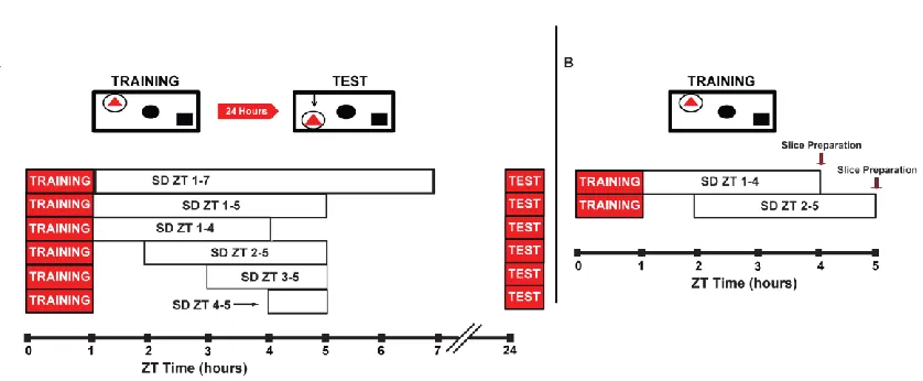
Figure 2.1. Schematic depicting the behavioral and LTP experimental design.... 53
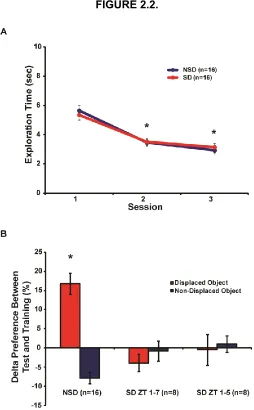
Figure 2.2. 4 hours and 6 hours of immediate sleep deprivation impairs spatial memory.
... 54
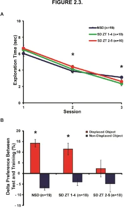
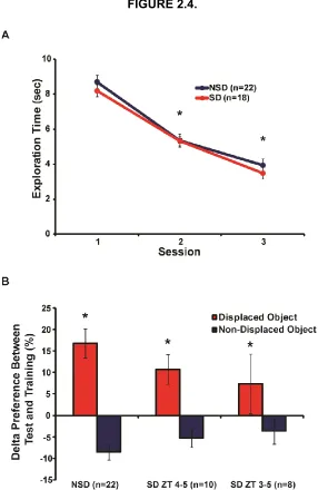
Figure 2.3. A specific 3-hour sleep deprivation period impairs spatial memory. ... 55
Figure 2.4. A delayed 2-hour and 1-hour sleep deprivation period does not impair spatial
memory. ... 56
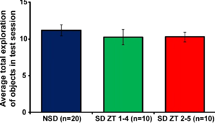
Figure 2.5. 3 hours of immediate or delayed sleep deprivation does not alter total
exploration time of objects during test session. ... 57
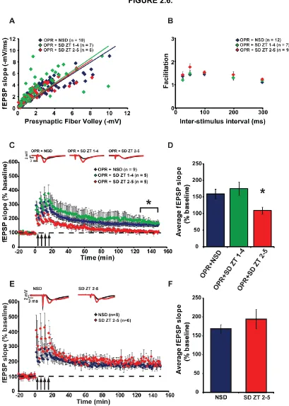
Figure 2.6. Delayed sleep deprivation after object-place recognition training disrupts
LTP. ... 59
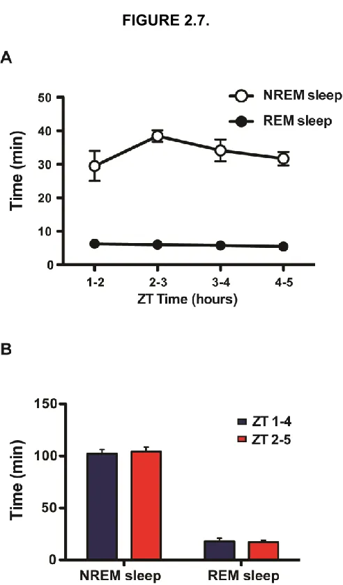
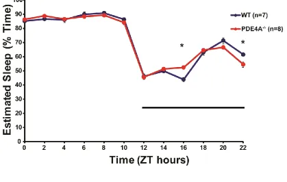
Figure 2.7. Mice exhibit similar sleep patterns during periods ZT 1-5. ... 60
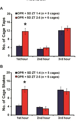
Figure 2.8. The early sleep deprivation group requires less disturbance to achieve a
wakeful state in the 1st hour of SD than the SD ZT 2-5 group. ... 61
Chapter Three
Figure 3.1. PDE4A deletion modulates activity during the dark the dark period. ... 80
Figure 3.2. 5 h sleep deprivation did not impair contextual fear conditioning in PDE4A
ix
Figure 3.3. Deletion of PDE4A rescues object-place recognition memory after sleep
deprivation. ... 82
Figure 3.4. Deletion of PDE4A does not affect synaptic transmission or synaptic
plasticity. ... 83
Figure 3.5. 5 h sleep deprivation does not affect cAMP levels in whole hippocampus
tissue. ... 84
Appendix
Figure A.1. Loss of p75NTR impairs contextual fear memory. ... 120
Figure A.2. Loss of p75NTR does not affect spatial memory in the OPR task. ... 121
Figure A.3. Preliminary Results: Loss of p75NTR does not appear to alter the effects of
1
CHAPTER ONE
2 OVERVIEW
Chronic sleep loss is a widespread problem in our society (Strine and Chapman,
2005). According to the Center for Disease Control, about 7-19% of adults in the US
report inadequate sleep, and an estimated 50-70 million Americans suffer from chronic
sleep disorders. Insufficient sleep is co-morbid with chronic problems such as heart
disease, kidney disease, high blood pressure, diabetes, obesity and mental illness (Ford,
1989; Gillin, 1998; Knutson and Van Cauter, 2008; Hirotsu et al., 2010; Vijayan, 2012;
Engeda et al., 2013; Najafian et al., 2013; Palagini et al., 2013). Sleep loss also
contributes to irritability, aggression, inattentiveness and diminished psychomotor
vigilance (Rajaratnam and Arendt, 2001; Van Dongen et al., 2003; Kamphuis et al.,
2012). The negative impact of sleep loss on physical and mental health places a strain
on our healthcare system (Kapur et al., 2002) and a large financial burden on our
economy (Durmer and Dinges, 2005). Unfortunately, the common myth is that people
can function on little sleep with no consequences, even though studies employing both
human and animal models demonstrate that mental and physical health requires
sufficient sleep (Banks and Dinges, 2007). Because so many people are unable to
obtain sufficient sleep on a daily basis, it is critical to understand the molecular and
cellular impact of sleep loss in an effort to identify novel therapeutic approaches to
counteract these effects.
In the introduction to my thesis, I bridge what is known about critical periods of
molecular signaling post-learning with our understanding of the role of sleep and sleep
deprivation in memory consolidation. This is followed by my study focusing on the time
windows when sleep deprivation can disrupt hippocampal function in Chapter 2. I
3
Fig. 1 for schematic overview). Finally, I discuss the implications of my findings from
these studies in Chapter 4.
Section 1. Interplay between sleep and memory
1.1. Molecular signaling consolidates memory
Memory consists of at least 3 stages; encoding, consolidation, and retrieval.
Each phase requires specific molecular machinery (Abel and Lattal, 2001). Because the
focus of this dissertation is memory consolidation, we elaborate on the molecular
signaling underlying this stage of memory processing, termed synaptic consolidation
(Dudai, 2004). A large body of work suggests that sleep is particularly beneficial to the
consolidation stage of memory storage, and that this stage is vulnerable to sleep
manipulation (Sagales and Domino, 1973; Buzsáki, 1998; Stickgold et al., 2000; Graves
et al., 2003a). Learning induces a transient increase in calcium (Ca2+) and adenylyl
cyclase, an enzyme responsible for production of the second messenger cyclic
adenosine monophosphate (cAMP) (Xia and Storm, 2012). cAMP activates 3
downstream targets important for protein synthesis and eventual memory consolidation.
These targets include protein kinase A (PKA), exchange protein directly activated by
cAMP (Epac) and hyperpolarization-activated cyclic nucleotide-gated channels (HCN
channels) (Arnsten, 2007). Activation of these downstream targets, along with
neurotrophins and other kinases such as calmodulin dependent protein kinase (CaMKI,
CaMKII, and CaMKIV), mitogen activated protein kinase (MAPK), and extracellular
signal-regulated kinase (ERK 1/2), leads to phosphorylation of transcription factors
(Enslen et al., 1994; Matthews et al., 1994; Roberson et al., 1999; Ahmed and Frey,
2005). Transcription factors such as cAMP response element binding protein (CREB)
4
1997) to promote up-regulation of gene expression for proteins that will consolidate
labile memories into long-term memories (Roberson and Sweatt, 1999; Roberson et al.,
1999). Previous time course studies identified time windows where inhibition of these
signaling components in the hippocampus impairs memory consolidation. Two periods
for activation of cAMP downstream signaling and protein synthesis, immediately after
learning and 4 hours after learning, are required for consolidation (Grecksch and
Matthies, 1980; Bourtchouladze et al., 1998). Three peaks in cAMP levels have also
been observed after training at 0.5 hours, 3 hours and 6 hours during consolidation
(Bernabeu et al., 1997a). Two early sensitive periods during consolidation also exists for
mRNA synthesis, ERK1/2 activity, and phosphorylation of CREB (Igaz et al., 2002;
Trifilieff et al., 2006). These critical signaling periods appear to occur within the first few
hours of sleep post-learning (Graves et al., 2001; Hernandez and Abel, 2011). When
sleep loss overlaps with these critical time windows for molecular signaling,
consolidation is impaired (Smith and Rose, 1996; Graves et al., 2003a; Palchykova et
al., 2006). These sensitive periods of molecular signaling give an approximation of when
sleep deprivation could disrupt memory through disruption of molecular signaling
processes. I isolate this critical period in which sleep deprivation impairs memory
consolidation in Chapter 2.
1.2. Distinction between sleep states
To discuss the consequences of sleep or sleep loss on memory, it is crucial to
understand the pattern of neuronal electrical activity that occurs during different sleep
states. Sleep is measured by polysomnographic recordings that combine
electroencephalography (EEG), electro-oculogram (EOG), and electromyography (EMG)
5
eye movement (NREM) and rapid eye movement (REM) sleep (Stickgold, 1998). In
humans, NREM sleep can further be dissected into 4 stages. Rechtshaffen and Kales
developed the standard scoring guidelines to determine sleep stage in humans
(Rechtschaffen and Kales, 1968). Rolling eye movements are prominent during stage 1
sleep, while slow oscillations, fast sleep spindles (12-15 Hz), and k-complexes are
prominent during stage 2. Recordings during stages 3 and 4 of NREM sleep, termed
slow wave sleep (SWS), are characterized by high amplitude, low-frequency delta waves
(also known as slow waves, 1-4 Hz) and spindle activity (Mölle et al., 2011). NREM
sleep is accompanied by decreased muscle tone as assessed by EMG recordings
(Steriade et al., 1993a, 1993b). REM sleep EEG exhibits features similar to wake EEG
with cortical activation characterized by low voltage fast EEG activity (Hobson and
Steriade, 2011). Other common features of REM sleep include complete loss of muscle
tone (atonia) (John et al., 2004) and characteristic rapid eye movements detected by
EOG (Aserinsky and Kleitman, 1953; Jouvet, 1962). Hippocampal theta rhythms,
associated with wake, occur during REM sleep as well (Buzsáki et al., 1983; Greenstein
et al., 1988). Interestingly, this oscillatory pattern can be observed in hippocampal and
cortical regions during learning in spatial navigation and episodic retrieval (Klemm, 1976;
O’Keefe and Burgess, 1999; Klimesch et al., 2001). NREM and REM sleep have been
examined in depth in the context of memory to determine the contribution of each stage
in the consolidation process.
Sleep patterns differ across species, and this difference needs to be taken into
account when reviewing rodent sleep literature. While humans are diurnal and sleep
approximately 8 hours/night in a mono-phasic pattern, rodents are nocturnal mammals
that exhibit poly-phasic sleep patterns. In humans, a full cycle between NREM and REM
6
cycle is much shorter, lasting approximately 10 minutes (Trachsel et al., 1991; Benington
et al., 1994). Scoring rules in rodents also differ slightly from humans. While rodents
exhibit NREM and REM sleep, NREM can be further broken down to SWS stage I or
SWS stage II. SWS I is identified by the presence of sleep spindles in the cortical EEG.
SWS II is identified by the presence waves in the range of 0.1-4.0 Hz in the cortical
EEG. (Datta and Hobson, 2000; Datta and Maclean, 2007). Despite the differences in
human and rodent sleep, the neurobiology regulating sleep/wake states is similar and
much has been learned about how sleep affects memory by studying animal models.
1.3. Sleep enhances memory consolidation
Evidence from various human and rodent studies supports the importance of
sleep in learning and memory. In 1924, Jenkins and Dallenbach were the first to discover
that sleep facilitates long-term memory formation. In this seminal study, human subjects
displayed improved memory of nonsense syllables when they slept during the period
between learning and recall (Jenkins and Dallenbach, 1924). Since this pivotal discovery
termed the ‘sleep effect’, several other research groups have replicated the finding that
memory benefits from sleep (Smith, 2001; Born et al., 2006; Gais et al., 2007; Oudiette
et al., 2013; Stickgold and Walker, 2013).
Although researchers agree that sleep is more effective than wakefulness for
memory consolidation, debates have emerged as to what stage of sleep is beneficial for
memory. Initially, most research groups hypothesized that REM sleep enhanced memory
consolidation due to its similarity to waking EEG and its increased firing of hippocampal
neurons that had been active prior to sleep (Pavlides and Winson, 1989). Much of the
support for the role of REM sleep in memory came from animal studies that
7
1985a; Hennevin et al., 1995). The increase in REM sleep after learning has been
demonstrated in cats trained in the instrumental learning paradigm (Lecas, 1976).
Increases in REM sleep were also observed in rabbits after handling, which led to more
activity in successive open field behavior assessment (Denenberg et al., 1977). Rodent
studies also showed increases in REM sleep episodes after learning in associative fear
related learning tasks, such as the fear conditioning paradigm and an escapable shock
task (Sanford et al., 2010; Machida et al., 2013; Menz et al., 2013). REM sleep also
increased in rodents after training in the Morris water maze paradigm (Smith and Rose,
1996, 1997). Not only was an increase in REM sleep observed after training, but also
REM sleep-related processes such as theta rhythm and pontine-wave activity were
identified as important for memory consolidation in rodents trained in the two-way active
avoidance task (Datta et al., 2005; Datta and O’Malley, 2013). Many took these
observed increases in REM sleep after conditioning as evidence that REM sleep was
closely tied to enhancements in the memory process. This hypothesis was supported by
rodent studies that involved REM sleep deprivation during these same REM sleep time
windows, demonstrating memory impairments following these deprivation periods
(Smith, 1985a).
Until the late 1980s, REM sleep was considered to be the critical sleep stage in
memory improvement. However, later studies encountered difficulty demonstrating the
importance of REM sleep alone in consolidation of memory (Siegel, 2001). Human and
rodent studies observed an increase in NREM sleep, and NREM associated processes
such as slow wave activity and spindle density after training (Stickgold et al., 2001; Gais
et al., 2002; Huber et al., 2004; Hellman and Abel, 2007). A few studies also
demonstrated a NREM sleep-memory link in rodents through the discovery of neuronal
8
active during spatial learning fired in the same sequence during hippocampal
sharp-wave ripple events (SPW-R) that occur in NREM SWS (Wilson and McNaughton, 1994;
Lee and Wilson, 2002; Ji and Wilson, 2007; Ego-Stengel and Wilson, 2010; Bendor and
Wilson, 2012). The role of NREM sleep in memory consolidation was further cemented
by Rasch and colleagues who conducted a study where human subjects performed an
associative task consisting of card locations paired with a particular odor. The
researchers introduced these same smells during SWS, which activated neuronal replay
in the hippocampus. This manipulation resulted in enhanced recall of card location the
following day (Rasch et al., 2007). Spatial memory enhancement has also been
observed when a trial-unique auditory cue paired with an object was re-presented during
NREM sleep in humans (Rudoy et al., 2009). The presentation of the auditory cue during
SWS increased activation of the medial temporal lobe and altered
parahippocampal-medial prefrontal connectivity which has traditionally been associated with declarative
memory (van Dongen et al., 2012). Not only has inducing neuronal replay during SWS
been shown to enhance memory consolidation, but SPW-R disruption impairs
hippocampus-dependent memory (Girardeau et al., 2009; Nokia et al., 2012). In 2012,
Nokia and colleagues demonstrated the necessity of SPW-R hippocampal events that
are characteristic of SWS by disrupting SPW-R with electrical stimulation in rabbits. This
manipulation impaired trace eyeblink conditioning, a hippocampus-dependent learning
task (Nokia et al., 2012). Other studies in humans have shown that reactivation during
slow wave sleep can enhance not only spatial memory but procedural memories as well,
suggesting that reintroduction of cues during sleep may reactivate other brain regions as
well (Antony et al., 2012; Oudiette et al., 2013). These findings validate the hypothesis
that NREM SWS is important for learning and memory (Girardeau et al., 2009;
9
Reports from human studies suggest that there is a dissociation between
different sleep stages and the consolidation of different types of memory. In the ‘dual
hypothesis’ of sleep, NREM sleep is responsible for improvements in declarative
memory consolidation, while REM sleep plays a more significant role for procedural and
emotional memory consolidation (Gais and Born, 2004). Declarative memory relies on
the hippocampus, while procedural memory relies on striatal and cerebellar function
(Squire et al., 1993; Doyon et al., 2003). Early studies demonstrated that NREM sleep
improved declarative memories in humans. In the first set of studies to examine the role
of NREM sleep in a declarative task, humans learned a verbal paired associates task
before a sleep period known for high percentage of NREM sleep and low percentage of
REM sleep. These subjects displayed superior memory in comparison to subjects
trained before a high percentage of REM sleep. (Yaroush et al., 1971; Barrett and
Ekstrand, 1972; Fowler et al., 1973). A later study by Plihal and Born found that subjects
trained on a declarative task before sleep (predominantly composed of NREM sleep)
and awakened 3 hours later exhibited higher rates of retention compared to individuals
trained on the same task who slept during a period known for high REM sleep
composition (Plihal and Born, 1997, 1999).
While the work mentioned above demonstrates the importance of NREM sleep to
declarative memory, researchers have also observed that procedural memory (a type of
non-declarative memory) benefits from REM sleep. Plihal and Born also examined the
effects of REM sleep for procedural memory consolidation. Subjects who slept during a
period dominated by REM sleep displayed more procedural memory gains than those
who slept during a predominantly NREM sleep period (Plihal and Born, 1997, 1999).
Although these studies established the importance of NREM sleep for declarative
10
that has the ability to interfere with learning and recall (Ackermann et al., 2013; Goerke
et al., 2013), differed between those who were trained right before a high period of
NREM sleep and those who were trained right before a high period of REM sleep (Plihal
and Born, 1997, 1999). However, other studies have confirmed their findings by
demonstrating this phenomenon of REM sleep-related memory enhancements after
learning on various procedural tasks including priming and visuo-motor tasks (Mandai et
al., 1989; Buchegger et al., 1991; Smith and Lapp, 1991; Smith and Smith, 2003;
Wagner et al., 2003).
This ‘dual hypothesis’ paints a simplistic picture of how a particular stage of sleep
potentially benefits one type of memory, while another sleep stage mediates the
consolidation of other forms of memory. However, the sleep period known for a high
percentage of REM sleep also contains a high percentage of stage 2 sleep and thus
spindles. Therefore, procedural tasks could be benefiting from high spindle density,
which have been associated with improvements in procedural memory, and not solely
REM sleep (Tamaki et al., 2008, 2009; Rasch et al., 2009). Furthermore, other findings
contradict this REM sleep-procedural memory link and NREM sleep-declarative memory
link assertion. For instance, REM sleep has been shown to be important for emotional
declarative memories (Wagner et al., 2001; Wagner, 2002). NREM sleep has also been
found to play a role in procedural tasks further weakening the “dual hypothesis”
argument (Walker et al., 2002, 2003). However, many of the procedural tasks developed
for these experiments seem to require hippocampal involvement as well (Poldrack et al.,
2001; Schendan et al., 2003). This hippocampal component of the task may require
stages of NREM sleep to enhance procedural memory consolidation. This suggests that
the design of the task matters to a great extent in order to control for activation of certain
11
Clearly, the “dual hypothesis” dissociation between REM and NREM sleep in
memory consolidation is more complex than the previously described straightforward
examples. Other groups have posed alternative hypotheses that may account for the
sleep stage-memory complexities, one such alternative is the “sequential hypothesis”
(Giuditta et al., 1995). In this hypothesis, neither REM nor NREM sleep alone can
account for memory consolidation, but the order of NREM and REM sleep after training
as well as the transitions between NREM and REM sleep are essential for memory
(Giuditta et al., 1995; Ambrosini and Giuditta, 2001).
Section 2. Sleep deprivation disrupts hippocampal function
2.1. Sleep deprivation disrupts memory consolidation
Many studies have utilized sleep deprivation to examine the role of sleep in
memory consolidation. Researchers have developed several techniques for sleep
deprivation in rodents to assess how sleep loss impairs memory. Some of the main
methods of sleep deprivation include the “rotating platform” technique, the “flower-pot”
technique, gentle handling, novel object introduction, and optogenetic stimulation. Each
method has associated positive and negatives and has been discussed more thoroughly
previously (see Havekes et al. 2012). Sleep deprivation administered after learning
disrupts the consolidation period and impairs memories (Fishbein, 1971; Leconte et al.,
1974; Linden et al., 1975). The hippocampus, in particular, appears to be vulnerable to
this manipulation as demonstrated in hippocampal-dependent memory tasks after sleep
deprivation. The first set of experiments to demonstrate this sensitivity used the Morris
water maze task, which can be configured to either a hippocampus-dependent version
or a hippocampus-independent version (Morris et al., 1982). Previously sleep deprived
12
task. However, sleep deprived animals subjected to the hippocampus-independent
version did not demonstrate a memory impairment (Smith and Rose, 1996, 1997). This
interesting dissociation between dependent and
hippocampus-independent memory tasks was not restricted to the Morris water maze, but has been
demonstrated with fear-conditioning tasks as well (LeDoux, 2000). Mice sleep deprived
post-training exhibit memory impairments in the hippocampus-dependent configuration
of this task, but not the hippocampus-independent configuration of the task (Graves et
al., 2003a). Other studies examined sleep deprivation prior to learning in fear
conditioning tasks and observed similar results (Bueno et al., 1994; McDermott et al.,
2003; Ruskin et al., 2004; Ruskin and LaHoste, 2009). The Y-maze or T-maze is another
type of dissociation task where researchers can examine the learning strategy animals
employ to perform the task (Oliveira et al., 1997). This task allows researchers to assess
whether sleep deprived animals shift from employing a spatial strategy
(hippocampus-dependent) to a response strategy (hippocampus-in(hippocampus-dependent) (Hagewoud et al.,
2010b). Daily 5-hour sleep deprivation after training induced a shift from using a spatial
strategy to a response strategy to navigate the maze (Hagewoud et al., 2010b). Watts
and colleagues also demonstrated that decreased REM sleep as well as decreased
spindle-rich transition to REM sleep by a norepinephrine reuptake inhibitor, desipramine
(DMI), impaired performance in a hippocampus-dependent spatial task, while REM sleep
suppression actually enhanced striatal-dependent configuration of the T-maze. This
enhancement in striatal learning was likely due to the increased SWS that accompanied
the pharmacological inhibition of REM sleep (Watts et al., 2012). These results confirm
that the hippocampus is susceptible to the negative effects of insufficient sleep,
13
Studies have examined the effect of sleep deprivation during specific time
windows of consolidation. Memory appears most sensitive to sleep deprivation when
sleep is delayed after acquisition. However, if sleep occurs immediately after acquisition
then long-term memory remains intact even if sleep is prevented at later time points,
suggesting a sensitive period for sleep early during consolidation (Smith and Rose,
1996; Graves et al., 2003a; Gais et al., 2006; Palchykova et al., 2006). Based on rodent
studies, this immediate window coincides with sensitive periods of molecular signaling,
protein synthesis, and mRNA synthesis required for memory consolidation (Bernabeu et
al., 1997a; Bourtchouladze et al., 1998; Igaz et al., 2002; Trifilieff et al., 2006). For
instance, delaying sleep for 5 hours after acquisition impaired long-term memory in the
contextual fear condition paradigm. However, immediate sleep followed by a later 5-hour
period of sleep deprivation had no effect on long-term hippocampus-dependent memory
(Graves et al., 2003a). This finding has also been observed within the
hippocampus-dependent version of the water maze task (Smith and Rose, 1997). Subgroups of
animals were sleep deprived during different times after training. Delaying sleep for the
first 4 hours after training impaired memory in this task, whereas immediate sleep after
learning did not impair memory (Smith and Rose, 1997). The effect of immediate versus
delayed sleep after acquisition was also examined using the novel object recognition
task (Palchykova et al., 2006). The beneficial effect of immediate sleep on memory has
also been documented in humans performing a hippocampus-dependent declarative
memory task (Gais et al., 2006). In a study by Gais and colleagues, one group was
allowed immediate sleep after task acquisition at night, while sleep was delayed in the
other group. Delaying sleep after task acquisition impaired performance in this task
(Gais et al., 2006). These findings suggest the existence of a critical period for sleep
14
acquisition, and overlaps with molecular signaling-sensitive time windows during the
consolidation period. In Chapter 2, I explore the critical time window for sleep
deprivation to disrupt consolidation-related processes.
2.2. Sleep deprivation impairs hippocampal synaptic plasticity
Sleep deprivation is detrimental to hippocampus-dependent memory. As the
neural correlate of learning and memory, it is not surprising that sleep deprivation
disrupts synaptic plasticity in the hippocampus as well. Long-term potentiation (LTP), a
form of synaptic plasticity, is a long-lasting change in the strength of synaptic
connections through the involvement of various molecular signaling cascades and, in
some cases, protein synthesis (Bliss and Lomo, 1973; Whitlock et al., 2006). Campbell
and colleagues examined LTP in area CA1 in vitro after 12 hours of total sleep
deprivation, and found that the procedure inhibited induction of LTP in the hippocampus
of rodents (Campbell et al., 2002). Since this study, follow-up studies have given us an
in depth perspective on the effects of sleep deprivation on LTP. Researchers
demonstrated that, similar to behavioral studies, LTP is vulnerable to total sleep
deprivation, as well as REM-specific sleep deprivation and fragmented sleep
(McDermott et al., 2003; Tartar et al., 2006; Ravassard et al., 2009; Florian et al., 2011).
Similar LTP deficits occurred after sleep deprivation in vivo in dentate gyrus-CA3 region
(Romcy-Pereira and Pavlides, 2004; Marks and Wayner, 2005; Ishikawa et al., 2006;
Alhaider et al., 2011). The in vivo studies demonstrated that this LTP deficit was not an
artifact of slice preparation but was a result of the influence of sleep deprivation on the
intact hippocampal circuitry. The ability to induce LTP in vivo allowed researchers to
investigate the effect of sleep deprivation on the maintenance phase of LTP., which
15
paradigm (Romcy-Pereira and Pavlides, 2004; Ishikawa et al., 2006). This suggests that
sleep deprivation perturbs molecular signaling pathways underlying both the induction
phase and the maintenance phase of LTP.
A limited number of studies have examined disrupted signaling pathways that
underlie the deficit in LTP. Of those, even fewer have tried to rescue the phenotype.
Work from our research group demonstrated that acute sleep deprivation by gentle
handling specifically disrupted LTP requiring cAMP signaling. Our group went on to
show that the LTP deficit induced by sleep deprivation could be rescued by increasing
cAMP signaling (Vecsey et al., 2009). More recent work showed that LTP was resistant
to sleep deprivation if extracellular adenosine was attenuated either using a
pharmacological approach or genetic approach (Alhaider et al., 2010a; Florian et al.,
2011). These studies established cAMP and adenosine as playing a role in the LTP
deficit caused by sleep deprivation. There are additional cellular signaling mechanisms
that are also altered by sleep loss. Other studies have examined the contribution of
N-methyl-D-aspartate (NMDA) receptor function in the impairment of LTP after sleep
deprivation. An extended period of sleep deprivation for 24-72 hours affected NMDA
receptor composition and attenuated receptor function, leading to a disruption in both
induction and maintenance of LTP. This LTP deficit was reversed by treatment with an
NMDA receptor NR1 subunit agonist, glycine (McDermott et al., 2006). This finding
suggests disturbances in NMDA receptor function can lead to the LTP deficits observed
after chronic periods of sleep deprivation.
The effects of sleep deprivation have also been examined in long term
depression (LTD), which is another form of hippocampal synaptic plasticity requiring
signaling components different from LTP. In contrast to the attenuation of LTP by sleep
16
sleep deprivation in the hippocampus (McDermott et al., 2003; Tadavarty et al., 2009,
2011; Yang et al., 2012). This discrepancy in the effect of sleep deprivation on LTD
facilitation is likely due to experimental design differences in LTD induction protocol as
well as different sleep deprivation manipulations. For instance, while Tadavarty and
colleagues used gentle handling to sleep deprive animals allowing them to examine the
circadian contribution, McDermott and colleagues used the multiple platform method to
have a 72-hour period of REM sleep deprivation (McDermott et al., 2003; Tadavarty et
al., 2009). Similarly, Yang and colleagues examined circadian contribution along with
sleep pressure on the resulting LTD (Yang et al., 2012). Additionally, in vitro preparations
were used in earlier cases of observed LTD facilitation after sleep deprivation (Tadavarty
et al., 2009, 2011). However, facilitation of LTD in vivo after sleep deprivation has
recently been observed with elevated sleep pressure due to a combination of sleep
deprivation and time of day, further validating the effect of sleep deprivation on LTD
facilitation (Yang et al., 2012). Tadavarty and colleagues examined the signaling
pathways underlying facilitation of LTD in response to sleep deprivation. They found
increased reliance on the GABA B receptor and metabotropic Glutamate 1α receptors,
while NMDA receptors did not play a role (Tadavarty et al., 2011). This suggests that
sleep deprivation has different effects on the signaling pathways underlying these two
opposing forms of plasticity.
LTP induced in vitro and in vivo displays a graded sensitivity to sleep deprivation.
Extended periods of sleep deprivation for 24-72 hours appear to eliminate or reduce LTP
induction in vitro (Campbell et al., 2002; McDermott et al., 2003). However, brief periods
of sleep deprivation only appear to disrupt signaling underlying LTP maintenance while
induction remains intact (Vecsey et al., 2009; Florian et al., 2011). No studies have
17
and colleagues have demonstrated that as little as 4 hours of sleep deprivation can
impair LTP in vitro (Kopp et al., 2006). In terms of in vivo experiments that have
examined the time course of sleep deprivation, Marks and Wayner found that 3, 6, and
9 hours of sleep deprivation impaired LTP, demonstrating that even an acute 3
hour-period of sleep deprivation is sufficient to impair synaptic plasticity (Marks and Wayner,
2005). Critically, however, the effect of sleep deprivation on hippocampal LTP during a
period of active memory consolidation has not previously been examined. In Chapter 2,
I explore the time window during consolidation for sleep deprivation to disrupt LTP. By
assessing hippocampal LTP after training, we can determine the effects of sleep loss on
hippocampal plasticity during the period of consolidation. Prior to these experiments, the
contribution of sleep deprivation to LTP during the consolidation stage of memory had
yet to be examined. In conclusion, sleep deprivation perturbs hippocampal plasticity
even after brief bouts of sleep deprivation. These studies suggest that specific
disruptions in molecular signaling by sleep deprivation impair LTP, as well as related
behavioral phenotypes observed. Some of the known sleep deprivation-induced
changes in signaling will be discussed in the following section.
2.3. Sleep deprivation disrupts hippocampal signaling necessary for memory
Sleep deprivation disrupts multiple signaling pathways in the hippocampus in
parallel that lead to plasticity and memory impairments. This section outlines some of the
more well-known signaling targets sensitive to sleep loss.
N-methyl-D-aspartate (NMDA) Receptor and
α-amino-3-hydroxy-5-methyl-4-isoxazolepropionic acid (AMPA) Receptor
NMDA receptor activity plays a significant role in all 3 stages of memory, most
18
more stable permanent form (Hernandez and Abel, 2011). These receptors allow the
expression of LTP through increased influx of Ca2+ (Xia and Storm, 2012). Sleep
deprivation has been shown to impair proper activation of this glutamate receptor-type
through altering receptor subunit composition, surface expression, and reduced Ca2+
influx (Chang et al., 2012). McDermott and colleagues found that prolonged sleep
deprivation (72 h) reduced the NMDA/AMPA ratio in CA1 pyramidal cells in response to
Schaffer collateral stimulation. NMDAR-mediated currents from the distal dendrites of
CA1 cells had reduced amplitude after sleep deprivation manipulation. This was most
likely due to the reduced surface expression of NMDA receptors after sleep deprivation
(McDermott et al., 2006). The same research group also observed a higher proportion of
NR1 and NR2A subunits of the NMDA receptor located intracellularly compared to
surface level after sleep deprivation (McDermott et al., 2006). This disruption in NMDA
receptor trafficking to the cell surface and reduction in NMDAR-mediated current was
also observed with 24 hours of sleep deprivation (Chen et al., 2006). Other groups have
also observed decreases in NR1 protein expression in the hippocampus after sleep
deprivation, supporting the hypothesis that the NMDA receptor is an important molecular
target for sleep deprivation (Ravassard et al., 2009; Chang et al., 2012). NR1 subunit
decrease was accompanied by synaptic plasticity and memory deficits that could be
rescued with pharmacological treatment of glycine, an NR1 agonist (McDermott et al.,
2003; Chen et al., 2006). The NR2B subunit of the NMDA receptor has also been
observed to decrease in the hippocampus as a result of REM sleep deprivation (Lopez
et al., 2008; Park et al., 2012). Although both of these studies observed clear differences
in trafficking and NMDAR-mediated current using extended periods of sleep deprivation,
these findings conflict with findings from briefer periods of sleep deprivation (Vecsey et
19
novel environment and introduction to new nesting material increased the NR2A/NR2B
NMDA-receptor subunit ratio as well as total NR2A subunits in the hippocampus using
electron microscopy (Kopp et al., 2006; Longordo et al., 2009). This finding was
correlated with a shift in the frequency dependence needed to elicit LTD and LTP,
decreasing the threshold frequency to induce LTD and increasing the threshold
frequency to induce LTP. This group also observed that removal of NR2A subunits
prevented the synaptic plasticity changes induced by sleep deprivation (Longordo et al.,
2009). However, these differences in subunit ratios and mediated current after sleep
deprivation have not been observed by other research groups. After 5 hours of sleep
deprivation by the gentle handling procedure (Ledoux et al., 1996), no difference was
observed in NMDA receptor-mediated current or in the NMDAR/AMPAR ratio in CA1
(Vecsey et al., 2009). These differences in NMDA receptor-mediated current could be
attributed to sleep deprivation techniques used in these two experimental designs. In
conclusion, NMDA receptor function is needed for plasticity and memory. Longer periods
of sleep deprivation disrupt NMDA receptor function, impairing both plasticity and
memory. Meanwhile, more acute sleep deprivation may or may not have this effect on
receptor function depending on the sleep deprivation technique.
Sleep-wake homeostasis also alters the expression of AMPA receptors, another
class of glutamate receptors involved in memory, in cortical regions (Cirelli and Tononi,
2000). Cortical and hippocampal AMPA receptor levels increase during waking hours
and decrease over the sleeping period (Vyazovskiy et al., 2008). Although AMPA
receptors appear to be under the influence of sleep homeostatic processes, sleep
deprivation was previously not thought to interfere with hippocampal AMPA receptor
function (McDermott et al., 2006). However, more recent studies showed that sleep
20
and hippocampus. Ravassard and colleagues observed that multiple days of REM sleep
deprivation reduced AMPA receptor function in the hippocampus. Specifically, they
found decreased protein expression of the AMPA receptor GluA1 subunit as a result of
sleep deprivation (Ravassard et al., 2009). Other work has shown similar results, finding
a reduction in GluA1 in the hippocampus after REM sleep deprivation (Lopez et al.,
2008). This subunit in particular has been linked to spatial memory (Schmitt et al., 2005),
suggesting that reduced GluA1 expression due to sleep deprivation could possibly
explain the impaired spatial memory. To further assess this subunit in sleep deprivation,
Hagewoud and colleagues examined the effect of a 12-hour sleep deprivation period on
total hippocampal GluA1 protein expression and phosphorylation of GluA1-serine 845
site, an important step for incorporation of the receptor into the membrane. In this study,
12 hours of sleep deprivation did not decrease total protein levels of hippocampal GluA1,
however this sleep deprivation manipulation decreased phosphorylation of the serine
845 site (Hagewoud et al., 2010a). Although these studies demonstrated reduced AMPA
receptor function, other contradictory findings from Vyazovskiy and colleagues observed
increased levels of total GluA1 in the hippocampus and cortex after enforced
wakefulness (Vyazovskiy et al., 2008). This difference could be a result of animal strain.
In their study, they chose to use the Wistar Kyoto rat strain that is a well-known genetic
animal model of depression. This may have confounded their study because short-term
sleep loss has been shown to improve symptoms of depression and increase
hippocampal neurogenesis (Grassi Zucconi et al., 2006). Overall, there is a disruption in
AMPA receptor function after sleep deprivation due to either altered protein expression
of GluA1 and in some cases reduced phosphorylation of a site on GluA1 necessary for
AMPA receptor membrane insertion. Disruption of AMPA receptor function contributes to
21
Glutamate
As described in the previous section, both glutamate NMDA receptors and AMPA
receptors fluctuate through the sleep-wake cycle. Not surprisingly, the ligand for these
receptors, glutamate, has also been implicated in sleep-wake homeostasis (Disbrow and
Ruth, 1984; Mukherjee et al., 2012). Limited studies have examined glutamate levels in
the hippocampus following sleep deprivation. One study that examined the effects of
sleep deprivation on glutamate in the hippocampus found that glutamate levels increase
after sleep deprivation (Cortese et al., 2010). Of the studies that have examined effects
of sleep on glutamate, most concentrate on the effects of sleep and sleep loss in the
cortex. These studies have observed that glutamate levels fluctuate progressively
through sleep/wake states in the cortex (Jasper et al., 1965; Lopez-Rodriguez et al.,
2007). Few have examined the effects of sleep deprivation on cortical glutamate levels.
Bettendorff and colleagues found that after 12-24 hours of REM sleep deprivation
glutamate levels increased (Bettendorff et al., 1996). Contradicting these findings, Wang
and colleagues found that 96 hours of REM sleep deprivation did not affect glutamate
levels in the cortex (Wang and Li, 2002). While these studies used microdialysis to
obtain glutamate measurements, another study using in vivo amperometry observed
increased glutamate levels in cortical areas during extended wakefulness and during
REM sleep and decreased glutamate levels during SWS (Dash et al., 2009). These
findings become more complex as previous sleep-wake history of the animals also
factors into these results. The authors also examined the effects of sleep deprivation on
cortical glutamate levels. Initially, if the animal is sleep deprived, glutamate levels will
increase in the first hour of sleep deprivation. However, as the sleep deprivation
continues, glutamate levels will begin to decline after 3 hours. During this period of
22
Adenosine and Astrocytes
Different research groups have hypothesized that increased sleep pressure
correlates with elevated adenosine tone, resulting in increased intensity of future sleep
episodes (Bjorness and Greene, 2009; Porkka-Heiskanen and Kalinchuk, 2011).
Adenosine is a key neuromodulator highly implicated in the sleep-wake literature
(Basheer et al., 2004). Adenosine levels are known to fluctuate throughout the day,
peaking during the height of the active period and then diminishing over the animal’s
resting period in both the hippocampus and the neostriatum (Huston et al., 1996). This
fluctuation in adenosine over the sleep-wake cycle has also been observed in the
forebrain of animals (Porkka-Heiskanen, 1997). Studies have observed both this natural
homeostatic oscillation of adenosine as well as an increase in adenosine signaling with
extended periods of wakefulness (Porkka-Heiskanen, 1997; Basheer et al., 2007;
Elmenhorst et al., 2007). The increase in adenosine after periods of wakefulness may
contribute to the increased drive for sleep. The effect of heightened sleepiness as a
result of elevated adenosine levels can be reproduced by pharmacologically increasing
adenosine (Porkka-Heiskanen, 1997). Increased adenosine due to sleep deprivation
contributes to activation of the adenosine A1 receptor, which inhibits synaptic
transmission through attenuation of activity from neighboring excitatory neurons
(Brundege and Dunwiddie, 1996; Haas and Selbach, 2000; Hargus et al., 2009).
Activation of the A1 receptor also inhibits cAMP signaling through Gi protein coupling,
intersecting with another disrupted pathway (Haas and Selbach, 2000; Fredholm et al.,
2005). The actions of adenosine on the A1 receptor within the hippocampus could
account for the negative contribution of sleep deprivation to memory. To examine this
possibility, adenosine A1 receptors were pharmacologically inhibited with
23
Florian et al., 2011). CPT infusion into the hippocampus rescued memory and plasticity
impairments induced by sleep deprivation (Florian et al., 2011). Blocking release of
transmitters from astrocytes, a source of adenosine, also rescued memory and plasticity
impairments (Halassa et al., 2009). These studies support the involvement of adenosine
and A1 receptor signaling in the negative consequences induced by sleep deprivation.
cAMP-PKA-PDE4
Hippocampal activation of cAMP and PKA signaling are known to be important
for memory consolidation as previously discussed in the “Molecular signaling
consolidates memory” section of this Chapter. Both of these molecular targets have also
previously been implicated in sleep processes (Graves et al., 2001; Hendricks et al.,
2001; Hellman et al., 2010; Luo et al., 2013). Sleep deprivation interferes with this
hippocampal signaling pathway in electrophysiology studies (Vecsey et al., 2009). The
observation that shorter periods of sleep deprivation disrupt LTP maintenance
suggested that sleep deprivation perturbs signaling pathways specific to the
maintenance phase of LTP (Romcy-Pereira and Pavlides, 2004; Vecsey et al., 2009;
Florian et al., 2011). Vecsey and colleagues examined molecular signaling required for
late-phase LTP (L-LTP) maintenance and observed that brief sleep deprivation reduced
both cAMP and PKA activity (Vecsey et al., 2009).
As a result of cAMP and PKA disruption by sleep deprivation, hippocampal
downstream targets in this pathway are also perturbed. For instance, the
aforementioned phosphorylation of serine 845 in the AMPA receptor subunit GluA1 by
PKA has been shown to be altered in 3 studies examining the contribution of sleep
deprivation to molecular signaling (Vyazovskiy et al., 2008; Ravassard et al., 2009;
24
reduced phosphorylation of serine 845 (Ravassard et al., 2009; Hagewoud et al.,
2010a), Vyazovskiy and colleagues found an increase in phosphorylation of serine 845
after 4 hours of enforced wakefulness (Vyazovskiy et al., 2008). This difference could be
due to both the length of sleep deprivation as well as the sleep deprivation technique
used in these experiments. Another downstream target in the cAMP-PKA pathway is the
transcription factor CREB, which has already been described as important for memory
and plasticity and is also affected by sleep deprivation. Phosphorylation of CREB by
PKA at Serine 133 is reduced in the hippocampus as a result of sleep deprivation
(Vecsey et al., 2009; Zhao et al., 2010; Alhaider et al., 2011). Sleep deprivation
decreased phosphorylation of CREB in the amygdala, a brain region that receives
contextual inputs from the hippocampus, and is important for emotionally-laced
memories (Pinho et al., 2013). In this experiment, in contrast to the experimental design
used in the study by Vecsey and colleagues, animals were sleep deprived for 72 hours
by multiple platform method and then trained in the fear conditioning task. This extensive
sleep deprivation procedure along with fear conditioning may elevate stress, which is
known to affect phosphorylation of CREB, to a greater extent than gentle handling alone
(Xu et al., 2006).
Phosphodiesterase 4 (PDE4) is the enzyme responsible for degradation of
hippocampal cAMP, thereby reducing PKA activity. Vecsey and colleagues also found
increased activity of PDE4 and protein expression of the specific PDE4 isoform,
PDE4A5, after sleep deprivation. They found that blocking PDE4 signaling during sleep
deprivation rescued not only LTP deficits due to sleep deprivation, but also the sleep
deprivation induced deficits in hippocampus-dependent memory consolidation (Vecsey
et al., 2009). These experiments provided further evidence that the cAMP-PKA pathway
25
Moreover, rescue by PDE4 inhibition demonstrates that this molecular disruption by
sleep deprivation produces the functional deficits in behavior and plasticity. I further
investigate PDE4A as a mediator for the effects of sleep deprivation on hippocampal
function in Chapter 3. In addition, it appears that PDE4A5 may regulate cAMP through
interaction with the neurotrophin receptor p75NTR (Sachs and Akassoglou, 2007; Sachs
et al., 2007). I examine the effect of this partnering receptor on
sleep-deprivation-induced impairments in the Appendix.
Conclusion
Within our society, working around the clock is applauded and seen as a form of
dedication. It is commonly viewed as a driving force for success. Due to this mindset,
sufficient sleep often falls by the wayside. One recurring theme that has surfaced from
sleep research is that even minimal sleep loss contributes to less than stellar cognitive
performance. Time windows exist where sleep deprivation disrupts hippocampal
function. As mentioned previously, the time period most critical for memory formation
appears to occur somewhere within the first six hours following learning. Sleep
deprivation constrained to this period after learning is sufficient to disrupt consolidation
dependent on the hippocampus (Smith and Rose, 1997; Graves et al., 2003a;
Palchykova et al., 2006). However, the exact onset and end of these windows where
sleep is necessary for hippocampus-dependent memory have not been clearly defined.
This is the focus of Chapter 2, as identification of the temporal parameters of sleep
necessary for memory consolidation will enable a better understanding of the effects of
both sleep and sleep loss on memory.
So how do we currently counter the effects of sleep deprivation, which affects so
26
is severely lacking in the ability to battle the negative cognitive effects that accompany
insufficient sleep. Many of the drugs that exist have uncharacterized mechanisms of
action. Caffeine, one of the most common stimulants, is still considered the top over the
counter drug to combat tiredness. Caffeine enhances alertness by antagonism of
adenosine A1 receptor and increases cAMP signaling through inhibition of
phosphodiesterase (Fredholm et al., 1999; Wu et al., 2009). Chronic caffeine
administration has been shown to prevent sleep loss-induced impairment of cognitive
function and synaptic plasticity (Alhaider et al., 2010b). Another stimulant, modafinil,
prescribed for treatment of excessive daytime sleepiness prevents sleep deprivation
induced cognitive impairments (Moreira et al., 2010). This drug has not been as well
characterized, but is thought to involve orexinergic neurons that project to many areas
that regulate wakefulness (Chemelli et al., 1999a; Scammell et al., 2000). Although the
orexinergic system is involved in the stimulant effects of modafinil, this system is not the
only pathway that modafinil works through to increase alertness and rescue cognition
(Gerrard and Malcolm, 2007). Other neurotransmitter systems affected by modafinil
include histamine, norepinephrine, serotonin, dopamine and gamma-aminobutryic acid
(GABA). The GABA neurotransmitter system has come to the forefront as a possible
target for therapeutic treatment. Zolpidem (Ambien), a GABA A receptor agonist, is a
drug recently assessed for its effect on sleep features and resulting
hippocampus-dependent memory consolidation. This drug seemingly enhances memory consolidation
by increasing sleep spindle density, and decreasing REM sleep, (Mednick et al., 2013).
Although these drugs aid in treating the cognitive impairments induced by sleep
deprivation, none of the current treatment options effectively treat the underlying
physiology of sleep deprivation, but instead seem to mask the issue by treating
27
molecular pathways involved in sleep deprivation will increase the likelihood of creating
more effective treatment options in the future. In this vein, I examine some potential
molecular targets of sleep deprivation in Chapter 3 and the Appendix.
Acknowledgements
We thank Dr. Robbert Havekes, Dr. Jennifer H.K. Choi, and Christopher Angelak
os for input on the manuscript, and Dr. Pepe Hernandez and Dr. Jennifer H.K. Choi for
help with the illustration. The research was supported by the Systems and Integrative
Biology Training Grant GM07517 (To T.N.P.; Principal Investigator Michael Nusbaum),
NIH (P01AG017628 to T.A.; Principal Investigator Allan Pack) and NHLBI Training in
Sleep and Sleep Disorders (T32HL007953; Principal Investigator Allan Pack).
Author Contributions
This chapter was modified from a review originally published in Learning & Memory.
Prince, T-M, Abel, T. 2013.The Impact of sleep loss on hippocampal function. Learning
& Memory. 20: 558-569 PMID: 24045505 with comments and editing by Dr. Jennifer
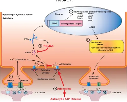
28 FIGURE 1.
Figure 1. A schematic overview of hippocampal signaling pathways following sleep deprivation.
29
CHAPTER TWO
SLEEP DEPRIVATION DURING A SPECIFIC 3-HOUR TIME WINDOW POST