Neurosarcoid Presents Differently in Children Than in Adults
Robert J. Baumann, MD, and William C. Robertson, Jr, MD
ABSTRACT. Background. Neurosarcoid is seldom
recognized in children. In the absence of any large pedi-atric series, it has been assumed that the presenting signs and symptoms are identical in adults and children.
Objective. To test the hypothesis that childhood neu-rosarcoid differs in presenting signs and symptoms from neurosarcoid in adults.
Methods. We tabulated the initial neurologic signs and symptoms in all reported cases of childhood sarcoid with evidence of central nervous system involvement. These data then were compared with published studies of adult neurosarcoid.
Results. Twenty-nine cases (from the English, French, and German literature) had descriptions of pre-senting signs and symptoms. Ages were 3 months to 18 years; 48% (14 of 29) presented before 13 years. Seizures were the most common presenting symptom (38%, 11 of
29), and 73% of these children (8 of 11) were<13 years
old at presentation. Twenty-one percent (6 of 29) had cranial nerve involvement at presentation, and all were
>12 years old. Twenty-one percent (6 of 29) had
hypo-thalamic dysfunction. Five children presented with head-ache, 4 with motor signs, and 3 with papilledema. Twen-ty-four percent (7 of 29) had mass lesions on imaging.
Conclusions. Children with neurosarcoid present dif-ferently than do adults. Children are more likely to have seizures, less likely to have cranial nerve palsies, and perhaps more likely to have a space-occupying lesion. Our analysis of the cases available for review in the published literature suggests that children evolve to an
adult pattern as they progress through adolescence.
Pe-diatrics 2003;112:e480 –e486. URL: http://www.pediatrics.
org/cgi/content/full/112/6/e480;adolescence, child, cranial
nerve injuries, manifestations neurologic, sarcoidosis, sei-zures.
ABBREVIATIONS. CNS, central nervous system; CSF, cerebrospi-nal fluid; MRI, magnetic resonance imaging; ACE, serum angio-tensin-converting enzyme; CT, computed tomography.
P
hysicians seldom include sarcoid in the differ-ential diagnosis of progressive encephalopa-thies of childhood.1,2 This apparently occursbecause sarcoid is seldom diagnosed in the young and is therefore considered rare.3,4 The omission is
surprising, because sarcoid is both treatable and known to have protean manifestations. Moreover, if
untreated, CNS sarcoid can result in death or perma-nent disability.
In the absence of any large pediatric series, it has been assumed that the presenting signs and symp-toms of CNS sarcoid are identical in children and adults.3,5We recently cared for a child who did not
fit the usual adult pattern and decided to reexamine this premise. We searched for all reported cases in the world literature and analyzed them for present-ing signs and symptoms. Our review suggests that prepubertal children with neurosarcoid present dif-ferently than adults, and this pediatric pattern evolves to an adult pattern during puberty.
CASE REPORT
After 3 days of headache and malaise, a previously healthy 9-year-old white girl from rural Kentucky had a generalized tonic-clonic seizure. Cerebrospinal fluid (CSF) examination revealed a white blood cell count of 350 per mL (52% lymphocytes and 48% polymorphonuclear cells), normal glucose, and pro-tein of 68 mg per dL. Routine cultures were sterile. After intravenous phenytoin there were no addi-tional seizures. Her hospital course was character-ized by fever to 101°F, confusion, and headache. Electroencephalograms showed symmetrical slow-ing compatible with a nonspecific, diffuse encepha-lopathy. Precontrast magnetic resonance imaging (MRI) of the head was unremarkable, but gadolini-um-enhanced images revealed 2 irregular areas of enhancement involving the anterior aspect of the left frontal lobe. After 10 days she was afebrile, symp-tom-free, and discharged on phenytoin with a pre-sumptive diagnosis of viral encephalitis.
After 3 medically uneventful months (though marred by poor school performance), the child be-came acutely confused and had another generalized tonic-clonic seizure. On readmission, routine labora-tory studies were unremarkable. Gadolinium-en-hanced MRI showed diffuse, fine punctuate lesions with increased signal from cortical gyri on the late T2 sequence. Magnetic resonance angiogram was unre-markable. The erythrocyte sedimentation rate was 11, and the phenytoin level was therapeutic. Numer-ous laboratory studies including serum aldolase, an-tineutrophil cytoplasmic and perinuclear antibodies, antinuclear antibodies, anti-Smith antibodies, factor V Leiden, smooth muscle antibodies, and anti-cardiolipin antibodies were negative. Homocysteine, protein C, and protein S were unremarkable. Spinal fluid analysis revealed 4 mononuclear cells, 1 red blood cell, glucose of 64 mg per dL, and protein of 53 mg per dL. CSF cultures including those for fungi, From the Departments of Neurology and Pediatrics, University of
Ken-tucky, Lexington, Kentucky.
Received for publication May 15, 2003; accepted Jul 30, 2003.
tuberculosis, and viruses were sterile. Other CSF studies including venereal disease research labora-tory test, pyruvate, oligoclonal bands, and myelin basic protein were unremarkable. Valproic acid was added to the patient’s medication program. She im-proved and was discharged after 6 days of hospital-ization.
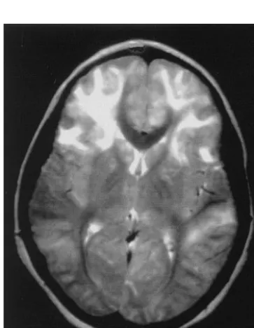
After discharge the patient had headaches daily. Over the next 3 weeks she became unsteady and had difficulty remembering numbers. After 2 generalized seizures the child was readmitted. Examination re-vealed confusion, a right 6th-nerve palsy, extensor plantar responses, and truncal ataxia. Multiple labo-ratory studies again were normal. Brain MRI now showed large areas of increased T2 signal outlining multiple, swollen cerebral cortical gyri and the cau-date nuclei (Fig 1). The dot-like enhancement previ-ously seen with gadolinium enhancement was more prominent and again corresponded to areas of in-creased T2 signal (Fig 2).
The child’s headache was unrelenting. She devel-oped bilateral 6th-nerve palsies, left lower facial weakness, and slow speech. Brain and meningeal biopsy revealed discrete noncaseating epithelioid granulomas consistent with sarcoid (Fig 3). The se-rum angiotensin-converting enzyme (ACE) level was 15 international units (normal: 41–126), and chest radiograph was normal.
The child had marked clinical improvement within 48 hours of high-dose steroid therapy. After 5 months of tapering doses of prednisone, MRI find-ings had completely resolved, and steroids were dis-continued. There were no cranial nerve findings or focal neurologic deficits; however, the child did have frontal lobe indifference and mild cognitive difficul-ties. Four years later these cognitive deficits have persisted, but there has been no recurrence of
sei-zures or focal neurologic signs and repeat MRI stud-ies have been normal.
METHODS
We attempted to identify all reported childhood cases of neu-rosarcoid. We searched PubMed for the common manifestations of
Fig 1. Axial T2-weighted image demonstrates large areas of in-creased signal involving primarily frontal cortex and subcortical white matter.
Fig 2. Coronal MRI with gadolinium shows enhancing punctate lesions within sulci and cortical gray matter.
sarcoid in childhood (pulmonary and arthritic) as well as for neurosarcoid. Each reported case was examined for evidence of CNS manifestations, and reference lists were scanned for reports of additional cases. We also reviewed child neurology and neu-roradiology textbooks for cases described in the texts but not reported otherwise.
RESULTS
Including our patient, there were 29 cases of child-hood neurosarcoid with descriptions of presenting signs and symptoms (Table 1). An additional 15 cases lacked sufficient data about presenting signs and symptoms and were omitted. Because on rereview of the original articles we could not find evidence of neurologic involvement, we omitted 2 of the 23 chil-dren listed by Weinberg et al6in their 1983 review of
pediatric neurosarcoid. In the paper by Beier and Lahey7we could not find the child with a 7th-nerve
palsy referenced by Weinberg et al. In addition, in the 1956 series collected by McGovern and Merritt8
we were unable to identify the child tabulated by Weinberg et al as having “facial dysphagia.”
Cases were found in the English, French, and Ger-man language literature. Only 13 cases were specif-ically published in peer-reviewed journals as neuro-sarcoid. Eleven other children were reported as having systemic sarcoid, but our review of the pub-lished details indicated nervous system involvement. Two other cases were reported as examples of hypo-thalamic complications of sarcoid. Another was found in an article on the radiologic manifestations of sarcoid, 1 was reported as an ophthalmologic com-plication of sarcoid, and the final case was discov-ered in a child neurology textbook. Ages ranged from 3 months to 18 years with 48% (14/29) present-ing before 13 years. Seizures were the most common presenting symptom (38% of subjects, 11 of 29). Of the children presenting with seizures, 73% (8 of 11) were⬍13 years old at presentation. Twenty-one per-cent (6 of 29) had cranial nerve findings at presenta-tion that appeared to be caused by direct nerve in-volvement and not as a secondary effect such as 6th-nerve palsies associated with increased intracra-nial pressure. All children with craintracra-nial nerve palsies wereⱖ12 years old. Only 1 child had involvement of multiple cranial nerves (age 13). Among the others, 2 had optic nerve and facial nerve palsies, and 1 had a superior oblique palsy. Twenty-one percent (6 of 29) evidenced hypothalamic dysfunction with diabetes insipidus (4 of 6), short stature (3 of 6), and sexual immaturity (2 of 6). Hypothalamic dysfunction showed no age predilection. Five children presented with headache, 4 with motor signs, and 3 with pap-illedema.
Twenty-four percent (7 of 29) had mass lesions or focal edema on imaging: One 8-year-old child with seizures had a “ring cortical lesion” on computed tomography (CT)9; our case (age 9) had papilledema
and swollen frontal gyri on MRI; one 12-year-old child had nodular lesions in the optic chiasm and right sylvian fissure on MRI10; a 13-year-old child
had a pontine mass on MRI11; a 16-year-old child had
hemiparesis and a left frontal mass12; a 17-year-old
child with seizures had a temporal mass on MRI13;
and an 18-year-old child had hemiparesis and pap-illedema with a mass on angiogram.14
DISCUSSION
Our patient presented with an encephalopathy that lacked unique or distinguishing features, similar to many of the cases of childhood neurosarcoid re-ported in the literature. She lacked evidence of pul-monary sarcoid, had no adenopathy, and had a nor-mal ACE level. Neurosarcoid was not considered in the differential diagnosis, although there were hints as to the correct diagnosis on MRI (punctate, enhanc-ing lesions with gadolinium). She developed signs of increased intracranial pressure (papilledema and bi-lateral 6th-nerve palsies), leading to a biopsy of the mass-like, intracranial lesion and the correct diagno-sis. Her response to corticosteroid therapy was prompt and probably life-saving. She has been off medication and in remission for⬎4 years.
Sarcoid is a systemic disorder of unknown cause characterized by noncaseating granulomas.15 It is
well known in adults for its protean features includ-ing abnormal chest radiographs and progressive multiorgan failure.15 In adults, “the lungs and
tho-racic lymph nodes are almost always involved; most patients report acute or insidious respiratory prob-lems.”15Neuropathy, especially facial nerve palsy, is
the most common neurologic complication among adults.16 –19Among older children sarcoid is
primar-ily viewed as a pulmonary or ocular disorder,10with
the triad of skin, joint, and eye disease occurring in younger children.20 –22In younger children, systemic
sarcoid differs clinically from the adult form of the disease and can mimic various disorders unique to childhood.23Adult and childhood sarcoid also differ
epidemiologically. Children do not appear to show the female predominance noted in adults23;
addition-ally, in the United States, they do not seem to dem-onstrate the higher prevalence in African Americans seen in adult populations.15,22 Although our patient
lives in a rural area, it is unknown if American children as a group show the rural predominance noted in American adults with this disorder.15,24
In adults, neurosarcoid is estimated to occur in 5% to 10% of patients with systemic sarcoid, although this may be an underestimate.25,26 Adult
neurosar-coid characteristically presents with cranial nerve palsies, most commonly involving the 7th nerve.19,27
A recent review reported that 50% of adults pre-sented with cranial nerve palsies, 30% with head-aches, and 10% each with seizures, pituitary
dys-function, sensory deficits, neuropsychological
deficits, and cerebellar signs (patients could have⬎1 sign on presentation).19,27In adults, both seizures28,29
and CNS involvement26have been considered
mark-ers of poor outcomes.
Most surprising of all is the large number of children with mass-like lesions on imaging. We found 7 cases not including 2 children with papilledema who were reported in 193730and in 196931before the advent of
modern imaging. It is also striking that 1 of the 7 children had no neurologic symptoms, and the mass-like lesion was discovered by MRI done “for com-pleteness.”10 Although adults with seizures and
other intracranial lesions respond poorly to thera-py15 and have a higher rate of chronic handicap,32
this does not seem to be true of our 9-year-old patient or of other children. Previous authors have regarded mass-like lesions as rare.10 It is unclear if they are
wrong and this is an important and previously un-recognized feature of childhood neurosarcoid or if this is an artifact of a survey of reported cases, with unusual findings being more likely to be published. Sarcoid is characterized by noncaseating granulo-mas, and all cases that we accepted were biopsy- or autopsy-proven, although biopsy material was often not from the CNS. Other tests can be useful in estab-lishing the diagnosis, but biopsy still provides the strongest evidence for this disease.33 Elevated ACE
levels are not specific for sarcoid but are elevated in the serum of 70% to 80% of patients with systemic disease and in the CSF of ⬃50% of patients with neurosarcoid.19 Analysis of CSF may show a mild
lymphocytosis (10 –100 cells perL), mildly elevated protein (to 70 mg%), mildly decreased glucose, and increased immunoglobulins with oligoclonal band-ing.19When available, the Kveim test can be positive
in 85% of patients.33 MRI has become increasingly
useful. Findings in neurosarcoid include periven-tricular high-signal lesions on T2-weighted imag-es,13,33,34leptomeningeal enhancement,33,34and
soli-tary masses.34 Many of the reported pediatric cases
antedate the availability of MRI, and whether there are different patterns in children than in adults is unclear.
The most appropriate treatment for pediatric neu-rosarcoid is uncertain. Corticosteroids are widely used and are often effective.10For adult patients for
whom corticosteroid therapy fails, azathioprine or cyclosporin A have been prescribed.19
An important caveat to our analysis is that pub-lished case reports may not be representative of the population of patients with a disorder. Furthermore, many of these cases were published by nonneurolo-gists whose interests were in pulmonary, rheumatic, or other manifestations of the disease. The neuro-logic findings were presented for completeness. Al-though we omitted those cases where we could not confidently extract a detailed picture of the patients’ findings on presentation, it is always possible that if experienced neurologists rather than nonneurolo-gists had reported these patients, they would have conveyed different findings.
CONCLUSIONS
An analysis of 29 pediatric neurosarcoid cases in-dicates that compared with adults, children are more likely to have seizures, less likely to have cranial nerve palsies, and perhaps more likely to have a space-occupying lesion. Analyzing these children by
age group, preadolescents and adolescents, suggests that pediatric cases evolve to adult patterns of dis-ease, with more frequent cranial neuropathies and less frequent seizures as the patients progress to adulthood.
REFERENCES
1. Fenichel GM.Clinical Pediatric Neurology. A Signs and Symptoms Ap-proach.4th ed. Philadelphia, Pa: WB Saunders Company; 2001 2. Swaiman KE, Shevell MI. Intellectual and motor regression. In:Pediatric
Neurology. Principles & Practice.Swaiman KE, Ashwal S, eds. St. Louis, MO: Mosby; 1999:561–567
3. Shetty AK, Gedalia A. Sarcoidosis in children.Curr Probl Pediatr. 2000; 30:149 –176
4. Sheets RM, Ashwal S, Sizer IS. Neurologic manifestations of rheumatic disorders of childhood. In:Pediatric Neurology. Principles & Practice. Swaiman KE, Ashwal S, eds. St. Louis, MO: Mosby; 1999:1125–1146 5. Dyken PR. Neurosarcoidosis. In:Principles of Child Neurology. Berg BO,
ed. New York, NY: McGraw-Hill; 1996:889 – 899
6. Weinberg S, Bennett H, Weinstock I. Central nervous system manifes-tations of sarcoidosis in children. Case report and review.Clin Pediatr (Phila). 1983;22:477– 481
7. Beir FR, Lahey ME. Sarcoidosis among children in Utah and Idaho.J Pediatr.1964;65:350 –359
8. McGovern JP, Merritt DH. Sarcoidosis in childhood.Adv Pediatr. 1956; 8:97–135
9. Grossman H, Merten DF, Spock A, Kirks DR. Radiographic features of sarcoidosis in pediatric patients.Semin Roentgenol. 1985;20:393–399 10. Kone-Paut I, Portas M, Wechsler B, Girard N, Raybaud C. The pitfall of
silent neurosarcoidosis.Pediatr Neurol. 1999;20:215–218
11. Leiba H, Siatkowski RM, Culbertson WW, Glaser JS. Neurosarcoidosis presenting as an intracranial mass in childhood. J Neuroophthalmol. 1996;16:269 –273
12. Douglas AC, Maloney AF. Sarcoidosis of the central nervous system.
J Neurol Neurosurg Psychiatry. 1973;36:1024 –1033
13. Miller DH, Kendall BE, Barter S, et al. Magnetic resonance imaging in central nervous system sarcoidosis.Neurology. 1988;38:378 –383 14. Griggs RC, Markesbery WR, Condemi JJ. Cerebral mass due to
sarcoid-osis. Regression during corticosteroid therapy. Neurology. 1973;23: 981–989
15. Newman LS, Rose CS, Maier LA. Sarcoidosis.N Engl J Med. 1997;336: 1224 –1234
16. Turner RG, James DG, Friedmann AI, Vijendram M, Davies JP. Neuro-ophthalmic sarcoidosis.Br J Ophthalmol. 1975;59:657– 663
17. James DG, Siltzbach LE, Sharma OP, Carstairs LS. A tale of two cities. A comparison of sarcoidosis in London and New York.Arch Intern Med. 1969;123:187–191
18. Stern BJ, Krumholz A, Johns C, Scott P, Nissim J. Sarcoidosis and its neurological manifestations.Arch Neurol. 1985;42:909 –917
19. Nowak DA, Widenka DC. Neurosarcoidosis: a review of its intracranial manifestation.J Neurol. 2001;248:363–372
20. Hafner R, Vogel P. Sarcoidosis of early onset. A challenge for the pediatric rheumatologist.Clin Exp Rheumatol. 1993;11:685– 691 21. Clark WC, Acker JD, Dohan FC Jr, Robertson JH. Presentation of central
nervous system sarcoidosis as intracranial tumors.J Neurosurg. 1985;63: 851– 856
22. Lindsley CB, Petty RE. Overview and report on international registry of sarcoid arthritis in childhood.Curr Rheumatol Rep. 2000;2:343–348 23. Keesling CA, Frush DP, O’Hara SM, Fordham LA. Clinical and imaging
manifestations of pediatric sarcoidosis.Acad Radiol. 1998;5:122–132 24. Kajdasz DK, Lackland DT, Mohr LC, Judson MA. A current assessment
of rurally linked exposures as potential risk factors for sarcoidosis.Ann Epidemiol. 2001;11:111–117
25. Delaney P. Neurologic manifestations in sarcoidosis: review of the literature, with a report of 23 cases.Ann Intern Med. 1977;87:336 –345 26. Ferriby D, de Seze J, Stojkovic T, et al. Clinical manifestations and
therapeutic approach in neurosarcoidosis.Rev Neurol (Paris). 2000;156: 965–975
27. Scott TF. Neurosarcoidosis: progress and clinical aspects.Neurology. 1993;43:8 –12
28. Krumholz A, Stern BJ, Stern EG. Clinical implications of seizures in neurosarcoidosis.Arch Neurol. 1991;48:842– 844
29. Delaney P. Seizures in sarcoidosis: a poor prognosis.Ann Neurol. 1980; 7:494
[English translation: On the incidence of Schaumann’s benign lympho-granulomatosis (Boek’s benign milary lupoid) in children].Z Kinderhei-lkd. 1937;59:280 –302
31. Reed WG. Sarcoidosis: a review and report of eight cases in children.J Tenn Med Assoc. 1969;62:27–36
32. Ferriby D, de Seze J, Stojkovic T, et al. Long-term follow-up of neuro-sarcoidosis.Neurology. 2001;57:927–929
33. Zajicek JP, Scolding NJ, Foster O, et al. Central nervous system sarcoidosis– diagnosis and management.QJM. 1999;92:103–117 34. Pickuth D, Heywang-Kobrunner SH, Spielmann RP. Role of magnetic
resonance imaging in the diagnosis of neurosarcoidosis.Radiologe. 1999; 39:889 – 893
35. Naumann O. Kasuistischer Beitrag zur Kentnis der Schaumannschen “benignen Granulomatose” (Morbus Besnier-Boeck-Schaumann) [En-glish translation: Case report on recognition of Schaumann’s benign granulomatosis].Z Kinderheilkd. 1938;60:1– 8
36. Posner I. Sarcoidosis: a case report.J Pediatr. 1912;20:486 – 495 37. Pilz P, Harrer G. Zur Sarkoidose des Nervensystems [English
translation: On sarcoidosis of the nervous system].Wien Klin Wochenschr Jg. 1979;91:590 –592
38. Sharma OP, Anders A. Neurosarcoidosis. A report of ten patients
illustrating some usual and unusual manifestations.Sarcoidosis. 1985;2: 96 –106
39. Wiederhold WC, Siekert RG. Neurologic manifestations of sarcoidosis.
Neurology. 1965;15:1147–1154
40. Delaney P, Henkin RI, Manz H, Satterly RA, Bauer H. Olfactory sar-coidosis. Report of five cases and review of the literature.Arch Otolar-yngol. 1977;103:717–724
41. Shealy CN, Kahana L, Engel FL, McPherson HT. Hypothalamic-pituitary sarcoidosis.Am J Med. 1961;30:46 –55
42. Dressler M, Wagner H. Uber zwei Falle von Lymphogranulomatosis benigna (Schaumann) [English translation: Two cases of Schaumann’s benign granulomatosis].Acta Derm Venereol. 1941;15:511–544 43. Ford FR.Diseases of the Nervous System in Infancy, Childhood and
Adoles-cence. 5th ed. Springfield, IL: Charles C. Thomas; 1966
44. Pennell WH. Boeck’s sarcoid with involvement of the central nervous system.Arch Neurol Psychiatry. 1951;66:728 –737
45. Beardsley TL, Brown SV, Sydnor CF, Grimson BS, Klintworth GK. Eleven cases of sarcoidosis of the optic nerve.Am J Ophthalmol. 1984; 97:62–77
2003;112;e480
Pediatrics
Robert J. Baumann and William C. Robertson, Jr
Neurosarcoid Presents Differently in Children Than in Adults
Services
Updated Information &
http://pediatrics.aappublications.org/content/112/6/e480 including high resolution figures, can be found at:
References
http://pediatrics.aappublications.org/content/112/6/e480#BIBL This article cites 41 articles, 7 of which you can access for free at:
Subspecialty Collections
sub
http://www.aappublications.org/cgi/collection/hematology:oncology_
Hematology/Oncology
following collection(s):
This article, along with others on similar topics, appears in the
Permissions & Licensing
http://www.aappublications.org/site/misc/Permissions.xhtml in its entirety can be found online at:
Information about reproducing this article in parts (figures, tables) or
Reprints
2003;112;e480
Pediatrics
Robert J. Baumann and William C. Robertson, Jr
Neurosarcoid Presents Differently in Children Than in Adults
http://pediatrics.aappublications.org/content/112/6/e480
located on the World Wide Web at:
The online version of this article, along with updated information and services, is
by the American Academy of Pediatrics. All rights reserved. Print ISSN: 1073-0397.