ORIGINAL ARTICLE
IJPHY
ABSTRACT
Introduction: Nonspecific neck pain is a frequent cause for adults to consult health care providers. Therefore, the phys-ical therapist should always seek the most effective intervention(s) within the wide spectrum of treatments available, so this study was conducted to examine the effect of transcranial laser on somatosensory integration in nonspecific neck pain patients.
Methods: Thirty-two male subjects suffering from non-specific neck pain participated in the current study. They were randomly assigned via a balanced stratified assignment. The experimental group (N=16) received trans-cranial in-fra-red laser (50mw, 90 snm pulsed mode, and 1.2 J/cm 2) over sensory cortex whereas, the control group (N=16) received placebo laser. The assessment of N30 and P22-N30 components of somatosensory evoked potentials (SEPs) were done before and immediately after treatment session for both groups.
Results: There was a statistically significant difference between groups regarding N30 peak of SEPs after a single trans-cranial laser session (p=0.0062) with no significant difference between pre and post-treatment data in the exper-imental group (p=0.0803) and control group (p=0.5577) with a percentage of change (-2.38%) and (1.009%) respec-tively. In addition, post-treatment results revealed no statistically significant difference between groups regarding an a P22-N30 component of SEPs (p=0.0933).
Conclusion: According to the parameters used in the study, it was concluded that trans-cranial laser did not affect so-matosensory integration in nonspecific neck pain patients.
Keywords: Transcranial, laser, somatosensory, integration, neck, pain.
Received 15th December 2017, revised 18th March 2018, accepted 29th March 2018
www.ijphy.org
10.15621/ijphy/2018/v5i2/170748
CORRESPONDING AUTHOR
Int J Physiother. Vol 5(2), 80-86, April (2018) ISSN: 2348 - 8336
EFFECT OF TRANSCRANIAL LASER ON SOMATOSENSORY
INTEGRATION IN PATIENTS WITH NONSPECIFIC NECK
PAIN: RANDOMIZED CONTROLLED TRIAL
*1Roshdy M. kamel ²Ghada I. Mohamed ³Ibrahim M. Moustafa ⁴Ragia M.Kamel ⁵Omaima M. Kattabei
*1Roshdy M. kamel
Assistant Lecturer of Physical Therapy, Basic Science Department,
Faculty of Physical Therapy, Deraya University, Egypt. e-mail: roshdykamel @live.com ²Lecturer of Physical Therapy, Basic Science
Depart-ment, Faculty of Physical Therapy, Cairo University. ³Assistant Professor of Physical Therapy, Basic Sci-ence Department, Faculty of Physical Therapy, Cairo University.
⁴Professor of Physical Therapy, Basic Science Depart-ment, Faculty of Physical Therapy, Cairo University. ⁵Professor of Physical Therapy, Basic Science De-partment, Faculty of Physical Therapy, Cairo Uni-versity. Vice Dean for graduate studies and scientific research at Deraya University, Egypt.
INTRODUCTION
Neck pain is a common painful musculoskeletal condition that affects individuals worldwide. Prevalence data suggest that neck pain can affect children and elderly alike. Its in-cidence is ranging from 6% to 22% and up to 38% of the elderly population [1]. There is wide variability in identify-ing neck pain despite its high incidence. This is due to the presence of both physical and psychological factors predis-posing cervical spine pain. So, as a result of multiple factors presenting neck pain and the inability to precisely identify the cause of its symptoms [2], the term nonspecific neck pain has been assigned to any undiagnosed symptomatic disorder of the cervical spine [3].
Neck pain incorporates a significant result on individuals’ health quality of life and on society as a whole. So, high-er management approaches in medical aid required to be developed to stop patients from developing chronic pain and disability [4]. It is evident that the neuromuscular and proprioceptive functions impaired in patients with back and neck ache which give areas on why what starts in the form of a restricted range of motion and/or mild pain be-comes chronic as the neck is joined to the upper limb neu-rologically and biomechanically. A systematic review in 2012, by Haavik–Taylor, and Murphy [5], bestowed proof suggesting that altered sensory process from areas of joint dysfunction could lead to faulty somatosensory integration attribute to central neuroplastic changes.
The label somatosensory reflects all the sensory informa-tion, motor informainforma-tion, central integration and process-ing elements that sustain stability in the postural control system throughout intrinsic motor-control properties. So, somatosensory impairment can arise from altered cervical afferent input [6].
There is a great proof pointing to the idea that subjects with chronic pain might develop anatomical alterations in brain areas responsible for cognition and emotional modulation of pain, like dorsolateral and medial prefrontal cortex, the anterior cingulate cortex and the insula. Moreover, the results of the previous studies which used in vivo proton magnetic resonance spectrometry revealed rise in glu-tamate and/or reduction of N-acetyl aspartate in frontal cortices in subjects with a fibromyalgia and chronic back-ache; such results agreed that reduced grey matter might be related to possible excitotoxicity in patients with chronic pain [7,8].
Early somatosensory evoked potentials (SSEPs) as an ob-jective modality offer a mechanism for investigation of al-terations in somatosensory integration areas of the brain. It had been disclosed that N30 component of SSEPs peak is produced in post-central gyrus as primary sensory cortex is connected to a complex subcortical and cortical circuit connecting the pre-motor areas, primary motor cortex, basal ganglia, and thalamus. Therefore N30 peak is as-sumed to assess somatosensory integration [9,10,11,12,13]. There is a growing body of knowledge concerning the effect of transcranial laser therapy (TLT) on cognition [14], attention bias [15], depression [16], traumatic brain injury [17], and stroke[18], and considered as a promis-ing non-invasive modality for neuromodulation that can
produce biochemical changes including enhancing ade-nosine triphosphate synthesis by mitochondria, increase reactive oxygen species and intracellular calcium, nitric oxide release, and might include inhibition of apoptosis in ischemic penumbra and improvement of neuro recovery mechanisms [19].But, there is a gap of knowledge regard-ing its effect on somatosensory integration. So, the current study was designed to examine the effect of transcranial la-ser on somatosensory integration in nonspecific neck pain patients.
METHODS
The current research was performed in Faculty of physical therapy, Cairo University, from April 2016 to August 2017 to examine the effect of transcranial laser on somatosenso-ry integration in nonspecific neck pain patients.
Design of the study: prospective, randomized, placebo, controlled trial.
Flowchart 1: Diagram of the study design Subjects:
vascular disease , intravenous or intra-arterial thrombo-lytics, epilepsy, multiple sclerosis, dystonia, abnormal pe-ripheral nerve function, and skin infection [21]. Patients should have a history of chronic nonspecific neck pain > three months and their body mass index between 18 and 25 kg∕m2. Ethical approval was obtained from the insti-tutional review board Faculty of Physical therapy, Cairo university with number T. REC/012/001178 and registered with pan african clinical trial registry with number (PAC-TR 20181001399154).
Instrumentation:
Two types of instrumentation were used in this study: 1. Computerized electromyography (EMG) device. 2. Laser therapy unit.
(1) Computerized electromyography (EMG) device: Deymedtru trace flexi cart computerized electromyogra-phy system, manufacturer Deymed diagnostic s.r.o, coun-try of manufacturing was the Czech Republic, and the model of the device was true trace EMG CL4 its volt was 220-250 V and 50-60 Hz.
(2) Laser therapy unit:
Medical Italia LIS laser therapy unit was used, manufactur-er EME physio, country of manufacturing was Italy. It is a probe Infra-Red (IR) laser with one diode (MLA1/50) of 50 mW for 905 nm. It has the following technical param-eters; its volt was 230V, 50-60Hz, pulsed operating mode 10-100%, an automatic contact sensor, and automatic dose calculation. The pulse frequency can be adjusted between 200 – 10000 Hz with pulse duration 100 nsec.
PROCEDURE
Measurement of somatosensory evoked potential: The position of the patient: good relaxation of the patients is vital due to the very small size of the cortical potentials. Therefore, the patients were instructed to sit still while re-maining quiet with their eyes closed; room’s lights were turned off.
Skincare under stimulating electrodes; methylated alcohol was used to carefully wash the skin overlying the median nerve and then dried using dry clean cotton wool. This action was repeated until hyperemia of the skin occurred aiming to reduce skin resistance. Cautions were taken to avoid breaking or abrading skin under the stimulating electrode. When the impedance -across the recording elec-trodes preparation sites-was between 1,000 and 5,000Ω, abrasion is accepted and considered sufficient [22].
Parameters of SSEPs stimulation:
The stimuli were electrical square pulses, with 1ms pulse duration, and 2.47 Hz frequency, the stimulating electrode was Ag/AgCl ECG skin electrodes with resistance less than 5,000Ω. The median nerve was stimulated two: three centi-meters from the distal crease of the wrist, between tendons of Flexor pollicis longus and Palmaris longus with anode placed proximally, and cathode distally. The frequency, 2.47 Hz is optimum for N30 and didn’t lead to a decrease in SEP peak. For every patient, the amplitude of stimuli used was the motor threshold that is the most lower amplitude of stimuli that produces a noticeable contraction of
abduc-tor pollicis brevis muscle [23.24].
Recording parameters:
Recording electrodes were placed on the frontal Rossi site (six centimeters anterior and two centimeters contralateral to Cz) [25] according to (IFCN) guidelines [26]. The two recording electrodes were two millimeters gold cup EEG electrodes with an impedance less than 5,000Ω and the ground electrode placed on patients’ forehead. SSEPs sig-nal was amplified (gain 10,000), filtered (0.2–1000 Hz) and stored on a computer Far-field potentials demand a higher number of standard sweeps. The standard waveform was displayed in an analysis window from which amplitudes of the specific waveforms were measured [27].
N30 and P22-N30 components of SSEPs were measured before and after transcranial laser application according to the IFCN guidelines [9]. The following SSEPs components; N30, and P22-N30 complex were identified and analyzed. Experimental Protocol:
The patients were asked to attend one session of TLT. They were diagnosed by an orthopedic physician. All patients were screened for contraindications of TLT. One baseline SEP trial was carried out before treatment through stimu-lation of the median nerve and repeated immediately after transcranial laser application.
For treatment purpose, Medical Italia LIS laser therapy unit (230V, 50Hz) was used. It is a point probe Infra-Red (IR) laser delivers 50 mW for 905 nm. It has the following technical parameters, pulsed operating mode 10-100%, an automatic contact sensor, and automatic dose calculation. The frequency can be adjusted between 200 – 10000 Hz. Application of transcranial laser: The protocol of lamp and his colleagues in 2007 and Zivin and others in 2009 was followed using laser probe which was applied over the en-tire surface of the head according to 10/20 electroenceph-alographic system on sensory cortex for 4 minutes at every point. Infrared Laser power of 50mW, transmitting an en-ergy density of 1.2 J/cm 2, and pulsed mode 100% [19,28], with a frequency of 1000 Hz and a pulse duration 100 nsec. The transcranial laser was applied once to study its imme-diate effect on somatosensory integration.
The patient was seated in comfortable sitting position. Pa-tient and therapist wore protective glasses to avoid the pos-sibility of damage to retina if the laser beam enters through the lens of the eye and onto the retina. The laser probe is applied at a right angle to patient’s skin.
Statistics:
Statistical analysis was done using SPSS, version 20.0 for Windows; SPSS Inc., Chicago, Illinois, USA. Descriptive statistics for patients’ physical characteristics and the de-pendent variable were calculated as mean, and standard deviation. Paired t-test was conducted for comparison between the before and after treatment values in the same group. Unpaired t-test was conducted for comparing the results between groups. The alpha level of significance (α) was set less than 0.05.
Sample size:
estimation. In the pilot study, ten articipansing Lapist should5). patients were divided into two equal groups, study group (mean 28.452 and SD 3.5757) and control group (mean 31.64 and SD 1.8515) two-tailed hypothesis, alpha =0.05, power =80%. G-power test determined the sample size of each group with a total sample size of thir-ty-two patients.
Data Collection:
Patients were acclimated with the electrical stimulation re-quired to provoke SSEPs while the determination of mo-tor threshold, decreasing the possibility of rejection of any sweeps during data collection of SSEPs components. The regarded patient`s age and body mass index had been col-lected in information sheet before entry to study. Paired t-test and unpaired t-test were conducted for comparing values of dependent variables within and between groups respectively with (α) set less than 0.05.
RESULTS
Patients’ physical characteristics were illustrated in table (1). The unpaired t-test revealed nonsignificant differences between both groups concerning age, and body mass in-dex.
Table 1: physical characteristics of patients in both groups
Variables
Experimental
Group Control Group Comparison Mean ± S.D Mean ± S.D t-value Signifi-cance
Age
(Years) 25.13±3.1 26.88±1.63 2.0014 0.0545
Body mass index
(Kg/ m2) 22.194±1.886 23.019±1.749 1.2829 0.2094
Significant at an alpha level less than 0.05.
(1) N30 peak of somatosensory evoked potential results: As shown in tables (2and 3), unpaired t-test revealed a sta-tistically significant difference between groups regarding N30 peak of SSEPs after a single trans-cranial laser session (P=0.0062). Whereas, paired t-test revealed no significant difference between pre and post-treatment data in the ex-perimental group (P=0.0803) with a percentage of change (-2.38%) and control group (P=0.5577) with a percentage of change (1.009%).
Table 2: unpaired t-test of N30 peak SSEPs (ms) for both groups:
Unpaired
t-test ExperimentalMean±SD Mean±SDControl T value Significance
N30 pre 29.7244±2.4416 31.006±2.367 1.5078 0.1421
N30 post 29.0169±2.5579 31.319±1.798 2.9452 0.0062*
Significant at an alpha level less than 0.05.
Table 3: Paired t-test of N30 peak SSEPs (ms) for both groups:
Paired
t-test Mean± SD pre SD postMean± T value signifi-cance Mean differ-ence
Percent-age of change
Experi-mental 29.7244± 2.4416 29.0169± 2.5579 1.8754 0.0803 -0.7075 -2.38%
Control 31.006± 2.367 31.319± 1.798 0.5996 0.5577 0.313 1.009%
Significant at an alpha level less than 0.05.
(2)P22-N30 peak results
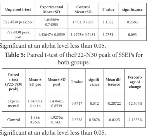
Post-treatment results showed no statistical significant dif-ference between groups regarding P22-N30 component of SEPs (P=0.0933), in addition to no significant difference between before and after treatment values in experimental and control groups with a percentage of change (-12.607%) in experimental group and (-1.1538%) in control group as presented in table (4and 5).
Table 4: Unpaired t-test of the P22-N30 peak of SSEPs for both groups:
Unpaired t-test ExperimentalMean±SD Mean±SDControl T value significance
P22-N30 peak pre 1.64369± 0.74303 1.95± 0.7607 1.1522 0.2583
P22-N30 peak
post 1.43647± 0.8539 1.9275± 0.7431 1.7351 0.093
Significant at an alpha level less than 0.05.
Table 5: Paired t-test of theP22-N30 peak of SSEPs for both groups:
Paired t-test (P22- N30
peak)
Mean ±
SD pre Mean± SD post T value signifi-cance Mean dif-ference
Percent-age of change
Experi-mental 1.64369± 2.4416 1.43647± 0.8539 0.6717 0.512 -0.20722 -12.607%
Control 0.76071.95± 1.9275± 0.7431 0.5538 0.5878 -0.0225 -1.1538%
Significant at an alpha level less than 0.05. DISCUSSION
consider-ation that latency gradually increases as the sensory nerve conduction velocity decreases [31].
Likewise, Taylor and Murphy in 2008 investigated alter-ations in intrinsic inhibitory interactions within the sen-sorimotor system after spinal manipulation session of dysfunctional cervical spines. Their study revealed no sig-nificant difference in N30 peak along with suppression of P22-N30 component of SEP [32].
The first explanation for the nonsignificant difference be-tween groups regarding dependent variables may be at-tributed to no current protocols were found in a healthy population, in which the parameters (intensity, power, du-ration, number of sessions, and fluency of the laser light) of the stimulation are settled. Therefore, there is no gener-al agreement regarding parameters that should be select-ed for stimulating the intact cortex in order to efficient-ly stimulate the target cortical area by near-infrared laser [33]. Similarly, the effects of transcranial laser application take time to start and it is possible that duration of tissue extraction was not enough to detect biochemical changes as well as the dosage [30].
Regarding the parameters, the review of the literature re-vealed that, there were contradictory results concerning the parameters of TLT as Pires de Sousa and others in 2016 applied transcranial laser in animal models with the following parameters (810 nm laser, 300mW/cm2, 7.2 or 36 J/cm2) to study its effect on peripheral nociception [30]. They found a decrease in metabotropic glutamate receptors in the tissue, leads to a reduction of conduction of painful stimuli due to the decreased action potentials’ frequency. Similarly, Salehpour and his colleagues used laser treat-ments for six weeks at wavelengths of 660 and 810 nm at 8 J/cm2 which have potential to ameliorate aging-induced mitochondrial dysfunction, apoptosis, and cognitive im-pairment. On the other hand, same wavelengths at the flu-ency of 4 J/cm2 had a poor effect on the behavioral and molecular indexes in the aging model [16].
In 2016, Hwang and others used transcranial low level la-ser therapy on two sites of forehead (total duration 8 min, 1064 nm, continuous mode, 250 mW/cm2, 60 J/cm2 per site of 13.6 cm2) in combination with acute aerobic exer-cise and found that TLT is capable of improving cognition, recommending that TLT augments pre-frontal cognitive functions [34].
In line with the other researchers (Xuan et al., 2014) inves-tigated the effect of transcranial laser in patients with trau-matic brain injury. They used 810-nm laser delivering 18J/ cm2 fluency and 25 mW/ cm2 and found TLT to be effective in improving neurological performance [35]. Similarly, (Konstantinovicet al., 2013) used (wavelength 905 nm, the frequency of 3 kiloHertz, the power density of 50mW/cm2, and single dosage of 3J/cm2) in the 1st part of their study to examine if the transcranial laser could change motor cortex excitability. They mentioned that the most promi-nent motor evoked potential (MEP) inhibition was within 10–25min after transcranial laser [36].
Laser irradiation produces neglected amount of heat with-out causing physical damage at the low power level was founded by (Schiffer et al., 2009). In their study, TLT had
been examined for its effect on depression. They recruited healthy subjects who received continuous near-infrared la-ser. The laser was delivered to the forehead using low-pow-er laslow-pow-er diode (wavelength 1064 nm). The powlow-pow-er density 250 mW/cm2, as well as the cumulative energy density 60 J/cm2showed antidepressant effect [37].
Hesse et al., 2016investigated efficacy of transcranial laser on alertness and awareness in patients with traumatic brain injury and severe disorders of consciousness. they stimu-lated the cortex of five subjects, four of them are chron-ic with unresponsive attentiveness or minimal awareness, and one patient subacute with a kinetic mutism, with the transcranial laser (785 nm, 10 mW/cm2, continuous wave mode, 21 emitting diodes) for ten minutes every workday for 6 weeks. Their results supported that TLT had been found to improve the patients’ alertness and awareness [38].
The second explanation for the nonsignificant difference between groups regarding P22-N30 may be attributed to that application of laser did not exert an analgesic effect as patients with neck pain had a significantly higher joint position sense error than healthy controls (De Vries and others in 2015) which in turn affects somatosensory inte-gration [39].
In 1997, Tinazzi and others recruited 10patients with a cervical disc prolapse compressing the C-6 nerve root and ten healthy age-matched controls. Unilateral radiating pain from the nerve root (C6) shows differences in SEP ampli-tude between impaired and unimpaired sides and between the impaired side and healthy controls. SEPs were recorded in a between-limb, and between-subjects design. Ampli-tudes of peaks N13, P14, N20, P27, and N30 were ampli-fied remarkably in the extremity with the presence of pain. This explains that there is a positive correlation between the presence of pain and SEP amplitude [40].
(Piresde Sousa et al., 2016) Proved that TLT is effective in decreasing pain sensation all over the body in response to different forms of stimulation. Application of laser precise-ly over somatosensory cortex was reinforced by proof of photoneuromodulation of neuromarkers related to noci-ception. Pain reduction could be related to the biochem-ical alterations that take place due to photons’ absorption. The release of prostatic acid phosphatase is regulated by adenosine triphosphate as its formation needs a sufficiency of energy. An increase in endogenous analgesic prostatic acid phosphatase could decrease the pain perception. The amount of Glutamate which is an excitatory neurotrans-mitter involved in nociception was remarkably high in the control group than in the groups received transcranial la-ser [30].
Limitations
There are several limitations of the study. The first one is the individual variability regarding the penetration depth of laser which is affected by the thickness of the skull and scalp that can be very different among healthy individuals, resulting in different penetration depths.
triggering photo neuromodulation might be hurtful as la-ser intensity has to be sufficient to penetrate skull without rising the temperature of the brain. The third point is the inability to deliver laser to deep structures of the central nervous system while maintaining a low intensity at the cortical surface.
CONCLUSION
According to the parameters used in the study, it was con-cluded that trans-cranial laser did not affect somatosenso-ry integration in patients with nonspecific neck pain Acknowledgments
The authors would like to acknowledge Prof. Omaima M.Kattabei for her valuable instructions throughout work-ing on this study and the subjects participated in the study. Financial support and sponsorship
Self-funded
Conflicts of interest Nil
REFERENCES
[1] Fejer R, Kyvik KO, Hartvigsen J. The prevalence of neck pain in the world population: a systematic critical review of the literature. Eur Spine J.2006;15(6): 834-48. [2] Borghouts JA, Koes BW,Bouter LM. The clinical course
and prognostic factors of non-specific neck pain: a sys-tematic review. Journal of Pain.1998; 77(1): 1-13. [3] Hogg-Johnson S, Van der Velde G, Carroll LJ, Holm
LW, Cassidy JD, Guzman J ,et al. The burden and deter-minants of neck pain in the general population: results of the Bone and Joint Decade 2000-2010 Task Force on Neck Pain and Its Associated Disorders. Spine (Phila Pa 1976). 2008 ;33(4 Suppl):S39-51.
[4] Jones R, Britten N, Culpepper L, Grass D, Grol R, Mant D, Silagy C. Oxford Textbook of Primary Medical Care. 2003.
[5] Jensen W, Andersen OK, Akay M. Replace, repair, re-store, relieve-briding clinical and engineering solution in neurorehabilitation. Proceedings of the 2nd Inter-national Conference on Neuro Rehabilitation. 2014. [6] Kristjansson E, Treleaven J. Sensorimotor function
and dizziness in neck pain: implications for assess-ment and manageassess-ment. J Orthop Sports PhysTher. 2009;39(5):364-67.
[7] Neugebauer V, Galhardo V, Maione S, Mackey SC. Forebrain Pain Mechanisms. Brain Res Rev. 2009; 60(1): 226–42.
[8] Obermann M, Nebel K, et al.Gray matter changes re-lated to chronic posttraumatic headache. Neurology journal. 2009; 73(12): 978–83.
[9] Cruccu G, Aminoff M, Curio G, Guerit J, Kakigi R, Mauguiere F, et al. Recommendations for the clinical use of somatosensory-evoked potentials. ClinNeuro-physiol. 2008;119(8):1705–19
[10] Toleikis JR, Carlvin AO, Shapiro DE, Schafer MF. The use of dermatomal evoked responses during surgical procedures that use intrapedicular fixation of the lum-bosacral spine. Spine journal. 1993; 18(16): 2401-07.
[11] Allison T, McCarthy G, Wood CC, Williamson PD, SpencerDD. Human cortical potentials evoked by stimulation of the median nerve. II. Cytoarchitectonic areas generating long-latency activity. J Neurophysiol. 1989; 62(3): 711–22.
[12] Aminoff MJ, Goodin DS, Parry GJ, Barbaro NM, Weinstein PR, Rosenblum ML. Electrophysiologic evaluation of lumbosacral radiculopathies: electro-myography, late responses and somatosensory evoked potentials. Neurology journal. 1985; 35(10): 1514-18 [13] Eisen A, Hoirch M, Moll A. Evaluation of
radiculop-athies by segmental stimulation and somatosensory evoked potentials. Can J NeurolSci. 1983; 10(3): 178-82.
[14] Vargas E, Barrett DW, Saucedo CL, Huang LD, Abra-ham JA, Tanaka H, et al. Beneficial neurocognitive ef-fects of transcraniallaser in older adults. Lasers Med Sci.2017;32(5):1153-62.
[15] Disner SG,Beevers CG, Gonzalez-Lima F. Transcra-nial Laser Stimulation as Neuroenhancement for At-tention Bias Modification in Adults with Elevated De-pression Symptoms. Brain Stimul. 2016; 9(5): 780-87. [16] Salehpour F, Rasta SH. The potential of transcrani-alphotobiomodulation therapy for treatment of ma-jor depressive disorder. Rev Neurosci journal. 2017; 28(4):441-53.
[17] Moges H, Vasconcelos OM, Campbell WW, Borke RC, McCoy JA, Kaczmarczyk L, et al. Light therapy and supplementary riboflavin in the SOD1 transgenic mouse model of familial amyotrophic lateral sclerosis (FALS). Lasers Surg Med. 2009;41(1):52–59.
[18] Thunshelle C, Hamblin MR. Transcranial Low-Level Laser (Light) Therapy for Brain Injury. PhotomedLa-ser Surg. 2016;34(12):587-98.
[19] Lampl Y. Laser treatment for stroke. 2007;7(8):961-965.
[20] Lapchak PA, De Taboada L. Transcranial near infra-red laser treatment (NILT) increases cortical adenos-ine-5′-triphosphate (ATP) content following embolic strokes in rabbits. Brain Res. 2010;1306:100–105. [21] Legrain V. Cognitive aspects of nociception and pain:
bridging neurophysiology with cognitive psychology. Clinical Neurophysiology.2012; 42(5):325–36. [22] Dumitru D, king JC. Median/Ulnar premotor
po-trential identification and localization. Muscle nerve. 1995;18(5):518-25.
[23] Murphy B, TaylorH, Wilson S, Knight J, Mathers K, Schug S. Changes in median nerve somatosensory transmission and motor output following transient deafferentation of the radial nerve in humans. Clini-cal Neurophysiology. 2003;114(8), 1477-88.
[24] Fujii M, Yamada T, Aihara M, Kokubun Y, Noguchi Y, Matsubara M, et al. The effects of stimulus rates upon median, ulnar and radial nerve somatosensory evoked potentials. electroencephalograpClin Neuro-physiol.1994; 92(6):518-26.
Bar-talini S, Spidalieri R, et al. Early somatosensory pro-cessing during tonic muscle path in humans: relation to loss of proprioception and motor ‘defensive’ strate-gies. Clinical Neurophysiology.2003;114(7), 1351-58. [26] Nuwer MR, Aminoff M, Desmedt J, Eisen AA, Good-inD, Matsukoa S, et al. IFCN recommended standards for short latency somatosensory evoked potentials. Report of an IFCN committee. ElectroencephalogrC-linNeurophysiol. 1994;91(1):6–11.
[27] Mauguiere F, Allison T, Babiloni C, Buchner H, Eisen A, Goodin D, et al. Somatosensory evoked poten-tials. The International Federation of Clinical Neuro-physiology. Electroencephalograpclin Neurophysiol. 1999;52, 79-90.
[28] Zivin JA, Albers GW, Bornstein N, Chippendale T, Dahlof B, Devlin T, et al. Effectiveness and safety of transcranial laser therapy for acute ischemic stroke. Stroke. 2009;40(4):1359–64.
[29] De LaTorre JC. Treating cognitive impairment with transcranial low levellaser therapy. Journal of Photo-chemistry and Photobiology B. 2017; 168:149-55 [30] Pires de Sousa MV, Ferraresi C, Kawakubo M,
Kaip-pert B, Yoshimura EM, Hamblin MR.Transcranial low-level laser therapy (810 nm) temporarily inhib-its peripheral nociception: photoneuromodulation of glutamate receptors, prostatic acid phophatase, and adenosine triphosphate. Neurophotonics. 2016; 3(1): 015003.
[31] Haseqawa O, Gondo G, Wada N, Matsumoto S, Mimura E. Study on the latency difference between compound muscle and sensory nerve action poten-tials. No to Shinkei journal. 2001;53(61):541-45. [32] Haavik-Taylor H, Murphy B. Altered central
integra-tion of dual somatosensory input after cervical spine manipulation. J Manipulative Physiol Ther. 2010; 33(3):178–88.
[33] Chaieb L,Antal A, MasuratF, Paulus W. Neuroplas-tic effects of transcranial near-infrared stimulation
(tNIRS) on the motor cortex. Front BehavNeurosci. 2015; 9: 147.
[34] Hwang J, Castelli DM, Gonzalez-Lima F.Cognitive en-hancement by transcraniallaser stimulation and acute aerobic exercise.Lasers Med Sci. 2016;31(6):1151-60. [35] Xuan W, Huang L, Hamblin MR. Repeated
transcra-nial low-level laser therapy for traumatic brain injury in mice: biphasic dose response and long-term treat-ment outcome. J Biophotonics. 2016;9(11-12):1263-72.
[36] Konstantinovic L M, Jelic MB, Jeremic A, Stevanovic VB, Milanovic SD, Filipovic SR. Transcranial appli-cation of near-infrared low-level laser can modulate cortical excitability. Lasers Surg Med. 2013;45(10): 648–53
[37] Schiffer F, Johnston AL, Ravichandran C,Polcari A, Teicher MH, Webb RH, et al. Psychological benefits 2 and 4 weeks after a single treatment with near in-frared light to the forehead: a pilot study of 10 pa-tients with major depression and anxiety. Behav Brain Funct. 2009;5:46.
[38] Hesse S, Werner C, Byhahn M. Transcranial Low-lev-el Laser Therapy may Improve Alertness and Aware-ness in Traumatic Brain Injured Subjects with Severe Disorders of Consciousness: A Case Series. J Neurol-Neurosci. 2016, 6:1.
[39] De Vries J, Ischebeck BK, Voogt LP, Van der Geest JN, Janssen M, Frens MA, et al. Joint position sense error in people with neck pain: A systematic review. Man Ther j. 2015; 20(6):736-44.
[40] Tinazzi M, Zanette G, Polo A, Volpato D, Manga-notti P, Bonato C, et al. Transient deafferentation in humans induces rapid modulation of primary senso-ry cortex not associated with subcortical changes: a somatosensory evoked potential study. NeurosciLett. 1997;223(1):21–4.
Citation