Iran J Public Health, Vol. 45, No.11, Nov 2016, pp.1473-1480
Original Article
Insertion/Deletion Polymorphisms and Serum
Angiotensin-converting Enzyme Levels in Iranian Patients with Sarcoidosis
Alireza JAVADI
1, *Masoud SHAMAEI
2, Masoud ZAREI
3, Lida REZAEIAN
4,
Arda KIANI
5, *Atefeh ABEDINI
51. Mycobacteriology Research Center, National Research Institute of Tuberculosis and Lung Diseases, Masih Daneshvari Hospital, Shahid Beheshti University of Medical Sciences, Tehran, Iran
2. Clinical Tuberculosis and Epidemiology Research Center, National Research Institute of Tuberculosis and Lung Diseases, Masih Daneshvari Hospital, Shahid Beheshti University of Medical Sciences, Tehran, Iran
3. Genetic Research Center, Molecular and Cellular Department, Ashkzar Azad University, Yazd, Iran
4. Tracheal Diseases Research Center, National Research Institute of Tuberculosis and Lung Diseases, Masih Daneshvari Hospital, Shahid Beheshti University of Medical Sciences, Tehran, Iran
5. Chronic Respiratory Diseases Research Center, National Research Institute of Tuberculosis and Lung Diseases, Masih Daneshvari Hospital, Shahid Beheshti University of Medical Sciences, Tehran, Iran
*Corresponding Author: Email: dr_shamaei@yahoo.com
(Received 12 Dec 2015; accepted 25 Jul 2016)
Introduction
Sarcoidosis is a multisystem inflammatory disease of unknown etiology, pathologically characterized by non-caseating epithelioid granuloma (1-3). A definitive diagnosis of sarcoidosis is still challeng-ing and no gold criteria for diagnosis have been established. A set of non-specific clinical features
and laboratory findings including hypercalcemia, hypergammaglobulinemia and high levels of an-giotensin-converting enzyme (ACE), along with pathologic findings such as non-caseating loma are all considered to exclude other granu-lomatous diseases (4). Genetic predisposition Abstract
Background: Sarcoidosis is a multisystem inflammatory disease of unknown origin with characterization of small
granulomas. Angiotensin-converting enzyme (ACE) is a pathophysiologic marker of sarcoidosis. We present the ACE insertion/deletion (I/D) polymorphism in correlation with serum ACE level in Iranian patients with sarcoidosis.
Methods:From Jan 2014 to Jan 2015, 102 Iranian patients who histopathologically diagnosed for sarcoidosis and 192
healthy age and sex-matched controls were recruited. PCR was used for detection of I/D polymorphism in ACE gene.
Results:Frequency of II/ID/DD genotype in sarcoidosis disease was 17%, 35.5%, and 47.1%, respectively. The
fre-quency of D allele was 0.65. A significant association between I/D genotypes and mean of sACE level was seen (DD=85.2±22.9, P<0.001). More frequent genotype in sarcoidosis patients was DD (47%), ID genotype (45.9%) was found more in controls. Logistic regression analysis adjusting age and sex showed that ID to II (OR=0.35, 95%CI=0.17-0.73, P=0.005) and DD to II (OR=2.11, 95%CI=0.98-4.54, P=0.05) could be considered as a predictor factor for the disease activity. No significant model for men in sarcoidosis group was seen, while women with II/ID were associated with a reduced risk for the disease.
Conclusion:Although more regional studies with appropriate statistical scale must be done to provide a better
diag-nosis and prognostic tool for this disease, this study demonstrates that ID and DD genotype could be predictive fac-tors for sarcoidosis.
and exposure to the environmental factors may cause the disease and begin granulomatous reac-tions (5). ACCESS study (A case controlled ethological study in sarcoidosis) has demonstrat-ed five exposures to sarcoidosis include farming, jobs raising birds, moulds and woodworking (6). In addition, exposure to Mycobacterium tuberculosis may trigger sarcoidosis (7, 8). However, these risk factors can vary in different races and geographi-cal regions due to the genetic predisposition and exposure factors such as M. tuberculosis and mould. For instance, HLA-DQB1 *06 has been associated with radiographic progression in an African-American cohort with advanced pulmo-nary disease (9) and uveitis in Dutch cohort (10, 11). However, HLA-DRB1 *07, *14 and *15 were closely related to progressive pulmonary disease in a Scandinavian cohort (12). Meta-analysis studies promote a better understanding of the disease ac-tivity and its predictive risk factors.
The serum ACE (sACE), known as a biochemical marker of sarcoidosis, as high level of sACE ap-pears to be associated with the active form of the disease (13). Relationship between sACE level and ACE genotype indicates that D/D genotype is related to the highest level and I/I with the lowest level of sACE (14-18). In African-American cohort study in 1998, the DD genotype was associated with increased risk of sarcoidosis, but was not confirmed by the next following stu-dies in the same population (19). Subsequently, there was no link between ACE gene polymor-phism and risk of sarcoidosis in German, Dutch, Italian, British, Finish and Czech populations (15, 18, 20, 21).
The aim of this study was to determine the dis-tribution of ACE I/D genotype among Iranian patients with sarcoidosis in comparison to healthy subjects and to investigate the relation-ship between serum ACE levels and the ACE I/D genotype in these patients.
Materials and Methods
SamplesThis descriptive cross-sectional study included
sarcoidosis patient registry, who registered or re-ferred to Masih Daneshvari Hospital, a third-level hospital in Tehran, Iran. Sarcoidosis was diag-nosed based on clinical and radiological evi-dences, along with histological and serological findings, and roll out other granulomatous dis-eases such as tuberculosis.
These patients were enrolled from Jan 2014 to Jan 2015. A control group of 192 non-dependent, age and sex matched individuals was recruited. In sarcoidosis patients and controls, those who suf-fered from diseases that increase or decrease sACE levels were excluded (22). The peripheral blood was collected from the rest of patients and control subjects.
The serum ACE level at the first visit (before tak-ing any medication) was measured. This assay was done by colorimetric method by Audit Diag-nostic Company (Ireland).
All participated patients signed a consent form prior to sampling. This study was approved by the Bioethics Committee protocol of Masih Da-neshvari Hospital, Tehran, Iran.
DNA extraction and PCR amplification PCR was used to identify I and D alleles, a seg-ment from the Intron 16 on ACE gene that the size fractionation was detected by electrophore-sis. Genomic DNA was extracted from peripher-al blood mononuclear cells (PBMCs) using Qia-gen DNA extraction kit (The QIAamp DNA blood Mini Kit, Germany) according to the man-ufacture structure. In each PCR experiment, mi-cro-tubes were contained of 25 µl PCR reaction consisted of 20 pmol/µl of each forward and re-verse primer: F 5'
CTGGAGAC-CACTCCCATCCTTTCT 3' and R 5'
GATGTGGCCATCAC-ATTCGTCAGAT3', 10 μl of
corresponding to 190bp as deletion and 490 bp as insertion were visualized by a Gel Doc Viber Transilluminator (Fig. 1A and 1B).
Statistical analysis
Characterization of the study population as re-leased as Mean±SD with 95% CI and absolute
frequencies for categorical variables were re-ported. Statistical analysis using SPSS software version 16.0 (SPSS Inc., Chicago, IL, USA) and ANOVA test were conducted to compare the genotypes sACE after normalization by homo-geneity of variance test.
(Fig. 1 A):Determination of genotypes ID, II, and DD in sarcoidosis patients on 2% agarose gel: From left to right, gels contain marker followed by seven patients with sarcoidosis. The single lower band in patients A, D, E, F, and G corresponding to 190 bp indicates the DD type. The genotype II in patient B is also found as a single upper band corresponding to 490 bp. The ID type as a double band, 490 and 190 bps is determined in C. NC= Negative control
The differences between the mean sACE levels in different genotypes based on the normal dis-tribution were analyzed using Student's t-test. Genotypic and allele distributions in different groups were compared using the Chi-square test. The Wald test was used to determine statistical significance for each of those independent va-riables. The Hosmer-Lemeshow test was used to determine the goodness of fit of the logistic re-gression model. In all cases, standard error less than 2 for the model was obtained. Allele fre-quencies were calculated using the Hardy– Weinberg's equilibrium. Genotype frequency dis-tribution of study group was in close agreement with Hardy–Weinberg's equilibrium (P value 0.02 by chi-square test).
Results
A 190bp fragment with the deletion (allele D) and a 490bp fragment with the insertion (allele I) for genotypes II, ID and DD were analyzed in 102 patients and 192 controls (Fig. 1A and 1B).
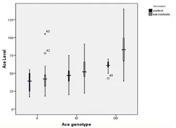
Serum ACE activity in patients and controls with genotypes separation is described in Table 1. The association between sACE activity in three geno-types of sarcoidosis patients and the control sub-jects was significant. DD and ID genotypes in controls and the patients with a maximum of mean value were observed. The significant differ-ence between the mean of sACE activity in sar-coidosis patients versus control groups (P<0.0001), and ID genotype (P=0.002) and DD genotype (P<0.0001) was observed by t-test. Fig. 2 show the relationship between the ACE and three genotypes in a clustered Box plot diagram. Genotypes and allele frequencies in sarcoidosis and control are shown in Table 2. In total, the genotype frequencies of II, ID and DD in the patients with sarcoidosis was 17%, 35.5% and 47.1%, respectively, and the frequency of D allele was 0.65. The frequency of both DD and ID ge-notypes in sarcoidosis and control among female and male generally showed no significant differ-ence (P=0.05) by chi-square test.
Table 1: Association of the ACE I/D polymorphism with sACE level in patients and controls
Variables Total Genotype
No
(mean±SD) II ANOVAID DD P value
n (%)
(mean±SD) (mean±SD) n (%) (mean±SD) n (%)
Sarcoidosis 102
(67.7±26.8) (44.4±21) 18 (17.6) 36 (35.2) (55.9±17.8) 48(47) (85.2±22.9) <0.001
Control 192
(47.5±12.6) (37.4±13.5) 25 (13) 135(70.3)(45.9±10.5) 32(16.6)(61.6±7.6) <0.001
95%CI 14.6-25.7 -4.5-18.5 3.73-16.26 16.4-30.7
<0.0001 0.22 0.002 <0.0001
Table 2: ACE genotype and allele frequencies in sarcoidosis patients compared with healthy controls
Variables Women Men Total
Case Ctrl P value† Case Ctrl P value Case Ctrl P-value
Genotype
II 12(21.1) 17(13.2) 0.17 6(13.3) 8(12.7) 0.9 18(17.6) 25(13) 0.2
ID 19(33.3) 90(69.8) <0.001 17(37.8) 45(71.4) <0.001 36(35.3) 135(70.3) <0.001 DD 26(45.6) 22(17.1) <0.001 22(48.9) 10(15.9) <0.001 48(47.1) 32(16.7) <0.001
IDorDD 45(78.9) 112(86.8) 0.17 39(86.6) 55(87.3) 0.9 84(82.3) 167(86.9) 0.2
P value‡ 0.2520 0.9228 0.3707
Allele
I 0.37 0.48 0.32 0.48 0.35 0.48
D 0.63 0.52 0.68 0.52 0.65 0.52
P value 0.11 0.02 0.06
Total 57 129 45 63 102 192
†P value of Genotype between case and control groups, ‡ P-value of Genotype between II and (ID or DD) genotypes
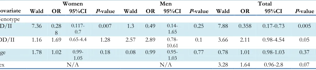
Logistic regression analysis, adjusting age and gender (Table 3) showed that the ID genotype to genotype II (OR = 0.35, 95% CI = 0.17-0.73, P=0.005) and the DD genotype to genotype II (OR = 2.11, 95% CI = 0.98-4.54, P<0.05) for sarcoidosis disease could be considering as a pre-dictor factor. Male gender does not reach statis-tical significance, and for the female gender-genotype II / ID has been associated with a re-duced risk of disease. (OR = 0.28, 95% CI = 0.11-0.7- P=0.007).
Discussion
Several studies have analyzed the relationship between ACE gene I/D polymorphism and sar-coidosis disease. The various regional and ethical studies have shown a significant correlation be-tween ACE genotypes and serum ACE level (15, 17, 25). The present study with the statistical
analysis of the relationship between angiotensin-converting enzyme (ACE) genotype and sACE level shows a significant association between ACE gene polymorphism and sACE activity in both patients with sarcoidosis and control sub-jects. In a Japanese study, sACE in comparison to controls showed a significant difference (DD = 29, 14%, ID = 101, 48.8%, II = 77, 37.2%) (18). The maximum sACE level was found in DD group among both sarcoidosis patients and controls.
Genotype distribution was the same in both pa-tients and controls group. In the American co-hort, Detroit Michigan, the ACE genotype fre-quency was according ID> DD> II order in African-American population patients with sar-coidosis.
Table 3:Binominal logistic regression covariate analysis of sarcoidosis
Covariate Wald OR 95%CI Women P-value Wald OR Men 95%CI P-value Wald OR Total 95%CI P-value
Genotype
ID/II 7.36 0.28
8
0.117-0.7 0.007 1.3 0.49 0.14-1.65 0.25 7.88 0.358 0.17-0.73 0.005
DD/II 1.16 1.69 0.65-4.4 1.28 2.57 2.89
0.78-10.61 0.1 3.66 2.11 0.98-4.54 0.05
Age 1.78 1.02
0.99-1.05 0.18 0.08 0.99 0.95-1.03 0.77 0.78 1.01 0.98-1.03 0.37
Sex N/A N/A 3.28 1.64 0.96-2.8 0.07
Another study from the UK and Czech popula-tion, the genotype frequencies of ID, DD and II was 47%, 28% and 25%, respectively in UK population. In Czech population, the genotype frequency order was ID>II>DD in the patients with sarcoidosis (20).
In Turkey cohort, as the same as our study, the predominant genotype was DD (45.7%), I/D genotype (42.8%), and I/I genotype was only seen in 11.4% of the patients. The frequency of D allele is 0.65 (P-value=0.06) that is much more comparable with the present study. Thus, the re-sults of current study are very close to Turkey report due to genetic affinity in this region, but the genotype frequency varies because of differ-ent populations. On the other hand, in Turkey study, the frequency of I/D genotype (53.6%) is higher in control subjects (27). Therefore, in oth-er studies (16) genotype distribution in patients and controls did not differ, whereas this frequen-cy varies in our study and other studies such as the American cohort in African-American popu-lation.
A study analyzed the families with more than one case of sarcoidosis. The over-representation of D/D genotype in sarcoidosis patients and their families can be seen in comparison to controls (28). On the other hand, attempts to show poly-morphism and severity of chronic disease have been done (20). Meanwhile, due to the nature of this disease, racial and regional differences in the polymorphism frequency, an association between sACE activity and sarcoidosis exists.
Therefore, to better understanding of the disease activity, its diagnosis and treatment, studies with
should be separately performed. In the current study, we tried to address ACE gene polymor-phism, serum ACE levels and its relationship to sarcoidosis with appropriate sample size. The difference in sACE levels in patients with sarcoi-dosis and controls was significant that is in ac-cordance with previous findings in this field. This level also in DD and ID genotypes showed sig-nificant differences between patients and con-trols. The level of sACE was higher in the DD genotype of both patient and control groups (Table 1). In a systematic review (29) that is ac-cordance with the present findings, the frequency of ID and DD genotypes in all cases and sepa-rately in males and females shows a significant difference (Table 2). In this study, weighted mean DD/II ratio was 1.85 for all studies, 2.01 for Caucasians and 1.64 for Asians (29). Besides, this general finding is theoretically justified. The var-iation in the results of photometric measure-ments is always seen. Polymorphism analysis can be considered in cases that sACE will be higher than 97.5 percentile for DD or II genotypes (29). Moreover, in the present study, the cases with the high level for sACE in II genotype patients are seen (Table 1). The results can be helpful to ob-tain the sensitivity, specificity and cut-off value for sACE and this outcome should be assessed, according to the region and race.
geno-not significant in men. However, in women with the ID genotype versus II, the risk of disease has decreased 0.3-fold.
However, this study has some limitations such as no clinical relationship with the sACE level and ACE genotype exists. This can be helpful in the interpretation of the relationship between the disease and the sACE level, meaning that how disease severity in different genotypes distributed and this fact that either ACE genotype only ef-fects on study result or severity of disease is im-portant too. We have not analyzed the cut off value of sACE level for this disease.
Conclusion
Although results of Insertion/Deletion polymor-phisms and sACE levels in different geographical areas do not follow the same pattern, these stu-dies improve our knowledge about the I/D po-lymorphism, s sACE levels and sarcoidosis In the future research, we attempt to to determine a cut off value for sACE level and sensitivity and spe-cificity of sACE level, according to I/D geno-type. Finally, we will introduce better models for diagnosis and prognosis of sarcoidosis by ex-amining other aspects of genetics and cytokines.
Ethical considerations
Ethical issues (Including plagiarism, informed consent, misconduct, data fabrication and/or fal-sification, double publication and/or submission, redundancy, etc.) have been completely observed by the authors.
Acknowledgments
The authors thank all colleagues who supported the current study.
References
1. James DG, Sharma OP (2002). From Hutchinson to now: a historical glimpse. Curr Opin Pulm Med,
8:416-423.
2. Gungor S, Ozseker F, Yalcinsoy M, Akkaya E, Can G, Eroglu H, Genc NS (2015). Conventional
markers in determination of activity of sarcoidosis. Int Immunopharmacol, 25:174-179. 3. Crommelin HA, Vorselaars AD, van Moorsel CH,
Korenromp IH, Deneer VH, Grutters JC (2014). Anti-TNF therapeutics for the treatment of sarcoidosis. Immunotherapy, 6:1127-1143. 4. Smith G, Brownell I, Sanchez M, Prystowsky S
(2008). Advances in the genetics of sarcoidosis.
Clin Genet, 73:401-412.
5. Sarcoidosis So (1999). Joint Statement of the American Thoracic Society (ATS), the European Respiratory Society (ERS) and the World Association of Sarcoidosis and Other Granulomatous Disorders (WASOG) adopted by the ATS Board of Directors and by the ERS Executive Committee, February 1999. Am J Respir Crit Care Med, 160:736-755.
6. Newman LS, Rose CS, Bresnitz EA, Rossman MD, Barnard J, Frederick M, Terrin ML, Weinberger SE, Moller DR, McLennan G (2004). A case control etiologic study of sarcoidosis: environmental and occupational risk factors. Am J Respir Crit Care Med, 170:1324-1330.
7. Song Z, Marzilli L, Greenlee BM, Chen ES, Silver RF, Askin FB, Teirstein AS, Zhang Y, Cotter RJ, Moller DR (2005). Mycobacterial catalase– peroxidase is a tissue antigen and target of the adaptive immune response in systemic sarcoidosis. J Exp Med, 201:755-767.
8. Dubaniewicz A, Dubaniewicz-Wybieralska M, Sternau A, Zwolska Z, Iżycka-Świeszewska E, Augustynowicz-Kopeć E, Skokowski J, Singh M, Zimnoch L (2006). Mycobacterium tuberculosis
complex and mycobacterial heat shock proteins in lymph node tissue from patients with pulmonary sarcoidosis. J Clin Microbiol, 44:3448-3451.
9. Iannuzzi MC, Maliarik MJ, Poisson LM, Rybicki BA (2003). Sarcoidosis susceptibility and resistance HLA-DQB1 alleles in African Americans. Am J Respir Crit Care Med, 167:1225-1231.
sarcoidosis with HLA DQB1* 0602. Hum Im-munol, 65:S34.
12. Grunewald J, Brynedal B, Darlington P, Nisell M, Cederlund K, Hillert J, Eklund A (2010). Different HLA-DRB1 allele distributions in distinct clinical subgroups of sarcoidosis patients. Respir Res, 11:25.
13. Society AT (1999). Statement on sarcoidosis. Am J Respir Crit Care Med, 160:736-755.
14. Furuya K, Yamaguchi E, Itoh A, Hizawa N, Ohnuma N, Kojima J, Kodama N, Kawakami Y (1996). Deletion polymorphism in the angiotensin I converting enzyme (ACE) gene as a genetic risk factor for sarcoidosis. Thorax,
51:777-780.
15. Kruit A, Ruven H, Grutters J (2010). Angiotensin II receptor type 1 1166 A/C and angiotensin converting enzyme I/D gene polymorphisms in a Dutch sarcoidosis cohort. Sarcoidosis Vasc Dif-fuse Lung Dis, 27:147-152.
16. Biller H, Ruprecht B, Gaede K (2009). Gene polymorphisms of ACE and the angiotensin receptor AT2R1 influence serum ACE levels in sarcoidosis. Sarcoidosis Vasc Diffuse Lung Dis,
26:139-146.
17. Arbustini E, Grasso M, Leo G, Tinelli C, Fasani R, Diegoli M, Banchieri N, Cipriani A, Gorrini M, Semenzato G (1996). Polymorphism of angiotensin-converting enzyme gene in sarcoidosis.
Am J Respir Crit Care Med, 153:851-854.
18. Tomita H, Ina Y, Sugiura Y, Sato S, Kawaguchi H, Morishita M, Yamamoto M, Ueda R (1997). Polymorphism in the angiotensin-converting enzyme (ACE) gene and sarcoidosis. Am J Respir Crit Care Med, 156:255-259.
19. Rybicki B, Maliarik M, Poisson L, Iannuzzi M (2004). Sarcoidosis and granuloma genes: a family-based study in African-Americans. Eur Respir J, 24:251-257.
20. McGRATH DS, Foley PJ, Petrek M, Izakovicova-Holla L, Dolek V, Veeraraghavan S, Lympany PA, Pantelidis P, Vasku A, Wells AU (2001). Ace gene I/D polymorphism and sarcoidosis pulmonary disease severity. Am J Respir Crit Care Med,164:197-201.
21. Pietinalho A, Furuya K, Yamaguchi E, Kawakami Y, Selroos O (1999). The angiotensin‐converting enzyme DD gene is associated with poor prognosis in Finnish sarcoidosis patients. Eur Respir J, 13:723-726. 22. Mihailovic-Vucinic V, Sharma OP (2006). Atlas of
Sarcoidosis: Pathogenesis, Diagnosis and Clinical Features. ed. Springer Science & Business Media. 23. Saiki RK, Gelfand DH, Stoffel S, Scharf SJ,
Higuchi R, Horn GT, Mullis KB, Erlich HA (1988). Primer-directed enzymatic amplification of DNA with a thermostable DNA polymerase.
Science, 239:487-491.
24. Rigat B, Hubert C, Corvol P, Soubrier F (1992). PCR detection of the insertion/deletion polymorphism of the human angiotensin converting enzyme gene (DCP1)(dipeptidyl carboxypeptidase 1). Nucleic Acids Res, 20:1433. 25. Alia P, Mana J, Capdevila O, Alvarez A, Navarro M
(2005). Association between ACE gene I/D polymorphism and clinical presentation and prognosis of sarcoidosis. Scand J Clin Lab Invest,
65:691-697.
26. Maliarik MJ, Rybicki BA, Malvitz E, Sheffer RG, Major M, Popovich Jr J, Iannuzzi MC (1998). Angiotensin-converting enzyme gene polymorphism and risk of sarcoidosis. Am J Respir Crit Care Med, 158:1566-1570.
27. Yilmaz D, Karkucak M, Coskun F (2012). ACE gene I/D polymorphism and risk of sarcoidosis development in Turkish patients. Tuberk Toraks,
60:201-206.
28. Schürmann M, Reichel P, Müller‐Myhsok B, Dieringer T, Wurm K, Schlaak M, Müller‐Quernheim J, Schwinger E (2001). Angiotensin‐converting enzyme (ACE) gene polymorphisms and familial occurrence of sarcoidosis. J Intern Med, 249:77-83.
29. Fløe A, Hoffmann HJ, Nissen PH, Møller HJ, Hilberg O (2014). Genotyping increases the yield of angiotensin-converting enzyme in sarcoidosis–a systematic review. Dan Med J,
61:A4815.