Themed Section: Science and Technology
A Review : Leaf Disease Detection Using Image Processing
Prof. Neha Chourasia, Mayuri Mahalle, Shubhangi Daduriya, Surbhi Lanjewar, Urvashi Giri
ETC Department, RTMNU, Nagpur, Maharashtra, India
ABSTRACT
Identification of the plant diseases is the key to preventing the losses in the yield and quantity of the agricultural product. The studies of the plant diseases mean the studies of visually observable patterns seen on the plant. Health monitoring and disease detection on plant is very critical for sustainable agriculture. In this paper we present an automatic detection of plant diseases using image processing techniques. The presented system is a software solution for automatic detection and computation of texture statistics for plant leaf diseases. The processing system consists of four main steps, first a color transformation structure for the input RGB image is created, then the green pixels are masked and removed using specific threshold value, then the image is segmented and the useful segments are extracted, finally the texture statistics is computed. From the texture statistics, the diseases, if present on the plant leaf are evaluated.
Keywords : ImageAcquisition, Pre-Processing, FeaturesExtraction, Classification, NeuralNetwork.
I.
INTRODUCTION
India is an agriculture country. 70% of India economy depends on agriculture. Due to environmental changes like huge rain fall, drastic changes in temp, the crops get infected. And that can be characterized by spots on the leaf, dryness of leaf, colour changes in leaf and defoliation. The maximum people can not be able to identify the disease easily and accurately. The proposed project leaf infection detection is made through image processing technique image because image from important data and information in biological science digital image processing and image analysis technology based on advance in micro electronics and computer has many applications in biology. The method for detection and classification of leaf diseases is based on masking and removing green pixels, applying a specific threshold extract to the infected region and computing the texture statistics to evaluate the disease using MATLAB.
II.
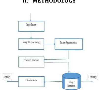
METHODOLOGY
Figure 1. Block Diagram of Proposed Method
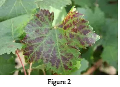
A. Input Image:
Figure 2 B. Image Preprocessing:
Image pre-process tasks are the initial stage before feature extraction. There are three steps of image preprocessing processing, i.e., image cropping, image converting and image enhancement. The image is cropped on leaf diseases area, and then converted to gray levels. To enhance the image we used laplacian filter.
Figure 3 C. Image Segmentation:
Image segmentation is one of the most important precursors for disease detection and has a crucial impact on the overall performance of the developed systems. The K-Means clustering technique is a well-known approach that has been applied to solve low-level image segmentation tasks. This clustering algorithm is convergent and its aim is to optimize the partitioning decisions based on a user-defined initial set of clusters Paper proposed k-means segmentation method to segment target areas. The area affected by the disease is the target area.
D. Feature Extraction:
Proposed method include two features color texture and space features. These features are total 17 in numbers including 13 color features and 4 shape features. Shape features including area, perimeter, circularity and complexity were extracted from the binary segmentation images.
Figure 4. Block Diagram
A. Color Transformation Structure :
First, the RGB images of leaves are converted into Hue Saturation Intensity (HSI) color spacerepresentation. The purpose of the color space is to facilitate the specification of colors in some standard, generally accepted way. HSI (hue, saturation, intensity) color model is a popular color model because it is based on human perception.
B. Hue Color Attribute:
It refers to the dominant color as viewed by a person. Saturation refers to the relative Purity or the amount of white light added to hue and intensity refers to the amplitude of the light. Color spaces can be converted from one space to another easily. After the transformation process, the H component is taken into account for further analysis. S and I are dropped since it does not give extra information.
C. Masking Green Pixels:
intensity is less than the precomputed threshold value, the red, green and blue components of the this pixel is assigned to a zero value.
D. Segmentation:
From the above steps, the infected portion of the leaf is extracted. The infected region is then segmented into a number of patches of equal size. The size of the patch is chosen in such a way that the significant information is not lost. In this approach patch size of 32*32 is taken. The next step is to extract the useful segments. Not all segments contains significant amount of that information. So the patches which are having more than fifty percent of the information are taken into account for the further analysis.
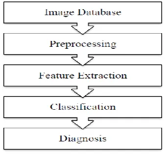
Figure 5. Flow Chart
A. Image Acquisition:
The images of the plant leaf are captured through the camera. This image is in RGB (Red, Green And Blue) form. Color transformation structure for the RGB leaf image is created, and then, a device-independent color space transformation for the color transformation structure is applied.
B. Image Pre-processing:
To remove noise in image or other object removal, different pre-processing techniques is considered. Image clipping i.e cropping of the leaf image to get the interested image region. Image smoothing is done using the smoothing filter. Image enhancement is carried out for increasing the contrast.the RGB images
into the grey images using color conversion using equation f(x)=0.2989*R + 0.5870*G +
0.114.*B - - - (1)
Then the histogram equalization which distributes the intensities of the images is applied on the image to enhance the plant disease images. The cumulative distribution functions used to distribute intensity values.
C. Image Segmentation:
Segmentation means partitioning of image into various part of same features or having some similarity. The segmentation can be done using various methods like otsu” method, k-means clustering, converting RGB image into HIS model etc.
1. Segmentation using Boundary and spot detection algorithm:
The RGB image is converted into the HIS model for segmenting. Boundary detection and spot detection helps to find the infected part of the leaf as discussed. For boundary detection the 8 connectivity of pixels is consider and boundary detection algorithm is applied.
2. K-means clustering:
The K-means clustering is used for classification of object based on a set of features into K number of classes. The classification of object is done by minimizing the sum of the squares of the distance between the object and the corresponding cluster. The algorithm for K –means Clustering:
1. Pick center of K cluster, either randomly or based on some heuristic.
2. Assign each pixel in the image to the cluster that minimizes the distance between the pixel and the cluster center.
III.
FUTURE SCOPE
The proposed method uses mobile cams for capturing the diseased images and does not require any kind of special training and sophisticated capturing devices. The proposed method is
(i) fully automatic for ROI calculation, background separation and parameter evaluation disease independently, (ii) low cost and possibility for the wide usability in field conditions, (iii) simpler segmentation method and more advanced parameters are used. We have developed a fully automatic color image sensing based system for classifying the four most dangerous soya bean foliar infections, namely bacterial blight, frog’s eye, brown spot, and soya bean rust. All four infections have similar color shades and are confusing for a non-plant pathologist. An algorithm was developed to find there fined lesion texture histogram and apply the DCT on statistical features of RLTH, followed by a normalization process. We develop a ST-NDCT based hybrid feature descriptor for lesion areas, and proved the suitability of using the same for classifying the infections under consideration.
The methodology has been implemented successfully and performance tested on a real set of soya leaf data. The result is quite convincing and wide adaptability in developing countries, where such information plays an important role for improvement in yield.
IV.
CONCLUSION
Main approach of our paper is to recognize diseases on the leaf. At first preprocessing is done which include two steps gray conversion Second stage is k-means based Image segmentation which eventually does image analysis. Third stage is feature extraction that include color features and shape features. And after that classification of diseases is performed victimization our projected formula. The goal of this
analysis work is to develop Advance automatic data processing system which will determine the illness affected on a part of a leaf spot by victimization the image analysis technique. Prediction of the diseases and cuss recommendation is finished. The producers will amend the Yield and scale back the loss. Through this projected system the farmers' burden has been reduced and saves their life. perform better than others. Accuracy of detection can be increased when using SVM classifier with more number of features included to it.
V.
REFERENCES
[1].Sandeep B. Patil& Santosh Kumar Sao „"AN IMPROVED LEAF DISEASE DETECTION USING COLLECTION OF FEATURES AND SVM CLASSIFIERS".
[2].RenukaRanjedraKajale „"DETECTION & RECOGNIZATION OF PLANT LEAF DISEASES USING IMAGE PROCESSING AND ANDROID SYETEM".
[3].Ms. Chithra J L (Government college) „"LEAF INFECTION DETECTION AND DIAGNOSIS USING IMAGE PROCESSING TECHNIQUE. [4].Ms. Arti N. Rathod ,Computer Engineering
,Gujarat (Technical University) „"IMAGE PROCESSING TECHNIQUES FOR DETECTION OF LEAF DISEASES".
[5].Mrs. Jayme Garcia ArnalBarbedo Digital image processing techniques for detecting, quantifying and classifying plant diseases(SPRINGER PLUS-2013.
[6].SmitaNaikwadi, NiketAmoda, "advances in image processing for detection of plant diseases" international journal of application or innovation in engineering & management, PP: 168-175, November 2013.
Probabilistic Neural Network" IEEE ICACSIS 2013.
[8].Al-Bashish, D., M. Braik, and S. Bani-Ahmad. 2011. "Detection and classification of leaf diseases using Kmeans- based segmentation and neural networks based classification". Information Technology Journal, 10(2): 267-275.