O R I G I N A L A R T I C L E
The application of DNA sequence data for the identification
of benthic nematodes from the North Sea
Philipp Vogt•Maria Miljutina•Michael J. Raupach
Received: 13 May 2014 / Revised: 1 September 2014 / Accepted: 3 September 2014 / Published online: 19 September 2014 ÓSpringer-Verlag Berlin Heidelberg and AWI 2014
Abstract Nematodes or roundworms represent one of the most diverse and dominant taxon in marine benthic habi-tats. Whereas a morphological identification of many spe-cies is challenging, the application of molecular markers represents a promising approach for species discrimination and identification. In this study, we used an integrative taxonomic approach, combining both molecular and mor-phological methods, to characterize nematodes of distinct sex and ontogenetic stages from three sampling sites of the North Sea. Morphospecies were discriminated after first visual determination, followed by a molecular analysis of the nuclear 28S rDNA: D2–D3 marker. By linking each sequence to a morphological voucher, discordant mor-phological identification was subjected to a so-called reverse taxonomic approach. Molecular operational taxo-nomic units (MOTUs) and morphospecies were compared for all of the three sampling sites to assess concordance of methodology. In total, 32 MOTUs and 26 morphospecies were assigned, of which 12 taxa were identified as described species. Both approaches showed high concor-dance in taxon assignment (84.4 %) except for a cluster comprising variousSabatieriaspecies. Our study revealed the high potential of the analyzed fragment as a useful molecular marker for the identification of the North Sea
nematodes and highlighted the applicability of this com-bined taxonomic approach in general.
Keywords 28S rDNA: D2–D3Molecular operational taxonomic unitsMolecular species identification
Sabatieria Wadden Sea
Introduction
Nematodes dominate the marine meiobenthos both in abundance and biomass (Soetaert et al. 1995; Giere 2009). Occupying a broad spectrum of trophic positions, nematodes are of great ecological importance, featuring high production efficiencies (Heip et al. 1990) and rep-resenting a crucial component in food webs and nutrient cycles by linking the micro- and macrofauna (e.g. Chardy and Dauvin 1992). Given their high diversity and ubiq-uitous appearance, their total number of species is still unknown (Hugot et al. 2001; Coomans 2002; Blaxter 2003). As consequence of a conserved and quite simple body plan, morphological diagnostics are difficult and laborious and in most cases restricted to experienced taxonomists. Many species can only be identified as adult males or specific female structures (Floyd et al. 2002), whereas it is almost impossible to identify juveniles or damaged specimens, resulting in a disdain of nematodes in most infaunal studies (Warwick and Robinson 2000). Moreover, natural variation as consequence of phenotypic plasticity (Kiontke and Fitch 2010) as well as cryptic and/or sibling species (Derycke et al. 2008, 2013) com-plicates a correct species identification based on mor-phology in many cases.
In order to overcome these limitations, molecular methods may represent a useful alternative or at least Communicated by H.-D. Franke.
P. VogtM. MiljutinaM. J. Raupach (&)
German Centre for Marine Biodiversity, Senckenberg am Meer, Su¨dstrand 44, 26382 Wilhelmshaven, Germany
e-mail: [email protected]
Present Address: P. Vogt
Museum fu¨r Naturkunde Berlin, Invalidenstraße 43, 10115 Berlin, Germany
supplementary approach for the identification of nema-todes. In this context, the International Barcode of Life initiative introduced the concept of DNA barcoding (He-bert et al.2003a,b), using a small specific genomic region as so-called ‘‘DNA barcode’’. The term ‘‘DNA barcoding’’ sensu stricto is generally used for the application of the small fragment of the mitochondrial cytochromecoxidase subunit I gene (COI) for animals (e.g. Hebert et al.2003a, b; Blaxter 2004). In the case of nematodes, this marker showed only a low applicability due to high sequence variability in the primer regions (Bhadury et al.2006a,b; Elsasser et al. 2009). Nevertheless, some newly designed COI primers that promised better amplification quotes have been tested for some selected free-living marine species (Derycke et al.2010a). Consequently, the application of a number of alternative nuclear DNA sequence markers has become popular for the identification of nematodes. These markers include various hypervariable expansion regions of the nuclear 18S (e.g. Blaxter et al.1998; Bhadury et al. 2006a, b; Creer et al. 2010) and 28S rRNA genes (e.g. Litvaitis et al.2000; De Ley et al.2005; Pereira et al.2010) as well as internal transcribed spacers (ITS; Hugall et al. 1999; Elbadri et al.2002; Fe´lix et al.2014). Here, various studies showed that the D2–D3 region of the 28S rDNA represents one of the most suitable and promising markers for a valid nematode identification, which can be used to delimitate even closely related species (De Ley et al.2005; Pereira et al.2010).
However, the combination of molecular and morpho-logical approaches has been shown to be most effective in species identification (De Ley et al.2005) as well as dis-entangling species complexes within the Nematoda in general (e.g. Derycke et al.2008; Gutie´rrez-Gutie´rrez et al. 2013); for example, Pereira et al. (2010) successfully identified nematodes from Baja California using partial 18S and 28S rDNA sequence data in combination with morphological analysis. Other studies successfully used a variety of markers (partial 28S rDNA, 18S rDNA, ITS and COI) in combination with morphological analysis to reveal the cryptic character and taxonomic status of morphospe-cies (Derycke et al.2008,2010b). An even more integra-tive approach was performed by Fonseca et al. (2008), combining molecular, multivariate morphometric, typo-logical taxonomic and interbreeding data. All these inte-grative studies revealed the applicability and, to a certain degree, concordance between morphospecies which rely on morphological species concepts, and so-called sequence-based molecular operational taxonomic units (MOTUs) (Blaxter et al.2005).
The aim of our study was a combined application of morphological and molecular methods as part of an inte-grative taxonomic approach to identify free-living benthic
nematode species of the North Sea. We also tested the concordance of both approaches for the valid identification of male, female, juvenile and/or damaged specimens. Specimens were assigned to morphospecies, followed by a molecular analysis testing the quality of the D2–D3 region of the nuclear 28S rDNA for successful species identification.
Materials and methods
Sampling
All samples were taken between April 2012 and May 2012 from an intertidal mudflat in Wilhelmshaven (sampling site 1), a sublittoral site located in the Jade Bay (sampling site 2), and one sublittoral site near the island Wangerooge in the North Sea close to the German coast (sampling site 3; Table1). For sampling site 1, the topmost 2–3 cm of the sediment was removed with a spoon. All sublittoral sam-ples were taken by a multicorer (MUC, ø 105 mm) where the topmost 5 cm were removed from each tube. All sed-iment samples were immediately preserved in DESS (Yo-der et al.2006).
In total, 246 nematodes were extracted from the sedi-ment by a procedure of multiple sieving and subsequent centrifugations. A combination of LevasilÒ and Kaolin (Al2O32SiO22H2O) was added to separate the sediment
pellet from the organisms in the liquid phase (see McIntyre and Warwick1984). Organisms were rinsed with tap water Table 1 Sampling data and site properties
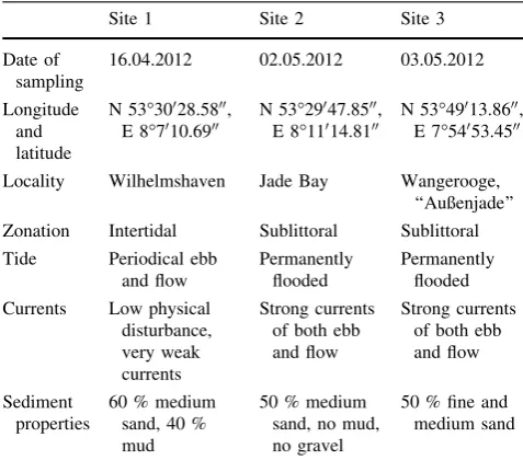
Site 1 Site 2 Site 3
Date of sampling
16.04.2012 02.05.2012 03.05.2012
Longitude and latitude
N 53°30028.5800, E 8°7010.6900
N 53°29047.8500, E 8°11014.8100
N 53°49013.8600, E 7°54053.4500
Locality Wilhelmshaven Jade Bay Wangerooge, ‘‘Außenjade’’ Zonation Intertidal Sublittoral Sublittoral Tide Periodical ebb
and flow
Permanently flooded
Permanently flooded
Currents Low physical disturbance, very weak currents
Strong currents of both ebb and flow
Strong currents of both ebb and flow
Sediment properties
60 % medium sand, 40 % mud
50 % medium sand, no mud, no gravel
50 % fine and medium sand
on a sieve of 40-lm mesh size and transferred into storage containers filled with DESS solution. Nematodes were hand-picked by use of a binocular (Leica M125) and rinsed three times with sterile water to remove traces of DESS. Single specimens were transferred into glycerin solution (79 % H2O, 20 % absolute ethanol (96 %), 1 % glycerin;
modified after Riemann1988) and followed by the method of slow evaporation used for further preparation.
Species identification, vouchering and DNA extraction Based on successful amplification and sequencing, 151 single nematode specimens were preserved in temporary slides using glycerin and paraffin. All specimens were identified to be morphospecies using a microscope (Leica DM 2500) and appropriate taxonomic literature (Platt and Warwick1983;1988; Warwick et al.1998) as well as with reference to the NeMys data base (Vanaverbeke et al. 2014). For subsequent vouchering and molecular studies, each specimen was removed from its slide and put onto a new slide provided with 5ll of so-called Worm Lysis Buffer (WLB, composed of 50 mM KCl solution, 10 mM Tris pH 8.3, 2.5 mM MgCl2, 0.45 % Tergitol solution
(NP40) and 0.45 % Tween 20, modified after Williams et al. 1992; and International Seabed Authority, 2011). Using two needles, the specimens were cut into several pieces. Anterior and posterior parts were isolated and kept in permanent slides. Additionally, high-quality prepara-tions of whole animals were kept for each morphospecies as vouchers to facilitate a closer subsequent inspection. For this second and more detailed identification, each specimen was sketched and measured using a camera lucida for the identification to species level, using the previously men-tioned literature. Gender (male, female) and, in the case of juveniles, the development stage was determined and documented. Total length, excluding the tail, and maxi-mum body diameter were measured for each nematode. For DNA extraction, another 15ll of WLB and 2ll of pro-teinase K (Macherey & Nagel, Du¨ren, Germany) were added to the remaining body fragments. Following an additional centrifugation, extracts were kept at -80°C overnight and incubated for 1 h at 65°C, followed by 10 min at 95°C. After a final centrifugation, all extracts were stored at-20°C.
PCR performance and sequencing
The primer pair D2A and D3B (De Ley et al.2005) was used for the amplification of the 28S rDNA: D2–D3 region. Amplicons were generated using PCR beads (illustra pu-ReTaq Ready-To-Go PCR Beads, GE Healthcare UK Limited, Buckinghamshire, United Kingdom) in 25-ll reactions, filled up with 2ll of DNA extract, 0.5ll of
primer D2A (20 pm/ll), 0.5ll of primer D3B (20 pm/ll) and 22 ll of ddH20. Negative and positive controls were
provided for each round of reactions. PCRs were per-formed by providing an initial denaturation at 95°C (5 min), followed by 40 cycles at 94 °C (denaturation, 30 s), 55°C (annealing, 1 min), 72°C (elongation, 2 min) and a final elongation at 72°C for 10 min. After verifica-tion in a 1 % agarose gel stained with GelRed (Biotium Inc., Hayward, USA), PCR products were purified for sequencing using the ExoSap purification kit (Thermo Scientific, Osterode, Germany). Tenll of the PCR product was mixed with 2ll of FastAp (Thermosensitive Alkaline Phosphatase, 1 U/ll) and 0.5ll of Exo I (Exonuclease I, 20 U/ll) on ice, and run on a thermocycler (37°C for 15 min, 85°C for 15 min). Purified amplicons were sequenced using the PCR primers in both directions at contract sequencing facilities (Macrogen Europe, Amster-dam, Netherlands, or GATC Biotech AG, Cologne, Ger-many). All morphological vouchers and DNA extracts were stored as part of the voucher collection of the AG ‘‘Molecular Taxonomy of marine Organisms’’ at the Ger-man Center of Marine Biodiversity (DZMB), Senckenberg am Meer, Wilhelmshaven. Chromatograms were assem-bled, edited and checked manually with the Geneious package (Geneious Pro 5.4.6, Biomatters, Auckland, New Zealand). Ambiguities were corrected according to chro-matogram properties, e.g. the presence and quality of peaks.
MOTU definition
All sequences were aligned in MEGA 5.2.2 (Tamura et al. 2011) using the implemented MUSCLE algorithm (Edgar 2004) with default settings. The alignment was checked manually and by eye for sequence differences as well as ambiguities by comparison with the corresponding chro-matograms in Geneious. Following previous studies, sequences differing by\3 base pairs (bp) were designated as a MOTU (see Floyd et al.2002for details). A neighbor joining phylogram (Saitou and Nei 1987) with nonpara-metric bootstrap replicates (n =1,000) (Felsenstein1985) using default settings (pdistances, pairwise deletions), and calculation of p distances within and between MOTUs of the same genera (default settings, pairwise deletions) were performed using MEGA. Using BLAST (Zhang et al.2000; Morgulis et al.2008), each MOTU was compared with the sequence library of GenBank (NCBI) to exclude contaminations.
Data analyses
congruence of both approaches. All results apart from congruence were classified as ‘‘splittings’’ and ‘‘lumpings’’ (see Thormann et al.2011). Whereas ‘‘lumping’’ describes a morphospecies that is represented by two or several MOTUs, ‘‘splitting’’ specifies a MOTU that is represented
by two or more morphospecies (e.g. Thormann et al.2011). A successful accordance was given if a specific MOTU corresponded to a specific morphospecies, indicating that one MOTU contained all members of one morphospecies, exclusively.
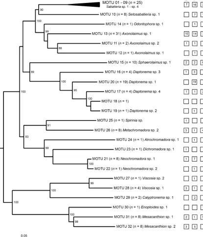
Fig. 1 Unrooted NJ-phylogram showing congruence of MOTU and morphospecies assignment. The phylogram was created by aligning single representative sequences from each MOTU cluster expect for MOTU 1–9 (see Fig.2 for a detailed subtree). Morphospecies are shown behind each MOTU. The number of sequences clustered into a
Results
Morphological assignment and species identifications The morphological identification of 151 specimens revealed 26 morphospecies. Out of these, 86 (57 %) were female, 36 male (23.8 %) and 28 juvenile (18.5 %). One of the vouchers was lost, so that no morphological assignment was performed. Each morphospecies com-prised between one and 31 individuals (Fig.1). It should be noted that the classification of some morphospecies was based on females or juveniles only, lacking impor-tant traits for a valid identification. In this context, the classification of Odontophora sp. 1, Axonolaimus sp. 1, Daptonema sp. 2, Atrochromadora sp. 1, Neochrom-adora sp. 2 and Viscosia sp. 2 was only based on one female specimen, whereas Calyptronema sp. 1 was rep-resented by two females, and Daptonema sp. 1 by 19 females. In the case of Enoploides, only one juvenile was given. Furthermore, most specimens of the found Sabatieria morphospecies were females: Four individu-als classified asSabatieriasp. 1 were females, one was a male and one was a juvenile. The morphospecies Sabatieriasp. 2 included six females whereasSabatieria sp. 3 was assigned by six female specimens and three males, and Sabatieria sp. 4 by three males and one juvenile.
Twelve morphospecies were determined as described species by morphological identification. The assignment of morphospecies to species is summarized in Table2. It should be noted that two species were found to bespecies affinis, indicating that species were related to the binomial species name indicated and showed a similar morphologi-cal appearance but were not identimorphologi-cal with it.
Consistency of approaches
The majority of the analyzed specimens showed congru-ence in MOTU and morphospecies assignment (n=126 or 80.8 %, Fig.1). The genus Daptonema Cobb, 1920 was represented by four morphospecies and corresponding MOTUS. Due to the loss of the corresponding voucher, it was not possible to correlate a fifth MOTU (MOTU 18) to a morphospecies. Consequently, this MOTU was classified as a member of theDaptonema morphospecies cluster on the basis of sequence properties only. However, it was possible to differentiate all remaining four morphospecies by a corresponding sequence (Fig.1). This was also true for the morphospecies of Axonolaimus de Man, 1889, Viscosiade Man, 1890,NeochromadoraMicoletzky, 1924 andMesacanthionFilipjev, 1927, which were represented by two morphospecies (Fig.1). In total, this study covered nine different families of five different orders of the Nematoda.
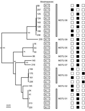
For the genusSabatieriaRouville, 1903, our molecular data revealed more MOTUs than previously defined four morphospecies (Fig.2). This is the case for MOTU 8 and MOTU 9 as well as MOTU 4 and MOTU 5. Both latter ones featured genetically distinct but closely related sequences and exhibited identical morphological assign-ment (Fig. 2). In contrast to this, a lumping was found for morphospecies 3, designated asSabatieria celtica (South-ern, 1914), whose sequences were differentiated as MOTU 8 and MOTU 9. Morphospecies 2 was represented within MOTU 1, MOTU 4 and MOTU 5. Furthermore, MOTU 1, MOTU 3, MOTU 6 and MOTU 7 contained sequences labeled as morphospecies 1, whereas morphospecies 4 was differentiated as MOTU 2 and MOTU 3 (Fig.2). Moreover, MOTU 1 contained sequences labeled as morphospecies 1 Table 2 Identified nematode
species based on a combination of morphological
(morphospecies) and molecular data (MOTU) to species level, with corresponding accession numbers of the 28S rDNA: D2– D3 fragment
Morphospecies Identified species MOTU Accession
number
Sabatieriasp. 3 Sabatieria celticaSouthern, 1914 MOTU 8 KC755203 Setosabatieriasp. 1 Setosabatieria hilarulade Man, 1922 MOTU 10 KC755205 Metachromadorasp. 2 Chromadoropsis viviparade Man, 1907 MOTU 26 KC755217 Spiriniasp. Spirinia parasitiferaBastian, 1865 MOTU 25 KC755216
Mesacanthionsp. 1 Mesacanthion diplechmaSouthern, 1914 MOTU 31 KC755226 Sphaerolaimussp. 1 Sphaerolaimus hirsutusBastian, 1865 MOTU 15 KC755210 Viscosiasp. 1 Viscosia elegansFilipjev, 1922 MOTU 28 KC755223 Calyptronemasp. 1 Calyptronema maxweberide Man, 1922 MOTU 29 KC755224 Axonolaimussp. 1 Axonolaimus paraspinosusSchuurmans,
Stekhoven & Adam, 1931
MOTU 12 KC755209
and 2 and MOTU 3 included so defined morphospecies 4 and 1.
Discussion
Choice of marker and implications
Our results clearly demonstrated a successful species dis-crimination using the 28S rDNA: D2–D3 marker for the analyzed nematode species. The amplification success of the used marker (about 62 %) is comparable to previous studies (i.e. 67 % in Pereira et al.2010). Earlier studies using the same fragment reported ‘‘unreliable’’ amplification results
(Bhadury et al.2006a) as well as very high success rates (De Ley et al.2005). Due to the fact that a marker for molecular species identification should be easy to amplify, PCR opti-mization represents an essential task for further studies. Problems in sample and specimen preservation and thus the degradation of DNA cannot be excluded as reasons for limited PCR success and should be optimized also. Fur-thermore, sequencing problems may be caused by the putative forming of primer dimers when using the PCR primer pair D2A and D3B for direct sequencing (GATC Biotech AG, European Custom Sequencing Centre, Cologne, Germany; personal communication).
A special characteristic of the D2–D3 region and ribo-somal expansion segments in general is given by the high Fig. 2 Subtree of the analyzed
Sabatieriaspecimens correlated with morphospecies information and corresponding MOTUs. Numbers at the tips of the branchesindicate specific sequence numbers.
Corresponding morphospecies assignment of each sequence is given inrectangles(sp. 1–4). Barsindicate specimens of the corresponding MOTU.Black boxesreveal the gender of the adult specimens (leftmales, centralfemales) or
proportion of variable sites as consequence of insertions and deletions, which are recognizable when observing the secondary structure of the complete 28S rRNA (Bae et al. 2010). Variation in secondary structures as stems, loops, single and double strands can affect the probability for mutations and therefore the variability within sites of the fragment significantly (Subbotin et al. 2007). Moreover, homology of expansion segments cannot be assumed between far-related species (e.g. Vogler et al. 1997; Gillespie et al.2004; Raupach et al.2010), making align-ments of these fragalign-ments difficult to impossible. Whereas this putative disadvantage of the rDNA genes limits their use in phylogeny drastically (e.g. De Ley et al. 2005; Schmidt et al. 2006; Xie et al. 2009), this fact can be interpreted as beneficial according for the identification of even closely related species (e.g. Sonnenberg et al. 2007; Raupach et al. 2010; Khalaji-Pirbalouty and Raupach 2014). In terms of marine nematodes, some studies pro-posed that the D2–D3 domain can be used for DNA species identification as well as phylogenetic analyses (e.g. Dery-cke et al.2008).
Based on sequence data, specimens can be identified and assigned to a known species when a close sequence match in a public sequence data base is given. For our data, only one match was found in GenBank [Calyptronema max-weberi(de Man 1922), Acc. No. AF210399], revealing the relatively low number of published nematode LSU sequences (n =11,300; NCBI, 21.08.2014) including many sequences of the same species (e.g.Caenorhabditis elegansMaupas, 1900=1,027, orUnicaria lucasi Stiles, 1901=141) in comparison with the total number of described nematode species ([25,000; Zhang2013). Even morphologically well-studied and common species of the intertidal and sublittoral areas of the North Sea are still seriously underrepresented.
Integrated taxonomy
Except for theSabatieriacluster it was possible to link all obtained MOTUs to a specific morphospecies of five dif-ferent orders. In case of the genusDaptonema, we were able to demonstrate the value of combining molecular results with a revision of corresponding vouchers: The most ana-lyzed specimens were female and showed almost no diag-nostic traits, e.g. spicules or a gubernaculum, and many specimens were also found in a bad preservation state. A comparison of these specimens with vouchers of a better condition and quality as result of identical sequences allowed assignment of these nematodes to the correspond-ing morphospecies. For our data, MOTUs have been shown not to be artificial units but to correspond to taxon assign-ment by morphological manners in most cases. Based on these considerations, referenced sequence libraries or
stepping-stone sequences (Blaxter 2004) will improve the quality of public databases and overcome the gap between large scale studies, which are restricted to molecular data only. Therefore, DNA sequence data should become part of new species descriptions. Such data can allow ecologists and taxonomists a more efficient handling of the over-whelming abundance and variety of nematode specimen (Fonseca et al. 2010) and may significantly advance knowledge and progress in meiofaunal research (Blaxter and Floyd 2003; Markmann and Tautz 2005; Creer et al. 2010) by revealing new perspectives to the understanding of ecological and taxonomic identity, rank abundance as well as concomitant trophic dynamics (Fonseca et al.2010). The Sabatieriaspecies complex
The molecular and morphological analysis of the analyzed Sabatieriaspecimens revealed a discrepancy between both approaches. MOTUs comprised sequences of individuals which were assigned to different morphospecies. Even an extensive morphological revision of the voucher specimens was not able to establish congruence between both approaches. This might be due to the variable character of many traits or difficulties in identifying them. Most ana-lyzed specimens of the genus Sabatieria were females (66.6 %) or juveniles (8.3 %). Furthermore, a majority of these individuals were in bad condition, complicating a closer inspection of taxonomically important morphologi-cal traits. In case that the given morphospecies classifica-tion was correct, our results clearly revealed the lack of diagnostic traits of many morphospecies. Many species of this genus are classified primarily by anatomical structures found in males, e.g. the gubernaculum or the precloacal supplements (Platt1985; Platt and Warwick1988), making the identification of juveniles or females impossible. Interestingly, the genus Sabatieria itself is defined by a combination of plesiomorphic traits as well as the lack of other characteristics that are present within other genera of the family of the Comesomatidae (Platt 1985). The genus itself is very diverse, including over 100 described species (WoRMS 2014). Specimens of Sabatieria represent some of the most common free-living nematodes in marine benthic habitats (Platt1985). Therefore it is not surprising that most species suffer from insufficient descriptions and diverse synonymizations (Platt 1985). As a consequence, this genus has been subjected to various more or less comprehensive revisions (Jensen 1979; Platt 1984, 1985; Vincx1986). Furthermore, Sharma et al. (2006) found in a study combining morphological and molecular methods that several characters used for classification of the Comesomatidae in general have to be reevaluated.
species delimitation and identification within this enig-matic genus. Specimens can be collected and identified to morphospecies on first sight. Subsequently, morphological vouchers of diagnostically important traits must be kept. In addition, digital vouchers can be made according to methods already described (De Ley et al. 2005). This method allows extensive revision of the available vouchers after molecular analysis of corresponding specimen.
Summary
Our results confirmed the effectiveness of integrative tax-onomic approaches for nematode species differentiation using a single molecular marker. The sequence analysis of distinct MOTUs facilitated the concept of reverse taxon-omy with corresponding vouchers, allowing the identifi-cation of juvenile specimen, females or damaged specimens. Furthermore, the given amplification and sequencing results of the 28S rDNA: D2–D3 marker underlined the need for PCR and sequencing optimization.
Acknowledgments We thank the crew of FS Senckenberg for their assistance in taking the sublittoral samples. We would also like to thank Prof. Dr. Pedro Martinez Arbı´zu (DZMB, Senckenberg am Meer) and Prof. Dr. Liliane Rueß (Institute for Biology, Humboldt Universita¨t zu Berlin) for their constructive ideas in data analysis as well as Dr. Adam Kubicki (Marine Sedimentology, Senckenberg am Meer) and Dipl.-Biol. Alexandra Markert (Actuopalaeontology, Senckenberg am Meer) for providing and discussing environmental data. We thank Dr. Thomas Knebelsberger, Dr. Silke Laakmann and Dr. Dmitry Miljutin (all DZMB, Senckenberg am Meer) for their assistance in statistical analysis and helpful comments. Finally, we are grateful to Valeska Borges and Corinna Girschik for helping in the laboratory. This study was funded by the Federal Ministry of Edu-cation and Research of Germany (Grant No. 03F0499A) and the Land Niedersachsen.
References
Bae CH, Robbins RT, Szalanski AL (2010) Secondary structure models of D2–D3 expansion segments of 28S rRNA for Hoplolaiminae species. J Nematol 42:218–229
Bhadury P, Austen MC, Bilton DT, Lambshead PD, Rogers AD, Smerdon GR (2006a) Development and evaluation of a DNA-barcoding approach for the rapid identification of nematodes. Mar Ecol Prog Ser 320:1–9. doi:10.3354/meps320001
Bhadury P, Austen MC, Bilton DT, Lambshead PD, Rogers AD, Smerdon GR (2006b) Molecular detection of marine nematodes from environmental samples: overcoming eukaryotic interfer-ence. Aquat Microb Ecol 44:97–103. doi:10.3354/ame044097 Blaxter M (2003) Counting angels with DNA. Nature 421:122–124.
doi:10.1038/421122a
Blaxter ML (2004) The promise of a DNA taxonomy. Philos Trans R Soc B 359:669–679. doi:10.1098/rstb.2003.1447
Blaxter M, Floyd R (2003) Molecular taxonomics for biodiversity surveys: already a reality. Trends Ecol Evol 18:268–269. doi:10. 1016/S0169-5347(03)00102-2
Blaxter ML, De Ley P, Garey JR, Liu LX, Scheldeman P, Vierstraete A, Vanfleteren JR, Mackey LY, Dorris M, Frisse LM, Vida JT, Thomas WK (1998) A molecular evolutionary framework for the phylum Nematoda. Nature 392:71–75. doi:10.1038/32160 Blaxter M, Mann J, Chapman T, Thomas F, Whitton C, Floyd R,
Abebe E (2005) Defining operational taxonomic units using DNA barcode data. Philos Trans R Soc B 360:1935–1943. doi:10.1098/rstb.2005.1725
Chardy P, Dauvin J-C (1992) Carbon flows in a subtidal fine sand community from the western English channel: a simulation analysis. Mar Ecol Prog Ser 81:147–161. doi:10.3354/ meps081147
Coomans A (2002) Present status and future of nematode systematics. Nematology 4:573–582
Creer S, Fonseca VG, Porazinska DL, Giblin-Davis RM, Sung W, Power DM, Packer M, Carvalho GR, Blaxter ML, Lambshead PJD, Thomas WK (2010) Ultrasequencing of the meiofaunal biosphere: practice, pitfalls and promises. Mol Ecol 19:4–20. doi:10.1111/j.1365-294X.2009.04473.x
De Ley P, De Ley IT, Morris K, Abebe E, Mundo-Ocampo M, Yoder M, Heras J, Waumann D, Rocha-Olivares A, Jay Burr AH, Baldwin JG, Thomas WK (2005) An integrated approach to fast and informative morphological vouchering of nematodes for applications in molecular barcoding. Philos Trans R Soc B 360:1945–1958. doi:10.1098/rstb.2005.1726
Derycke S, Fonseca G, Vierstraete A, Vanfleteren J, Vincx M, Moens T (2008) Disentangling taxonomy within the Rhabditis (Pel-lioditis)marina(Nematoda, Rhabditidae) species complex using molecular and morhological tools. Zool J Linn Soc Lond 152:1–15. doi:10.1111/j.1096-3642.2007.00365.x
Derycke S, Vanaverbeke J, Rigaux A, Backeljau T, Moens T (2010a) Exploring the use of cytochrome oxidase c subunit 1 (COI) for DNA barcoding of free-living marine nematodes. PLoS One 5:e13716. doi:10.137/journal.pone.0013716
Derycke S, De Ley P, De Ley IT, Holovachov O, Rigaux A, Moens T (2010b) Linking DNA sequences to morphology: cryptic diver-sity and population genetic structure in the marine nematode Thoracostoma trachygaster(Nematoda, Leptosomatidae). Zool Scr 39:276–289. doi:10.1111/j.1463-6409.2009.00420.x Derycke S, Backeljau T, Moens T (2013) Dispersal and gene flow in
free-living marine nematodes. Front Zool 10:1. doi:10.1186/ 1742-9994-10-1
Edgar RC (2004) MUSCLE: multiple sequence alignment with high accuracy and high throughput. Nucleic Acids Res 32:1792–1797. doi:10.1093/nar/gkh340
Elbadri GAA, De Ley P, Waeyenberge L, Vierstraete A, Moens M, Vanfleteren J (2002) Intraspecific variation inRadopholus similis isolates assessed with RFLP and DNA sequencing of the internal transcribed spacer region of the ribosomal RNA cistron. Int J Parasitol Res 32:199–205. doi:10.1016/S0020-7519(01)00319-8 Elsasser SC, Floyd R, Hebert PDN, Schulte-Hostedde AI (2009) Species identification of North American guinea worms (Nem-atoda: Dracunculus) with DNA barcoding. Mol Ecol Res 9:707–712. doi:10.1111/j.1755-0998.2008.02393.x
Fe´lix M-A, Braendle C, Cutter AD (2014) A streamlined system for species diagnosis in Caenorhabditis (Nematoda: Rhabditidae) with name designations for 15 distinct biological species. PLoS ONE 9:e94723. doi:10.1371/journal.pone.0094723
Felsenstein J (1985) Confidence limits on phylogenies: an approach using the bootstrap. Evolution 39:783–791
Floyd R, Eyualem A, Papert A, Blaxter M (2002) Molecular barcodes for soil nematode identification. Mol Ecol 11:839–850. doi:10. 1046/j.1365-294X.2002.01485.x
Fonseca VG, Carvalho GR, Sung W, Johnson HF, Power DM, Neill SP, Packer M, Blaxter ML, Lambshead JD, Thomas WK, Creer S (2010) Second-generation environmental sequencing unmasks marine metazoan biodiversity. Nat Commun 1:98. doi:10.1038/ ncomms1095
Giere O (2009) Meiobenthology: the microscopic motile fauna of aquatic sediments. Springer, Berlin
Gillespie J, Cannone JJ, Gutell RR, Cognato A (2004) A secondary structure model of the 28S rRNA expansion segments D2 and D3 from rootworms and related leaf beetles (Coleoptera: Chryso-melidae; Galerucidae). Insect Mol Biol 13:495–518. doi:10. 1111/j.0962-1075.2004.00509.x
Gutie´rrez-Gutie´rrez C, Cantalapiedra-Navarrete ER, Remesal E, Palomares-Rius JE, Navas-Corte´s JA, Castillo P (2013) New insight into the identification and molecular phylogeny of dagger nematodes of the genusXiphinema (Nematoda: Longidoridae) with description of two new species. Zool J Linn Soc Lond 169:548–579. doi:10.1111/zoj.12071
Hebert PDN, Cywinska A, Ball SL (2003a) Biological identifications through DNA barcodes. Proc R Soc Lond B Biol 270:313–321. doi:10.1098/rspb.2002.2218
Hebert PDN, Ratnasingham S, de Waard JR (2003b) Barcoding animal life: cytochrome c oxidase subunit 1 divergences among closely related species. Proc R Soc Lond B Biol 270(Sup-pl.):S96–S99. doi:10.1098/rsbl.2003.0025
Heip CHR, Vincx M, Vanreusel A, Smol N, Herman R, Herman PMJ (1990) Composition, distribution, biomass and production of North Sea meiofauna. Neth J Sea Res 26:333–342
Hugall A, Stanton J, Moritz C (1999) Reticulate evolution and the origins of ribosomal internal transcribed spacer diversity in apomictic Meloidogyne. Mol Biol Evol 16:157–164
Hugot JP, Baujard P, Morand S (2001) Biodiversity in helminths and nematodes as a field of study: an overview. Nematology 3:199–208
International Seabed Authority (2011) Marine benthic nematode molecular protocol handbook (nematode barcoding). Interna-tional Seabed Authority, Kingston
Jensen P (1979) Nematodes from the brackish waters of the southern archipelago of Finland. Benthic species. Ann Zool Fenn 16:151–168
Khalaji-Pirbalouty V, Raupach MJ (2014) A new species of Cymodoce Leach, 1814 (Crustacea: Isopoda: Sphaeromatidae) from the Persian Gulf based on morphological and molecular characteristics, with a redescription ofCymodoce tribullisfrom Queensland. Zootaxa 3826:230–254. doi:10.11646/zootaxa. 3826.1.7
Kiontke K, Fitch DHA (2010) Phenotypic plasticity: different teeth for different feasts. Curr Biol 20:R710–R712. doi:10.1016/j.cub. 2010.07.009
Litvaitis MK, Bates JW, Hope WD, Moens T (2000) Inferring a classification of the Adenophorea (Nematoda) from nucleotide sequences of the D3 expansion segment (26/28S rDNA). Can J Zool 78:911–922. doi:10.1139/z00-039
Markmann M, Tautz D (2005) Reverse taxonomy: an approach towards determining the diversity of meiobenthic organisms based on ribosomal RNA signature sequences. Philos Trans R Soc B 360:1917–1924. doi:10.1098/rstb.2005.1723
McIntyre AD, Warwick RM (1984) Meiofauna techniques. In: Holme NA, McIntyre AD (eds) Methods for the study of marine benthos, 2nd edn. Blackwell, Oxford, pp 217–244
Morgulis M, Coulouris G, Rayselis Y, Madden TL, Agarwala R, Scha¨ffer AA (2008) Database indexing for production Mega-Blast searches. Bioinformatics 24:17157–17164. doi:10.1093/ bioinformatics/btn322
Pereira TJ, Fonseca G, Mundo-Ocampo M, Guilherme BC, Rocha-Olivares A (2010) Diversity of free-living marine nematodes
(Enoplida) from Baja California assessed by integrative taxon-omy. Mar Biol 157:1665–1678. doi:10.1007/s00227-010-1439-z Platt HM (1984) The freeliving marine nematode genusSabatieria (Nematoda: Comesomatidae). II. Redescriptions of five Euro-pean species. Bull Br Mus Nat Hist Zool 46:355–375
Platt HM (1985) The freeliving marine nematode genusSabatieria (Nematoda: Comesomatidae). Taxonomic revision and pictorial keys. Zool J Linn Soc Lond 83:27–78. doi:10.1111/j.1096-3642. 1985tb00872.x
Platt HM, Warwick RM (1983) Free-living marine nematodes Part I: British Enoplids. Cambridge University, Cambridge
Platt HM, Warwick RM (1988) A synopsis of the free-living marine nematodes (Part II. British chromadorids). Brill and Backhuys, Leiden
Raupach MJ, Astrin JJ, Hannig K, Peters MK, Stoeckle MY, Wa¨gele JW (2010) Molecular species identification of Central European ground beetles (Coleoptera: Carabidae) using nuclear rDNA expansion segments and DNA barcodes. Front Zool 7:26. doi:10. 1186/1742-9994-7-26
Riemann F (1988) Nematoda. In: Higgins RP, Thiel H (eds) Introduction to te study of Meiofauna. Smithonian Institution Press, Washington, DC, pp 293–301
Saitou N, Nei M (1987) The neighbor-joining method: a new method for reconstructing phylogenetic trees. Mol Biol Evol 4:406–425 Schmidt S, Driver F, De Barro P (2006) The phylogenetic charac-teristics of three different 28S rRNA gene regions inEncarsia (Insecta, Hymenoptera, Aphelinidae). Org Divers Evol 6:127–139. doi:10.1016/j.ode.2005.07.002
Sharma J, Sun L, Hope WD, Ferris VR (2006) Phylogenetic relationships of the marine nematode family Comesomatidae. J Nematol 38:229–232
Soetaert K, Vincx M, Wittoeck J, Tulkens M (1995) Meiobenthic distribution and nematode community structure in five European estuaries. Hydrobiologia 311:185–206
Sonnenberg R, Nolte AW, Tautz D (2007) An evaluation of LSU rDNA D1–D2 sequences for their use in species identification. Front Zool 4:6. doi:10.1186/1742-9994-4-6
Subbotin SA, Sturhan D, Vovlas N, Castillo P, Tambe JT, Moens M, Baldwin JG (2007) Application of the secondary structure model of rRNA for phylogeny: D2–D3 expansion segments of the LSU gene of plant-parasitic nematodes from the family Hoplolaim-idae Filipjev, 1934. Mol Phylogenet Evol 43:881–890. doi:10. 1016/j.ympev.2006.09.019
Tamura K, Peterson D, Peterson N, Stecher G, Nei M, Kumar S (2011) MEGA5: molecular evolutionary genetics analysis using maximum likelihood, evolutionary distance, and maximum parsimony methods. Mol Biol Evol 28:2731–2739. doi:10. 1093/molbev/msr121
Thormann B, Raupach MJ, Wagner T, Wa¨gele JW, Peters MK (2011) Testing a short nuclear marker for inferring staphylinid beetle diversity in an African tropical rain forest. PLoS One 6:e18101. doi:10.1371/journal.pone.0018101
Vanaverbeke J, Bezerra TN, Braeckman U, De Groote A, De Meester N, Deprez T, Derycke S, Guilini K, Hauquier F, Lins L, Maria T, Moens T, Pape E, Smol N, Taheri M, Van Campenhout J, Vanreusel A, Wu X, Vincx M (2014) NeMys: world database of free-living marine nematodes.http://nemys.ugent.be. (Accessed Aug 2014) Vincx M (1986) Free-living marine nematodes from the Southern
Bight of the North Sea. I. Notes on species of the genera GonionchusCobb, 1920,NeochromadoraMicoletzky, 1924 and SabatieriaRouville, 1903. Hydrobiologia 140:255–286 Vogler AP, Welsh A, Hancock JM (1997) Phylogenetic analysis of
Oncholaimidae), with a description ofP. mediterraneasp. nov. J Nat Hist 34:641–662. doi:10.1080/002229300299327 Warwick RM, Platt HM, Somerfield PJ (1998) Free-living marine
nematodes Part III: Monhysterids. Field Studies Council Press, Shrewsbury
Williams BD, Schrank B, Huynh C, Shownkeen R, Waterston RH (1992) A genetic mapping system in Caenorhabditis elegans based on polymorphic sequence-tagged sites. Genetics 131:609–624
WoRMS Editorial Board (2014) World register of marine species. http://www.marinespecies.org. (Accessed 1 Feb 2014)
Xie Q, Tian X, Qin Y, Bu W (2009) Phylogenetic comparison of local length plasticity of the small subunit of nuclear rDNAs among
all Hexapoda orders and the impact of hyper-length-variation on alignment. Mol Phylogenet Evol 50:310–316. doi:10.1016/j. ympev.2008.10.025
Yoder M, De Ley IT, King IW, Mundo-Ocampo M, Mann J, Blaxter M, Poiras L, De Ley P (2006) DESS: a versatile solution for preserving morphology and extractable DNA of nematodes. Nematology 8:367–376
Zhang Z (2013) Animal biodiversity: an update of classification and diversity in 2010. Zootaxa 3703:5–11. doi:10.11646/zootaxa. 3703.1.3