R E S E A R C H
Open Access
Changes of hepatic biochemical parameters and
proteomics in broilers with cold-induced ascites
Yongwei Wang, Yuming Guo
*, Dong Ning, Yunzhi Peng, Hong Cai, Jianzhuang Tan, Ying Yang and Dan Liu
Abstract
Ascites syndrome is still a problem for chicken industry in various parts of the world. Despite the intensive
investigations of this syndrome for many years, its pathogenesis remains unclear. The objective of this study was to analyze the difference in hepatic proteomics between ascites and healthy broilers by two-dimensional
electrophoresis (2-DE) and matrix-assisted laser desorption/ionization-time of flight mass spectrometry (MALDI-TOF-MS). Changes of biochemical parameters of liver and blood were also determined. The results indicated that red blood cell counts (RBC), hematocrit (HCT) and haemoglobin (HGB) of ascites broilers were significantly greater than healthy broilers. Hepatic malondialdehyde (MDA) level of ascites broilers was significantly increased, and the activity of total superoxide dismutase (T-SOD) was significantly decreased. Hepatic lactic acid (LD) level of ascitic broilers were significantly lower than healthy ones. Serum glucose and cholesterol level of ascites broilers were significantly
increased, and serum globulin level was significantly decreased in ascites broilers. There was no significant difference in triglyceride (TG) and blood urea nitrogen (BUN) level. The activity of liver hexokinase (HK) and succinodehydrogenase (SDH) in ascites broilers was significantly decreased, and there was no significant difference in the activity of liver pyruvate kinase (PK) and Na+-K+-ATPase. The hepatic proteomics analysis showed that 18 proteins expression difference were identified between ascites and healthy broilers. These proteins were mainly involved in:
1) cytoskeleton; 2) glucose, lipids and amino acid metabolism; 3) cell secretion; 4) cell apoptosis; 5) signal transduction; 6) immune and inflammatory response; and 7) cellular redox homeostasis. Mitochondrial isoform phosphoenolpyruvate carboxykinase (M-PEPCK) mainly participates in gluconeogenesis of chicken liver. In conclusion, liver oxidative damage was significantly aggravated, but antioxidant capacity was decreased in cold-induced ascites broilers. Serum glucose level was significantly increased, with liver M-PEPCK expression higher in ascites broilers, which implied that some potential regulatory reagents may reduce ascites susceptibility and mortality under cold temperature by increasing liver gluconeogenesis level.
Keywords:Ascites, Broilers, Biochemical parameters, Proteomics analysis
Introduction
It is reported that 5% of broilers and 20% of roaster birds die of ascites, the economic loss due to ascites was significant [1]. Despite intensive investigations of the syndrome for decades, the pathogenesis and underlying mechanisms of ascites is yet to be understood [2-6]. It has been shown that histologic lesions [7], significant re-duction of hemoglobin content respective to red blood cell count [8,9], decreased production of vessel dilating active substances [10], abnormal metabolism of corticos-teroids and triiodothyronine [9], and down-regulation of
lung inflammatory chemokine genes [11], are involved in the occurrence of ascites.
The profound changes of liver glucose and lipids me-tabolism are associated with the metabolic abnormalities (such as obesity and diabetes), and differentially expressed proteins are responsible for the metabolic dis-order [12,13]. Cisar et al. (2005) studied the differentially expressed proteins in myocardial mitochondria matrix between ascites-resistant and ascites-susceptible line broilers with and without ascites using 2-DE, the results indicated that mitochondria of ascites broilers may in-appropriately respond to hypoxia [14]. However, hepatic differentially expressed proteins in ascites broilers were not yet identified.
* Correspondence:[email protected]
State Key Lab of Animal Nutrition, College of Animal Science and Technology, China Agricultural University, Beijing 100193, PR China
Cold temperature can increase ascites susceptibility by increasing metabolic oxygen requirements [15]. How-ever, there is little information about the changes of physiological parameters such as fasting blood glucose, serum protein, triglyceride, liver antioxidative capacity and metabolic enzymes activities between ascites and healthy broilers under a cold environment [16-18].
The objective of this study was to investigate the changes of hepatic proteomics profile, and changes of biochemical parameters of blood and liver tissue be-tween ascites and healthy broilers under a low temperature environment.
Materials and methods
Experimental design
A total of 196 day-old male broilers (Ross-308) were randomly assigned to fourteen replicate cages (2.4 × 0.6 × 0.6 m) of 14 birds each. The temperature in the house was 35°C during the first week, and was lowered by 1°C every other day till 30°C was reached on day 10. From day 11 to the end of experiment (42 days), all birds were exposed to a temperature regime of 17°C during the day and 14°C at night in order to increase ascites susceptibility. Diets (Table 1) were formulated to meet or exceed recommended requirements for all nutrients [19] and were pelleted. Birds had free access to feed and water, with 23 h fluorescent illumination per day throughout the experimental period. Birds were vacci-nated with newcastle disease-infectious bronchitis (ND-IB) vaccine at 7 and 21 days age, and infectious bursal disease (IBD) vaccine at 14 and 28 days age. The study protocol was approved and conducted in accordance with the Animal Ethics Committee guidelines of China Agricultural University.
Sampling and measuement
Blood biochemical parameters and organ index
On days 39 (the age at which a peak in the syndrome was demonstrated previously [20], 80 birds were selected and weighted after 8 h of feed deprivation. Whole blood samples were collected by venipuncture into EDTA-K3
anticoagulation tubes for the measurement of RBC, HCT and HGB (Sysmex KX-21 N Automatic blood analyzer, Kobe, Japan). Another set of blood samples were immediately collected into non-anticoagulant tubes to obtain serum for the determination of glucose, total protein, globulin, triglyceride, cholesterol (Unicel DXC 800, Beckman Coulter, California, America). Blood urea nitrogen (BUN) was measured using commercially avail-able colorimetric diagnostic kits as manufacturer’s instructions (Nanjing Jiancheng Bioengineering Institute, Nanjing, China).
Then the birds were killed by jugular bleeding. Liver, lung, and heart were separated to calculate liver index,
lung index, and heart index, respectively. Weights of right ventricle (RV) and total ventricle (TV) were recorded to calculate the ascites heart index (AHI). AHI was calculated as (RV/TV) × 100 [21].
Hepatic biochemical parameters
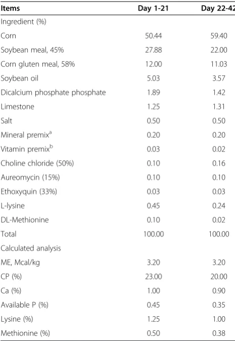
According to the following selection criteria, liver tissue of eight ascites and healthy broilers were collected for the determination of MDA and LD level, and the activity of T-SOD, HK, SDH, PK and Na+-K+-ATPase. Liver tis-sues were minced and homogenized (10%, wt/vol) in physiological saline water at 4°C, then centrifuged at 3,500 g for 15 min at 4°C. The supernatant was collected for the determination of biochemical parameters. MDA level was determined with the method of thibabituric acid (cat#: A003-1). The activity of T-SOD (cat#: A001-1), HK (cat#: A077), SDH (cat#: A022), PK (cat#: A076) and Na+-K+-ATPase (cat#: A016-2), and the level of liver LD (cat#: A019-2) were measured using commercially Table 1 Composition of experimental diets
Items Day 1-21 Day 22-42
Ingredient (%)
Corn 50.44 59.40
Soybean meal, 45% 27.88 22.00
Corn gluten meal, 58% 12.00 11.03
Soybean oil 5.03 3.57
Dicalcium phosphate phosphate 1.89 1.42
Limestone 1.25 1.31
Salt 0.50 0.50
Mineral premixa 0.20 0.20
Vitamin premixb 0.03 0.02
Choline chloride (50%) 0.10 0.16
Aureomycin (15%) 0.10 0.10
Ethoxyquin (33%) 0.03 0.03
L-lysine 0.45 0.24
DL-Methionine 0.10 0.02
Total 100.00 100.00
Calculated analysis
ME, Mcal/kg 3.20 3.20
CP (%) 23.00 20.00
Ca (%) 1.00 0.90
Available P (%) 0.45 0.35
Lysine (%) 1.25 1.00
Methionine (%) 0.50 0.38
a
supplied the following per kg complete diet: Cu, 8 mg; Zn, 75 mg; Fe, 80 mg; Mn, 100 mg; Se, 0.15 mg; I, 0.35 mg.
b
supplied the following (per kg complete diet): vitamin A, 12500 IU; vitamin D3, 2500 IU; vitamin E, 30 IU; vitamin K3, 2.65 mg; thiamine, 2 mg; riboflavin,
6 mg; vitamin B12, 0.025 mg; biotin, 0.0325 mg; folic acid, 1.25 mg;
available colorimetric diagnostic kits (Nanjing Jiancheng Bioengineering Institute, Nanjing, China) [22]. The pro-tein content of liver tissue was measured by Coomassie Brilliant Blue G-250 reagent with bovine serum albumin as a standard.
The selection criteria was based on the following three aspects: (1) HCT < 0.36, healthy; HCT≥0.36, ascites; (2) AHI < 0.28, healthy; AHI≥0.28, ascites; (3) Having effu-sion in abdominal cavity and pericardium, ascites, no ef-fusion, healthy.
Hepatic proteomic analysis
Another set of liver tissue were put in tubes, then snap frozen in liquid N2and stored at−80°C. Based on the
se-lection criteria mentioned above, liver tissue of four asci-tes and four healthy broilers were selected for the analysis of liver proteomics profile.
Protein sample preparation
Liver tissues were homogenized in a lysis buffer consist-ing of 40 mM Tris, 7 M Urea, 2 M Thiourea, 4% CHAPS, 1% Dithiothreitol (DTT), 1 mM EDTA-2Na, using a glass homogenization vessel in an ice bath. Tis-sues were ruptured using an ultrasonic cell disruptor at 0°C for 10 min with 2-s on and 8-s off cycles. Then the lysed cell suspension was kept at 4°C for 2 h to solubilize proteins. Subsequently, the homogenate was centrifuged at 4°C and 12,000 g for 5 min, finally the supernatant fluid was collected. Protein concentration for each of the final supernatants was determined by Bradford assay using bovine serum albumin as the standard. Protein extracts were stored in aliquots (1.5 mg/mL) at−80°C.
Two dimensional electrophoresis
This study consists of two groups with four replicates, so a total 8 gels were run for the 2-DE, using commer-cial IPG strips (pH 3-10 NL, 24 cm, GE Healthcare, Pis-cataway, NJ) for isoelectric focusing electrophoresis (IEF) and then standard vertical SDS-PAGE (13%) for second dimensional electrophoresis. Briefly, 1.5 mg pro-tein sample was loaded onto IPG drystrips using the in-gel sample rehydration technique, according to manu-facturer’s instructions. After rehydration for 12 h, the first-dimensional IEF was carried out at 20°C for 100,000 Vh in the Ettan IPGphorII IEF system (GE Healthcare, Piscataway, NJ) for 24 h. Sequentially, IPG strips were equilibrated for 15 min in 8 mL of equilibration buffer-1 (0.6 M urea, 2% DTT, 30% glycerol, and 50 mM Tris-Cl, 2% DTT, 0.07 M SDS, trace bromophenol blue, pH 8.8) and then in 8 mL of equilibration buffer-2 (0.6 M urea, 0.1 M iodoacetamide, 30% glycerol, and 50 mM Tris-Cl, 2% DTT, 0.07 M SDS, trace bromophenol blue, pH 8.8) for 15 min. The second dimensional electrophoresis was carried out on an Ettan DALTsix system (GE Healthcare)
at 20°C for 7 h (4 w/gel). Then the gels were stained with colloidal Coomassie Brilliant Blue R-250 (Amresco, Inc., Solon, OH).
Image analysis
The gel images were obtained using an UMAX Images-canner (Model Powerlook 2100XL, UMAX Technolo-gies, Atlanta, America) and image analysis was performed using Imagemaster Platinum Version 7.0 soft-ware (GE Healthcare). After normalizing the quantity of each spot by total valid spot intensity, differentially expressed protein spot with the point value (the relative expression volume ratio) over 2.0-fold were selected and subjected to identification by mass spectrometry (MS).
In-gel digestion
Protein spots were manually obtained and destained with 100 μL destaining solution (acetonitrile (ACN):100 mM ammonium bicarbonate = 3:7) for 1 h, then dehydrated with 100μL ACN for 15 min. The pro-tein samples were completely dried by vacuum centrifu-gation for 5 min, and subsequently digested with 10 μL of Trypsin (Amresco, Inc., Solon, OH) in 100 mM am-monium bicarbonate at 4°C for 45 min, and incubated at 37°C for 16 h. The resulting peptides were subjected to sequential extraction with 70% ACN and 0.1% trifluoroa-cetic acid (TFA) for 30 min (3 times at 37°C), then the three retain solutions were collected together and dried until 5-6 μL left with a vacuum centrifugation (Labconco).
Protein identification by MS and database queries
Protein identification was carried out on a Matrix Assisted Laser Desorption Ionization-Time of Flight MS (MALDI-TOF-MS) (Autoflex II, Bruker, Germany). MS fingerprinting data were performed by search engine Mascot (http://www.matrixscience.com), and Bony Ver-tebrates taxonomy against the NCBInr database. Search parameters include: (1) trypsin, as the enzyme of protein digestion; (2) MH+ and monoisotopic, as mass value; (3) unrestricted, as peptide mass; (4) ±0.3 Da, as peptide mass tolerance; (5) carbamidomethyl (C), as fixed modi-fications; oxidation (M), as variable modimodi-fications; and (6) 1, as maximum missed cleavages.
Statistical analysis
The biochemical parameters data were presented as Mean. The statistical analysis was performed with SPSS 16.0 software for Windows [23]. Independent sample
Results
Differences of body weight and organ index between ascites and healthy broilers
As shown in Table 2, body weight of ascites broiler was significantly lower than healthy broilers, and liver index, heart index and AHI of ascites broilers were significantly increased, but there was no difference in lung index be-tween ascites and healthy broilers.
Changes of blood biochemical parameters between ascites and healthy broilers
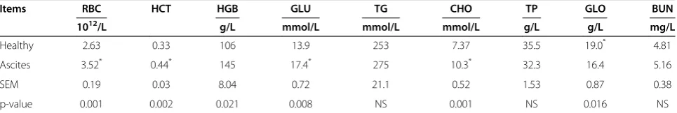
The data of blood biochemical parameters were shown in Table 3. RBC and HCT of ascites broilers were signifi-cantly greater than healthy broilers. Serum glucose, total cholesterol level of ascites broilers was significantly increased, and serum globulin level was significantly decreased, but there was no significant difference in tri-glyceride and BUN level.
Changes of liver biochemical parameters between ascites and healthy broilers
Liver MDA level of ascites broilers was significantly increased, and T-SOD activity was significantly lower than in healthy broilers. The activity of liver HK, SDH in ascites broilers was significantly decreased, and there was no significant difference in the activity of liver PK and Na+-K+-ATPase. Liver LD level of ascitic broilers were significantly lower than healthy ones (Table 4).
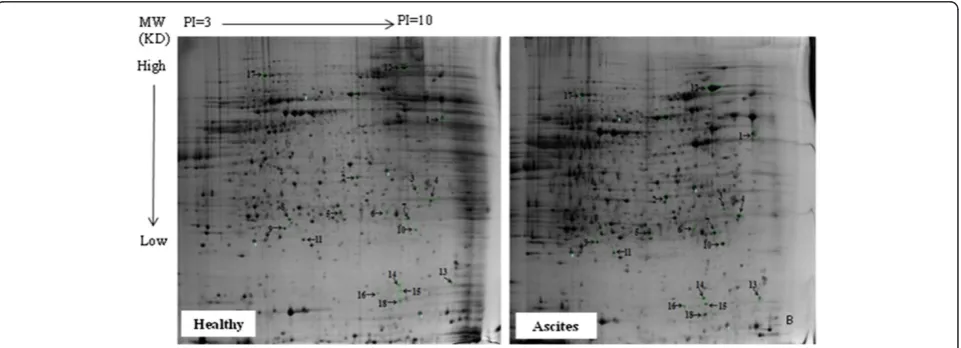
Changes of liver proteomics profiles between ascites and healthy broilers
The results of liver proteomics analysis indicated that 18 proteins expression difference were identified between
ascites and healthy broilers (Figure 1), including keratin, annexin, chicken M-PEPCK chain A, dihydropteridine reductase (DHPR), N-ethylmaleimide-sensitive factor (NSF), translationally-controlled tumor protein homolog (TCTP), ring finger protein 170, Rho GDP dissociation inhibitor alpha (GDI-α), ryncolin-1, natural killer cell en-hancing factor isoform 4 (NKEF), glia maturation factor beta (GMF), neutrophil cytosol factor 2 variant (NCF) and two unnamed proteins (Table 5). Chicken M-PEPCK was mainly involved in liver gluconeogenesis (Figure 2 and Figure 3). The result of the current study revealed that the expression of liver M-PEPCK chain A in ascites broilers was higher than in healthy broilers (Table 6).
Discussion
Organs index and blood parameters
The ascites syndrome in broilers is attributed to higher metabolic burdening, so one of the pathogenesis of the syndrome is metabolic hypoxia or low efficiency in oxy-gen utilization [2]. In the case of hypoxia, broilers have difficulties in fulfilling tissue demands for oxygen, and the birds exhibit the decreased blood saturation and increased production of RBC (partially immature) with decreased hemoglobin content relative to red blood cells, so the decrease in hemoglobin might have contrib-uted to enhanced development of hypoxemia and aggra-vation of ascites [9,24]. Luger et al. (2003) found RBC and HCT were significantly increased, and with no sig-nificant change in plasma volume in ascites chickens at wks 5, which was consistent with the current study. Be-sides, continually increased corticosterone level might be an inducer of erythropoiesis proliferation and differenti-ation arrest in ascites broilers [9]. Although increased haematrocrit values (and presumably increased haemo-globin) should be positive to alleviate hypoxaemia, birds dying of ascites all showed high haematocrits, which increased blood viscosity and augmented the inability of the failing right ventricle to pump blood through vas-constricted pulmonary blood vessels [8,25]. The liver and heart are the important organs to consume oxygen. Many studies take AHI > 0.27-0.30 as an indicator of as-cites occurrence, with right ventricular hypertrophy and Table 2 Changes of body weight and organ index
between ascites and healthy broilers (n = 8)
Items BW (kg) LI LUI HI AHI
Healthy 2.14* 2.62 0.24 0.49 22.2
Ascites 1.65 3.08* 0.25 0.60* 29.1*
SEM 0.09 0.09 0.01 0.02 1.45
p-value 0.003 0.006 NS 0.003 0.009
BW: Body weight, LI: Liver index, LUI: Lung index, HI: Heart index, AHI: Ascites heart index.*
P<0.05.
Table 3 Changes of blood biochemical parameters between ascites and healthy broilers (n = 8)
Items RBC HCT HGB GLU TG CHO TP GLO BUN
1012/L g/L mmol/L mmol/L mmol/L g/L g/L mg/L
Healthy 2.63 0.33 106 13.9 253 7.37 35.5 19.0* 4.81
Ascites 3.52* 0.44* 145 17.4* 275 10.3* 32.3 16.4 5.16
SEM 0.19 0.03 8.04 0.72 21.1 0.52 1.53 0.87 0.38
p-value 0.001 0.002 0.021 0.008 NS 0.001 NS 0.016 NS
pulmonary hypertension [26]. Hypertrophy of organs was a sign of abnormal function leading to the decrease in growth performance [7]. Results of the present study indicated that liver and heart of ascites broilers was sig-nificantly hypertrophic with the lower body weight, which increased the susceptibility to ascites and aggra-vated the occurrence of ascites syndrome.
Lipid antioxidant capacity and enzymes activity related to energy metabolism
Oxidative stress was aggravated in ascites broilers [27]. Ascites syndrome induced a large number of reactive oxygen species (ROS) and MDA production in many tis-sues, causing lipid peroxidation in mitochondrial mem-brane, leading to over-consumption of antioxidant enzymes and inadequately synthesize. The concentration of free radical and MDA were increased in serum and other tissues, but the activity of SOD and GSH-Px was significantly reduced [28]. The current study indicated that hepatic MDA concentration of ascites broilers was increased, and T-SOD activity was significantly decreased, which indicated that hepatic damage due to lipid peroxidation was significantly aggravated in ascites broilers. The scavenging agents of free radical, such as Vitamin E [29,30], or L-carnitine can enhance the
defense function of antioxidative system, and reduce the susceptibility to ascites [28].
Cold temperature can increase ascites susceptibility by increasing both metabolic oxygen requirements and pul-monary hypertension [15]. HK, SDH and PK are the im-portant enzymes in cell glycolysis and Krebs cycle. The activity of these enzymes is of importance to glucose ca-tabolism. Na+-K+-ATPase activity is an important indica-tor to measure mitochondria function for energy metabolism, and it has close relationship with the de-composition of ATP. The current study indicated that the activity of liver mitochondria enzymes was signifi-cantly decreased, which might block the ATP generation process in liver tissue of ascites broilers, so the energy requirement cannot be fulfilled under a cold environ-ment, which may aggravate the development of ascites.
Differentially expressed proteins indentified from proteomics profile
18 proteins expression difference were identified be-tween ascites and healthy broilers. These proteins were mainly associated with the following biological pro-cesses: 1) cytoskeleton; 2) glucose, lipids and amino acid metabolism; 3) cell secretion; 4) cell apoptosis; 5) signal transduction; 6) immune and inflammatory response; Table 4 Changes of liver biochemical parameters between ascites and healthy broilers (n = 8)
Items MDA T-SOD HK SDH PK Na+-K+-ATPase LD
nmol/mg protein U/mg protein U/mg protein U/mg protein U/g protein μmolPi/mg protein/h mmol/mg protein
Healthy 0.27 4.63* 65.14* 5.53* 34.9 6.29 0.20
Ascites 0.31* 3.74 41.35 3.99 29.8 8.01 0.16*
SEM 0.01 0.18 4.70 0.38 1.87 0.89 0.012
p-value 0.012 0.005 0.01 0.038 NS NS 0.044
MDA: Malondialdehyde, T-SOD: Total superoxide dismutase, HK: Hexokinase, SDH: Succinodehydrogenase, MDH: Malic dehydrogenase, PK: Pyruvate kinase, LD: Lactic acid.
and 7) cellular redox homeostasis. However, there are few reports on the relationship between the functions of differentially expressed proteins and ascites syndrome. It is reported that PEPCK, rather than being involved ex-clusively in gluconeogenesis, has a broader metabolic function in the cataplerosis of citric acid cycle inter-mediates (removal of citric acid cycle anions), which is required for gluconeogenesis and glyceroneogenesis [31]. So we are very interested in the PEPCK.
It is almost 60 years since the M-PEPCK (EC 4.1.1.32) from chicken liver was first reported by Utter and Kurahashi [32]. Most mammalian species display almost equal activity of the two isoforms of PEPCK in their liver, one in mitochondria (M-PEPCK) and another in cytosol (C-PEPCK), whereas chicken only express the
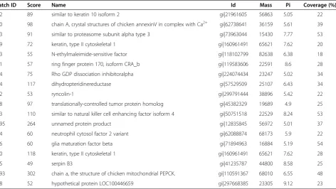
gene for M-PEPCK in their liver (The expression of liver C-PEPCK decreases to negligible levels 2 d before hatch-ing) [33-35]. In chicken, glucose synthesis in the liver is restricted to the Cori cycle, whereas net gluconeogenesis from amino acids occurs in the kidney, a tissue that con-tains both isoforms of PEPCK. Thus, birds recycle lac-tate generated by glycolysis in the muscle and red cells to the liver for gluconeogenesis [33,36]. These differ-ences in the regulation of gluconeogenesis may be related to the different ways of generating cytosolic NADH when specific metabolic precursors are used in gluconeogenesis [37]. In avian liver, the synthesis of phosphoenolpyruvate is restricted to the mitochondria, so that a continuous supply of NADH from oxidation-reduction reactions in the cytosol is needed. In tissues Table 5 Liver differentially expressed proteins of broilers identified by PMF query
Match ID Score Name Id Mass Pi Coverage (%)
882 89 similar to keratin 10 isoform 2 gi|21961605 56863 5.05 22
520 98 chain A, crystal structures of chicken annexinV in complex with Ca2+ gi|62738641 36159 5.61 39
443 91 similar to proteasome subunit alpha type 3 gi|73963044 15430 7.77 53
409 72 keratin, type II cytoskeletal 1 gi|160961491 65621 7.62 20
333 55 N-ethylmaleimide-sensitive factor gi|118102799 82638 6.38 18
311 57 ring finger protein 170, isoform CRA_b gi|119583606 22591 8.6 28
304 75 Rho GDP dissociation inhibitoralpha gi|224074434 23247 5.02 34
294 117 dihydropteridinereductase gi|57529509 25107 6.43 34
252 53 ryncolin-1 gi|299791444 38896 5.42 22
248 97 translationally-controlled tumor protein homolog gi|45382329 19689 4.9 25
213 110 similar to natural killer cell enhancing factor isoform 4 gi|50751518 22529 8.24 53
1895 264 unnamed protein product gi|12835845 56972 5.01 37
144 60 neutrophil cytosol factor 2 variant gi|62088874 68173 5.9 22
136 60 glia maturation factor beta gi|71894963 16884 5.19 54
130 118 keratin, type II cytoskeletal 1 gi|160961491 65621 7.62 28
125 49 serpin B3 gi|41235787 44800 8.58 25
1093 302 chain a, the structure of chicken mitochondrial PEPCK. gi|110591367 68010 6.55 48
108 52 hypothetical protein LOC100446659 gi|297668385 23305 9.12 23
that possess a C-PEPCK, malate is transported across the mitochondrial membrane, generating NADH in the cytosol where it is oxidized to oxaloacetate, then a var-iety of gluconeogenic precursors can be used (alanine, pyruvate and gluconeogenic amino acids) [34].
Daneshyar et al. (2009) reported that fasting blood sugar of cold-temperature treated birds at wks 4 and wks 6 was significantly greater than normal-temperature treated birds, and they attributed it to higher gluco-neogenesis level in ascites broilers [16,38]. Besides,
Yersin et al. (1992) and Biswas et al. (1995) observed decreased concentration of serum albumin in ascites broilers. It is suggested that deamidation process might contribute to the high rate of gluconeogenesis in hepa-tocytes of ascites broilers [17,18]. This study found that serum globulin level was significantly decreased in ascites broilers. In addition, the expression of liver M-PEPCK in ascites broilers was significantly up-regulated, with the significant decrease of liver lactic acid level and increase of serum glucose level in ascites broilers.
1182.734
1763.961
1574.809
2203.250 1374.809
1976.100 1530.765
2860.471
839.586 961.564
0 1000 2000 3000 4000 5000 6000
In
te
n
s
. [a
.u
.]
1000 1500 2000 2500 3000 3500 4000
m/z
Figure 3MALDI-TOF mass spectrum of M-PEPCK from a 2D gel.
Table 6 Expression volume of liver different protein spots in 2 DE map Spot
No.
Protein name Expression volume* p
-value Healthy Ascites
1 similar to keratin 10 isoform 2 0.141 0.150 0.081
2 chain A, crystal structures of chicken annexin v in complex with Ca2+ 0.042 0.057 0.076
3 similar to proteasome subunit alpha type 3 0.053 0.063 0.120
4 keratin, type II cytoskeletal 1 0.085 0.077 0.041
5 N-ethylmaleimide-sensitive factor 0.051 0.071 0.045
6 ring finger protein 170, isoform CRA_b 0.088 0.049 0.004
7 Rho GDP dissociation inhibitor (GDI) alpha 0.120 0.156 0.002
8 dihydropteridinereductase 0.122 0.128 0.053
9 ryncolin-1 0.080 0.121 0.004
10 translationally-controlled tumor protein homolog 0.131 0.160 0.014
11 similar to natural killer cell enhancing factor isoform 4 0.126 0.074 0.027
12 unnamed protein product 0.214 0.228 0.109
13 neutrophil cytosol factor 2 variant 0.129 0.150 0.216
14 glia maturation factor beta 0.046 0.046 0.039
15 keratin, type II cytoskeletal 1 0.193 0.387 0.177
16 serpin B3 0.171 0.231 0.057
17 chain A, the structure of chicken mitochondrial PEPCK. 0.084 0.130 0.006
18 hypothetical protein LOC100446659 0.432 0.894 0.114
*
It is implied that the gluconeogenesis level was increased during the development of broilers ascites under a low ambient temperature.
In summary, the study provides the first evidence for the changes of hepatic proteomics profile between ascites and healthy broilers. Eighteen differentially expressed pro-tein spots were identified between ascites and healthy broilers. It can be concluded that liver oxidation damage and energy generation obstruction occurred in ascites broilers. Hepatic M-PEPCK expression of ascites broilers was significantly increased-indicating that gluconeogenesis level was increased in ascites broilers under low ambient temperature. It is implied that some potential regulatory reagents may reduce ascites susceptibility by increasing gluconeogenesis level through up-regulating M-PEPCK protein expression.
Endnotes
Data in this paper were partially presented at the 2012 Poultry Science Association Annual Meeting, Athens, Georgia, America (p149).
Competing interests
The authors declare that they have no competing interests.
Authors’contribution
All authors read and approved the final manuscript.
Acknowledgements
This study was financed by Beijing Natural Science Foundation (6111002) and the Ear-marked Fund for China Agriculture Research System. Dong Ning, Yunzhi Peng, Hong Cai, Jianzhuang Tan participated in the analysis of biochemical parameters. We thank Ying Yang and Dan Liu for critical reading of the manuscript. We also thank Professor Jidong Feng and Mr. Jingqiang Zhang from the College of Biological Sciences of China Agriculture University for liver proteomics analysis.
Received: 14 September 2012 Accepted: 3 December 2012 Published: 11 December 2012
References
1. Balog JM:Ascites syndrome (pulmonary hypertension syndrome) in broiler chickens: are we seeing the light at the end of the tunnel?Avian Poultry Biol Rev2003,14:99–126.
2. Julian RJ:Ascites in poultry.Avian Pathol1993,22:419–454.
3. Currie RJW:Ascites in poultry: recent investigations.Avian Pathol1999, 28:313–326.
4. Crespo R, Shivaprasad HL:Diseases of poultry. 11th edition. Ames: Lowa State Press; 2003:1072–1075.
5. Moghadam HK, Mcmillan I, Chambers JR, Julian RJ, Tranchant CC: Heritability of sudden death syndrome and its associated correlations to ascites and body weight in broilers.Br Poult Sci2005,46:54–57. 6. Baghbanzadeh A, Decuypere E:Ascites syndrome in broilers: physiological
and nutritional perspectives.Avian Pathol2008,37:117–126.
7. Mohammadpour AA:Comparative histomorphological study of heart in healthy and ascites broiler chickens in Shahrekord district. Iran.Vet Res Commun2007,31:461–465.
8. Shlosberg A, Belaiche M, Berman E, Perk S, Deeb N, Neumark E, Cahaner A: Relationship between broiler chicken hematocrit-selected parents and their progeny with regard to hematocrit, mortality from ascites and body weight.Res Vet Sci1998,64:105–109.
9. Luger D, Shinder D, Wolfenson D, Yahav S:Erythropoiesis regulation during the development of ascites syndrome in broiler chickens: A possible role of corticosterone.J Anim Sci2003,81:784–790.
10. Hamal KR, Wideman RF, Anthony NB, Erf GF:Differential expression of vasoactive mediators in microparticle challenged lungs of chickens that differ in susceptibility to pulmonary arterial hypertension.Am J Physiol Regul Integr Comp Physiol2009,298:235–242.
11. Hamal KR, Wideman RF, Anthony NB, Erf GF:Differential gene expression of proinflammatorychemokines and cytokines in lungs of ascites-resistant and-susceptible broiler chickens following intravenous cellulose microparticle injection.Vet Immunol Immunopathol2010,133:250–255. 12. Shimomura I, Matsuda M, Hammer RE, Bashmakov Y, Brown MS, Goldstein
JL:Decreased IRS-2 and increased SREBP-1c lead to mixed insulin resistance and sensitivity in livers of lipodystrophic and ob/ob mice.Mol Cell2000,6:77–86.
13. Spiegelman BM, Flier JS:Obesity and the regulation of energy balance.
Cell2001,104:531–543.
14. Cisar CR, Balog JM, Anthony NB, Donoghue AM:Differential expression of cardiac muscle mitochondrial matrix proteins in broilers from ascites-resistant and susceptible lines.Poult Sci2005,84:704–708.
15. Stolz JL, Rosenbaum LM, Jeong D, Odom TW:Ascites syndrome, mortality and cardiological responses of broiler chickens subjected to cold exposure.Poult Sci1992,71(Suppl 1):4.
16. Diaz-Cruz A, Nava C, Villanueva R, Serret M, Guinzberg R, Pina E:Hepatic and cardiac oxidative stress and other metabolic changes in broilers with the ascites syndrome.Poult Sci1996,75:900–903.
17. Yersin AG, Huff WE, Kubena LF, Elissalde MA, Harvey RB, Witzel DA, Giroir LE: Changes in hematological, blood gas and serum biochemical variables in broilers during exposure to stimulated high altitude.Avian Disease
1992,36:189–197.
18. Biswas NK, Dalapati MR, Bhowmik MK:Ascites syndrome in broiler chickens: observations on certain biochemical and pathological changes.
Indian J Anim Sci1995,65:1068–1072.
19. NRC:Nutrition requirements of poultry. 9th edition. Washington DC: Natl Acad Sci; 1994.
20. Peacock AJ, Pickett CK, Morris KM, Reeves JT:Spontaneous pulmonary hypertension in rapidly growing broiler chickens reared at sea level [abstract].Am Rev of Respir Dis1988,137:s106.
21. Julian RJ, McMillan I, Quinton M:The effect of cold and dietary energy on right ventricular hypertrophy, right ventricular failure and ascites in meat-type chickens.Avian Pathol1989,18:675–684.
22. Tan GY, Yang L, Fu YQ, Feng JH, Zhang MH:Effects of different acute high ambient temperatures on function of hepatic mitochondrial respiration, antioxidative enzymes, and oxidative injury in broiler chickens.Poult Sci
2010,89:115–122.
23. SPSS:SPSS version 16.0 for windows. IIIth edition. Chicago: SPSS Inc; 2008. 24. Wideman RF, Forman MF, Hughes JD Jr, Kirby YK, Marson N, Anthony NB:
Flow-dependent pulmonary vasodilation during acute unilateral pulmonary artery occlusion in jungle fowl.Poult Sci1998,77:615–626. 25. Shlosberg A, Bellaiche M, Zeitlin M, Ya’akobi M, Cahaner A:Haematocrit
values and mortality from ascites in cold-stressed broilers from parents selected by haematocrit.Poult Sci1996,75:1–5.
26. Zerehdaran S, Grevehof EMV, Waaij EHVD, Bovenhuis H:A bivariate mixture model analysis of body weight and ascites traits in broilers.Poult Sci
2006,85:32–38.
27. Enkvetchakul B, Bottje W, Anthony N, Moore R, Huff W:Compromised antioxidant status associated with ascites in broilers.Poult Sci1993, 12:2272–2280.
28. Geng AL, Guo YM, Yuan JM:Effects of dietary L-carnitine and coenzyme Q10 supplementation on performance and ascites mortality of broilers.
Arch Anim Nutr2004,58:473–482.
29. Bottje W, Erf G, Bersi T, Wang S, Barnes D, Beers K:Effect of dietary DL-a-tocopherol on tissue andα- andγ–tocopherol and pulmonary hypertension syndrome (ascites) in broilers.Poult Sci1997, 75:1507–1512.
30. Villar-Patino G, Diaz-Cruz A, Avila-Gonzalez E, Guinzberg R, Pablos JL, Pina E: Effects of dietary supplementation with vitamin C or vitamin E on cardiac lipid peroxidation and growth performance in broilers at risk of developing ascites syndrome.Am J Vet Res2002,63:673–676.
31. Hanson RW:Thematic minireview series: a perspective on the biology of phosphoenolpyruvatecarboxykinase 55 years after its discovery.J Biol Chem2009,284:27021–27023.
33. Soling HD, Kleineke J, Willms B, Janson G, Kuhn A:Relationship between the intracellular distribution of P-enolpyruvatecarboxykinase, the regulation of gluconeogenesis and the energy cost of glucose formation.Eur J Biochem1973,37:233–243.
34. Weldon SL, Rando A, Kalonick PA, Matathias A, Hod Y, Savon S, Cook JS, Hanson RW:Nucleotide sequence of the cAMP for the mitochondrial form of P-enolpy-ruvatecarboxykinase (GTP) from the chicken [abstract].
J Cell Biol1988,107:412.
35. Savon S, Hakimi P, Hanson RW:Expression of the genes for the mitochondrial and cytosolic forms of PEPCK in avian liver during development.Biol Neonate1993,64:62–68.
36. Hanson RW, Garber AJ:P-enolpyruvatecarboxykinase: I. Its role in gluconeogenesis.Am J Clin Nutr1972,25:1010–1021.
37. Hod Y, Morris S, Hanson RW:Induction by cAMP of the mRNA encoding the cytosolic form of phosphoenolpyruvatecarboxykinase (GPT) from the chicken.J Biol Chem1984,259:15603–15608.
38. Daneshyar M, Kermanshahi H, Golian A:Changes of biochemical parameters and enzyme activities in broiler chickens with cold-induced ascites.Poult Sci2009,88:106–110.
doi:10.1186/2049-1891-3-41
Cite this article as:Wanget al.:Changes of hepatic biochemical parameters and proteomics in broilers with cold-induced ascites.Journal of Animal Science and Biotechnology20123:41.
Submit your next manuscript to BioMed Central and take full advantage of:
• Convenient online submission
• Thorough peer review
• No space constraints or color figure charges
• Immediate publication on acceptance
• Inclusion in PubMed, CAS, Scopus and Google Scholar
• Research which is freely available for redistribution