R E S E A R C H A R T I C L E
Open Access
Enhanced osteogenic commitment of
murine mesenchymal stem cells on
graphene oxide substrate
Jiyong Kim
1†, Hwan D. Kim
1†, Jungha Park
1, Eun-seo Lee
1, Eugene Kim
1, Seunghun S. Lee
2, Jin-Kyung Yang
1,
Yoon-Sik Lee
1and Nathaniel S. Hwang
1,2,3*Abstract
Background:Tissue engineering is an interdisciplinary field that attempts to restore or regenerate tissues and organs through biomimetic fabrication of scaffolds with specific functionality. In recent years, graphene oxide (GO) is considered as promising biomaterial due to its nontoxicity, high dispersity, and hydrophilic interaction, and these characteristics are key to stimulating the interactions between substrates and cells.
Method:In this study, GO substrates were fabricated via chemically immobilizing GO at 1.0 mg/ml on glass slides. Furthermore, we examined the osteogenic responses of murine mesenchymal-like stem cells, C3H10T1/2 cells, on GO substrates.
Results:C3H10T1/2 cells on GO substrates resulted in increased cell surface area, enhanced cellular adhesions, and instigated osteogenic differentiation. Furthermore, priming of C3H10T1/2 cells with chondrocyte-conditioned medium (CM) could further induce a synergistic effect of osteogenesis on GO substrates.
Conclusions:All of these data suggest that GO substrate along with CM is suitable for upregulating osteogenic responses of mesenchymal stem cells.
Keywords:Graphene, Osteogenesis, Mesenchymal stem cells, Graphene oxide
Background
Tissue engineering aims to provide biological living sub-stitutes for damaged tissues or organs from accidents, traumas or diseases [1–4]. As a key component for tis-sue engineering, stem cells are currently being actively utilized in tissue engineering and regenerative materials field. There have been great improvements for differenti-ating stem cells, in particular, mesenchymal stem cells, to induce tissue-specific differentiation. However, con-ventional methods may require several weeks to months of cell culture times for intended cell lineage differenti-ation [5]. Therefore, effective and facile methods for
stem cell differentiation are greatly needed [6]. Recently, modification of biomaterials surface to induce various cellular responses have been investigated [7–10]. These engineered biomaterials can elicit proliferation and dif-ferentiation control by selectively interacting with stem cells [11, 12]. Furthermore, it is reported that surface in-teractions caused by nanotopographies and patterned ar-rays of coated substrates also have significant effects on differentiation of stem cells [12–14]. As a result, devel-opments of new biomaterial would influence stem cell’s differentiation pathway and its therapeutic application with soluble proteins such as growth factors.
Due to its hydrophilic functional groups and pi-bond electrons on its surface [15], graphene oxide (GO) has been intensively researched for tissue engin-eering and its applications [16]. Its high electrical conductivity, flexibility, and one-atom-thick layer for-mation allow GO to be highly capable of interacting with cells, growth factors, and hydrophilic substrate
* Correspondence:nshwang@snu.ac.kr
†Equal contributors
1School of Chemical and Biological Engineering, Institute of Chemical Processes, Seoul National University, 1 Gwanak-ro, Gwanak-gu, Seoul 151-742, Republic of Korea
2Interdisciplinary Program in Bioengineering, Seoul National University, Seoul 152-742, Republic of Korea
Full list of author information is available at the end of the article
[17, 18]. These properties are attributed to not only functional groups such as carbonyl (CO), carboxyl (-COOH) and hydroxyl (-OH) groups but also pi-bond conjugation by sp2 hybridization. Mesenchymal stem cells cultured on GO-coated surface were re-ported to exhibit upregulated osteogenic differenti-ation [19–21]. Mesenchymal stem cells on GO-coated slide showed the increased expression of osteogenic markers, such as osteocalcin (OCN), but down-regulated other markers such as CD44, desmin, and MAP2 [21]. In addition, Nayak et al., has reported that the cells on GO substrate exhibited enhanced ad-hesion and proliferation [21]. As a result, calcium de-position level and alkaline phosphatase expression were raised. This suggests that GO can be used for osteogenic stimulation of mesenchymal stem cells; it is expected to be an outstanding material as a substrate in the application of not only tissue engin-eering but also dentistry field for advanced implant and clinical tests.
While it is important to understand the characteristics of GO, it should also consider understanding the back-ground of stem cells and their properties. Stem cells are affected by manipulating material-like mechanics and applying growth factor inducers [22, 23]. Especially dur-ing the endochondral ossification process, cartilage is presented by continuous cell division of chondrocytes, which affects the formation of bone tissue. With this phenomenon, Gerstenfeld et al. suggested that sub-stances that are secreted from chondrocyte promote osteogenesis [24]. Moreover, our group also demon-strated that C3H10T1/2 cells primed by bovine
chondrocyte-conditioned medium (CM) improved
osteogenic responses like expressions of osteogenic gene marker like osteocalcin (OCN), alkaline phosphatase (ALP), collagen type 1 (COL 1), and Runt-related tran-scription factor 2 (Runx2). In addition to the previous osteogenic gene markers, these primed C3H10T1/2 cells indicated calcium deposition when it is cultured with osteogenic factors like dexamethasone and ascorbic acid 2-phosphate (A2P) included osteogenic medium. During in vivo test, as a result, bone regeneration rate of critical defect on the mouse calvaria was increased [25].
Based on our previous studies, we hypothesized that GO could bring synergistic effects to C3H10T1/2 cells by priming with CM, complemented with bovine chon-drocyte secreted factors. With the characteristics of GO substrate previously stated, chondrocyte secreted factors can have an enhanced influence on stem cell differenti-ation, leading to up-regulated osteogenesis. These syner-gistic effects are anticipated to have a significant influence on the osteogenic differentiation experiments in the aspect of improved differentiation induction to the osteogenesis.
Methods
Cell priming with conditioned medium
Full-thickness articular cartilage was harvested from the patellofemoral groove and distal femoral condyle of bo-vine legs as previously described [26]. Collected cartilage tissues were cut into small pieces and incubated with 0.2% type II collagenase (Worthington Biochemical, USA) solution at 37 °C for 16 h. Isolated chondrocytes were filtered with 40 μm mesh and washed with phos-phate buffered saline (PBS). To make a conditioned medium, primary bovine chondrocytes were cultured with serum-free medium (15 ml DMEM including 1% Pen Strep) on culture plates (150 mm diameter) for 24 h. The chondrocyte-cultured serum-free medium was then filtered with a 0.2 μm syringe filter and supple-mented with 10% FBS (Gibco, USA) to make CM. In order to make a primed cell, C3H10T1/2 cells were cul-tured with CM for 14 days.
Graphene oxide (GO) substrate film preparation
Graphite flake and potassium permanganate (KMnO4)
were purchased from Sigma Aldrich (St. Louis, MO, USA). Sodium nitrate was obtained from Shimakyu’s pure chemical (Osaka, Japan). Sodium hydroxide and sulfuric acid were purchased from Dae-Jung Chemicals (Korea). (GO) was synthesized from graphite flakes by modified Hummers method as previously described [27]. Graphite was oxidized in H2SO4and KMnO4, then,
son-icated for 1.5 h at 12 W in an ice-bath. Subsequently centrifuged and GO was resuspended with H2O
inten-sively. Cover glass (1.5 × 1.5 cm) were treated with pi-ranha solution (H2SO4/H2O2 (70/30% (v/v))) for 1 h,
followed by 0.2 N NaOH solution treatment and coated with aqueous GO solution (1.0 mg/ml). GO solution loaded cover glass was kept at 60 °C oven for overnight as loosen lids for covalent bonding between hydroxyl group on the glass surface and epoxy group of GO and slowly evaporated the solution.
Cell culture and osteogenic differentiation
10 mM glycerol-2-phosphate (Sigma-Aldrich, USA), for 14 days. Osteogenic differentiation medium was replaced for every day.
Cell proliferation and viability analysis
Cell proliferation was examined by the Alama Blue Assay Kit (Invitrogen, USA) according to the manufac-turer’s instructions. Cells were seeded onto the slides at a density of 5000 cells / cm2and incubated for 4 h with 1:10 Alama blue solution in the medium. After incuba-tion, the Alama Blue solution containing medium is col-lected and the absorbance is measured using AT / Infinite M200 (TECAN, USA). Live / dead analysis was performed using live/dead viability kits (Invitrogen, USA) after 24 h of cell seeding on each slide. Twenty-four hours after seeding, the cells were incubated for 30 min with a live/dead solution containing calcein-AM and ethidium homodimer-1 (EthD-1). Images were then obtained using an LSM 720 confocal microscope (Zeiss).
Morphological analysis
After cells were cultured for 4 days with osteogenic dif-ferentiation medium (OM), cells were fixed with 10% (v / v) formalin and washed with PBS (3 times). Subse-quently, permeabilized using 0.1% Triton X-100 for 30 min. Then, samples were stained with a mixture of 1: 100 Phalloidin (Alexa Fluor 594, life technologies, USA) and 1: 500 vinculin (Abcam, USA) for 1 h. After another 1 h of second antibody treatment, the cells were further stained with DAPI (¢ 6-diamidino-2-phenylindole, Sigma-Aldrich) solution at a ratio of 1: 200 for 15 min. The fluorescence image was obtained with a confocal microscope (ZEISS LSM 720).
Real time-PCR
RNA samples were obtained from each GO and glass slides (n= 4) containing cells with the Trizol method (Trizol®, Life Technology, USA). Total RNA concentra-tion was measured with a NanoDrop spectrometer (ND-2000, NanoDrop Technologies, USA). Each sample was normalized to 1000 ng of total RNA and reverse transcribed into cDNA using the TOPscript ™ Reverse Transcriptase Kit (Enzynomics) according to the manu-facturer’s instructions. Real-time PCR was performed with ABI StepOnePlusTM real-time PCR system (Ap-plied Biosystems, USA) using the SYBR Green PCR Master mix. Expressions of genes related with osteogenic differentiation such as GAPDH, Runx2, ALP, and OCN were analyzed. cDNA samples were loaded and the data were analyzed by the –2ΔΔCt method. PCR primers se-quence was as follows: GAPDH (forward: 5′-GTA TGA CTC CAC TCA CGG CAA A-3′, reverse: 5’-CTA AGC AGT TGG TGG TGC AG-3′),RunX2(forward: 5′-GGA CGA GGC AAG AGT TTC A-3′, reverse: 5′-TGG TGC
AGA GTT CAG GCA G-3′), ALP (forward: 5′-GAA GTC CGT GGG CAT CGT-3′, reverse: 5’-CAG TGC GGT TCC AGA CAT AG-3′), OCN (forward: 5’-AGC AGG AGG GCA ATA AGG-3′, reverse: 5’-CGT AGA TGC GTT TGT AGG C-3′).
Calcium deposition analysis
To analyze the calcium deposition, Alizarin Red S stain-ing was performed. Cells were incubated with OM for 14 days, then the cells were fixed with 10% (v / v) forma-lin and washed three times with PBS. To make the ARS solution, 20 mg of Alizarin Red S powder (Sigma-Al-drich, USA) was dissolved in 1 ml of distilled water and the pH was adjusted to 4.1 ~ 4.2 with ammonium hy-droxide (NH4OH). Fixed cells were stained with ARS
so-lution for 20 min and washed 3 times with distilled water for 5 min. For ARS quantification, 800 μl of 10% (v / v) acetic acid per well was added and incubated at room temperature for 30 min. The cells were collected with a cell scraper, transferred to a 1.5 ml tube, and 500 μl of mineral oil was added. The samples were heated at 85 °C for 10 min and cooled with ice for 5 min. The solution was then centrifuged at 20,000 G for 15 min. After centrifugation, 500 μl of supernatant was collected. Then, 200 μl of 10% ammonium hydrox-ide was added to the supernatant to complete precipita-tion. To see the results, Absorbance values were measured with a spectrometer.
Field emission scanning electron microscopy
Cells seeded on GO / Glass slides were cultured in OM for 4 days, then fixed with 4% paraformaldehyde (Poly-sciences) for 15 min, subsequently dehydrated with 70– 100% ethanol (Daejung Chemical) and treated with Hex-amethyldisilazane (HMDS; Daejung Chemical) for 1 h. The sample was visualized with a field emission scan-ning electron microscope (FE-SEM; JSM-6701F, JEOL) at 20 mA for 100 s after platinum coating.
Western blotting
dilution for 1 h at room temperature. To visualize the protein expressions, the blots were developed by a chemiluminescence detection system (Amersham Bio-science, USA).
Data analysis
The quantitative data were expressed as the means ± standard deviations. The statistical significances were an-alyzed by one-way analysis of variance (ANOVA) (*p< 0.05, **p< 0.01).
Results
Preparation and characterization of Graphene oxide coated slide
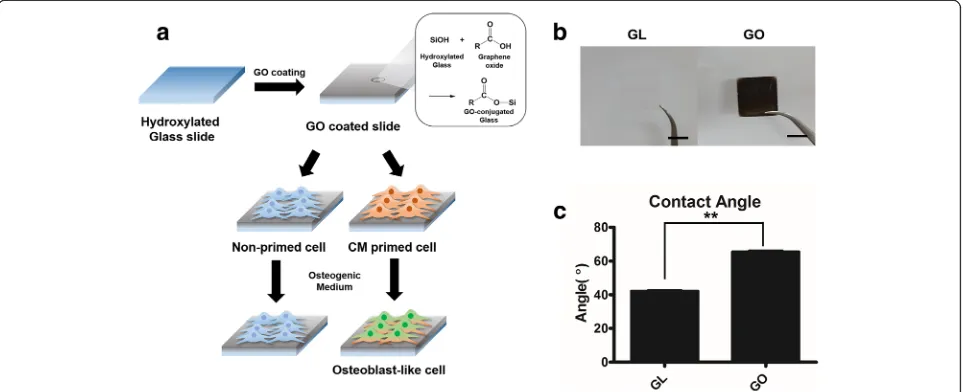
Figure 1a represents the overall scheme for graphene oxide coated slide and osteogenic differentiation of C3H10T1/2 cells. Firstly, C3H10T1/2 cells were ex-panded with either chondrocyte-conditioned medium (CM) or control growth medium (GM) were utilized. Next these cells were cultured on either control glass slide or GO coated slide (Fig. 1a).
GO flakes were made by modified Hummers method [27], and GO flakes were coated on the 18 × 18 mm glass slide. Covalent bonds were formed between hy-droxyl group from glass surface treated with piranha so-lution and epoxy group of GO as pH 10 water slowly evaporated at 60 °C. For the GO coated slides, we ob-served that the surface of the graphene oxide coated slide formed a dark, thin and opaque layer compared to a glass slide (Fig. 1b). Moreover, the water contact angle was altered due to the GO coating (Fig. 1c). After GO coating, water contact angle was increased about 24°,
indicating that the surface area of a GO coated slide was more hydrophobic than a glass slide.
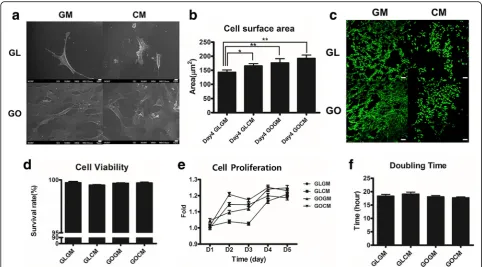
Cell proliferation, viability, and morphological change To observe the effect of chondrocyte-conditioned medium (CM) priming and GO coated slide, C3H10T1/ 2 cells were primed for 14 days with CM before seeding. Then, primed and non-primed cells were seeded at a same density on each glass slide or GO coated slide. After 4 days of seeding, cell surface area was altered by CM priming and GO coated slide. When compared to the non-primed cells, priming of C3H10T1/2 cells re-sulted in 1.18 fold increased in cell size (Fig. 2a and b). The cell size of C3H10T1/2 cells on GO coated slide was 174.3 um2 and primed C3H10T1/2 cells on gra-phene oxide coated slide was 189.2 um2, indicating that
the both CM expansion increased the cell coverage area. SEM analysis showed the substrate-dependent adhe-sion efficiencies (Fig. 2a). On the GO coated slide, there were more cells attached, and the surface area of a single cell was larger than the surface area of a cell on a glass slide. GO substrate is highly capable of interacting with cells due to its hydrophilic property and high adsorption properties for proteins and soluble growth factors. These properties may have resulted in increased cell surface area and adhesion efficiencies.
For the cell viability analysis (Fig. 2c and d), we seeded primed and non-primed C3H10T1/2 cells on either glass slide and GO coated slide. After 1 day of seeding, we measured cellular proliferation and assayed for live/dead. Regardless of the substrate and cell types, all groups showed a similar level of viability (Fig. 2d). All groups showed more than 98% of survival rate indicating that
GO or CM did not invoke any significant cytotoxicity to cells.
CM primed cells on glass slide showed decreased pro-liferation rate compared to the non-primed group while CM primed cells on GO coated slide exhibited improved proliferation rate, as indicated by Alamar Blue assay (Fig. 2e). This suggests that the proliferation of primed cell is enhanced on GO coated slide. The cell doubling time of CM primed cells on GO coated slide showed a statistically significant difference compared with CM primed cells on a glass slide (Fig. 2f ). Non-primed cells on glass slide showed a doubling time of 18.4 h, primed cells on the glass slide have a doubling time of 19.2 h, the longest doubling times among the groups. Non-primed cells on GO coated slides have a doubling time of 18.1 h and primed cells on GO coated slides showed a doubling time of 17.6 h, which was the shortest doub-ling time above all groups. In short, cell doubdoub-ling times of primed cells were shortened on the GO coated slides for 0.916 times compared to non-primed cells on a glass slide. This result showed that the CM priming had a synergistic effect on the GO coated slides in terms of cellular proliferation. Furthermore, these results demon-strated that a synergistic effect of GO substrate and CM priming could result in morphological change, improved viability, and cellular proliferation.
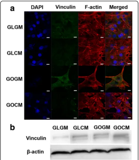
Improved focal adhesion of CM primed cells on GO slide Previously, increased cell size coupled with increased focal adhesion kinase (FAK)has been showed to activate cytoskeletal tension via RhoA/Rock signaling pathways, which also enhances osteogenic commitment of stem
cells [28]. We cultured primed or non-primed
through a FAK signaling pathway and ERK-dependent pathway [29, 30]. Hence, increased expression of vincu-lin meant improved focal adhesion and contact with the extracellular matrix protein of cells. With these results, we hypothesized that CM priming and GO coated slide both induced the osteogenic differentiation of C3H10T1/2, along with other synergistic effects when they were used together. To show the cellular morph-ology and expression of focal adhesion protein increased osteogenic differentiation of C3H10T1/2 cells, we inves-tigated the effect of CM priming and GO coated slide on osteogenic responses by confirming osteogenic gene ex-pressions and bone mineral deposition of each group.
Osteogenic differentiation of C3H10T1/2 cells
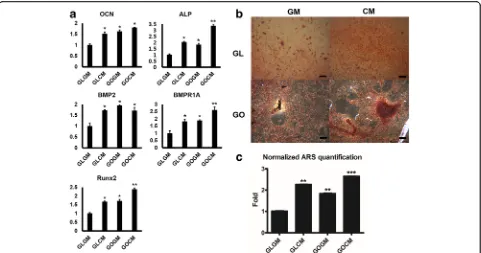
We cultured CM primed and non-primed C3H10T1/2 cells on each group of slides in the osteogenic medium for 14 days. Osteogenic medium contained ascorbic-2-phosphate, dexamethasone and β-glycerophosphate, which are required for osteogenic differentiation [31, 32]. After culturing cells, osteogenic gene expressions were analyzed by real-time PCR. Compared to those of control group (GLGM), CM primed cells on GO coated slide showed a substantially enhanced osteogenic gene
expressions including osteocalcin (OCN,
non-collagenous protein found in bone), alkaline phosphatase (ALP, protein promoting osteoblastic activity),
runt-related transcription factor 2 (Runx2, early osteogenic gene marker), bone morphogenetic protein 2 (BMP2, es-sential protein in the development of bone and cartil-age), and BMP receptor (BMPR1A) (Fig. 4a). Up-regulation of osteogenic gene expression in CM primed cells on glass slide (GLCM) group and non-primed cells on GO slide (GOGM) was observed, yet primed cells on GO coated slides group (GOCM) were in a higher range of expression.
To determine whether C3H10T1/2 cells differentiated into osteoblast cells, we measured the calcium contents from differentiated cells by Alizarin Red S staining (Fig. 4b and c). On the glass slide, the intensity of ARS stained cells (Red) was more distinguished on CM primed cell. It demonstrated that CM priming could augment the content of calcium deposition. The same phenomenon was observed on the GO slide as well. Fur-thermore, primed cells on the GO slide presented the greatest calcium deposition rate among all groups. We facilitated ARS quantification to measure the calcium deposition level quantitatively. In agreement with osteo-genic gene expression analysis by real-time PCR, cells under the condition of CM or GO slide could deposit more calcium contents than either non-primed cells or cells on glass side. The highest amount of calcium de-tected in primed cells on GO coated slides group (GOCM) further supported that CM and GO reinforced the synergistic effect in osteogenesis.
Discussion
Previous studies showed that GO substrate can enhance the osteogenic commitment of stem cells [16]. GO sub-strate usually functions as cell adhesive platform improv-ing cell adhesion and cell-protein interactions [8, 9]. Furthermore, in a separate study, bovine chondrocyte conditioned medium (CM) priming has been shown to enhance the osteogenic differentiation of mouse or hu-man mesenchymal stem cells [33, 34]. Conditioned medium treating also played a role by improving both chondrogenic and osteogenic responsibility of mesen-chymal stem cells through the interaction of cells with the secreted factors from bovine chondrocytes [19, 30].
In this study, we investigated the effect of conditioned medium priming and GO coated slide on osteogenic dif-ferentiation of C3H10T1/2 cells, and demonstrated that the synergistic effect of GO coated slide and CM prim-ing could facilitate osteogenesis of C3H10T1/2 (murine mesenchymal stem cells). GO coated surface had in-creased interaction of extracellular matrix and cells [35]. From our experiment, however, we saw a subtle increase in contact angle. This is due to the carbon components of GO. GO is basically composed of carbon atoms. Usu-ally, long carbon chain shows hydrophobic characters. However, GO coated slide shows hydrophilic features Fig. 3aNucleus, Vinculin and F-actin staining for GM/CM Cell
because of its pi bond conjugation and hydrophilic func-tional groups like -OH and -COOH [15], which may affect the focal adhesion of cell and cell proliferation. Furthermore, the interaction of cells with hydrophilic growth factors like TGF-B groups can be influenced by GO coating. As a result, these properties of GO sub-strate support that the GO coated surface are highly capable of interacting with cells due its high reactivity to the other proteins and growth factors.
Firstly, we observed that cell surface area from the GO coated slide groups was larger than that from the glass slide groups. Furthermore, the configuration of cell and adhesion rate was different. SEM image showed that there was a significant difference in the cell attachment rate between glass slide group and GO coated slide group, even though both groups had the same amount of days of cell culture with equal cell density. On the GO coated slide, attached cells were more apparent. GO coated slide presented enhanced cell interaction because of its hydrophilic property and these properties also af-fected the cell morphology such as increased cell surface area and cell adhesion rate.
In addition, CM priming also increased cell surface area. In this experiment, we used CM which contained chondrocyte secreted factors to prime C3H10T1/2 cells. Primed cells on GO slide showed larger surface area than non-primed groups. Among those groups, CM
primed cells on GO coated slide showed most stretched shapes to various directions and higher cell adhesion rate, which indicates higher potential and efficiency to be differentiated in direction of osteogenesis [28, 36]. In order to directly observe the cellular response of CM priming and GO substrate, primed C3H10T1/2 cells on GO slide with growth medium (i.e., without osteogenic factors) provide sufficient evidence for cellular commit-ment. However, our previous study has demonstrated the primed C3H10T1/2 cells cultured with growth medium resulted in stimulated chondrogenic response (i.e., not osteogenic response) [37]. Since our intention in this study was to observe the osteogenic response, we have utilized the osteogenic differentiation medium.
to various directions compared to non-primed cells. These morphological change on CM primed cells allows the cells to be effectively influenced through contacts with substrate. Moreover, hydrophilic properties of GO can increase the interaction of bovine chondrocyte se-creted factors with the cells, which have up-regulating effect on osteogenic differentiation of mesenchymal stem cells [38, 39].
With increased cell surface area, CM primed cell showed stretched F-actin in various directions, and vin-culin expression level showed enhancement of focal ad-hesion for primed cells. Also, increased cell surface area and stretched F-actin on GO slide due to the hydrophilic characters of GO indicates that the effect of focal adhe-sion protein and extracellular matrix interaction with cells were increased. Therefore, we can speculate that CM primed cells and GO coated slide could have syner-gistic effects because chondrocyte secreted factors, which had the influence to primed cells, had improved focal adhesion and enhanced the effect of GO substrate on cells. Moreover, it is known that mechanical stimulus on stem cell has a significant effect on its differentiation ability. As the stimulus level increases, the morphology of the cell stretches to various directions and the cell surface area is increases. Those morphological changes shows to have great influence on osteogenic differenti-ation of the mesenchymal stem cell [28]. In this regard, we demonstrated that the spread of F-Actin, the in-creased cell surface area of primed cells and, cell growth on GO coated slides enhanced the osteogenic differenti-ation of cells. In addition, the results of confirming the expression of vinculin, protein related with focal adhe-sion, through Western blotting had same tendency. The expression level of vinculin was highest at CM primed cells seeded on GO coated slides group.
In this study, furthermore, osteogenic gene marker ex-pressions and ARS staining results supported that the CM priming also to enhance osteogenic responses and calcium deposition. Gene expression level of osteogenic gene markers such asOCN, ALP, Runx2, BMP2, andBMPR1A were upregulated via CM priming and GO substrate cul-turing. Simlar to the study by Lee and colleagues, primed cells on GO coated slide showed enhanced osteogenic re-sponses and calcium deposition [40].
Conclusion
CM priming and GO coated slide affected proliferation, morphological change, and focal adhesions of the C3H10T1/2 cells. CM primed cell with GO slide showed substantially increased osteogenic responses. As a result, CM primed cells seeded on GO slides showed morpho-logical change like increased cell surface and stretched to various direction. In addition, cell proliferation also in-creased as GO slide was applied. Focal adhesion of
mesenchymal stem cells was enhanced as the result is shown at western blot image. This can induce osteogenic differentiation effectively. With the CM priming and GO coated slide, the gene expression level of the osteogenic markers likeOCN, ALP, Runx2, BMP2andBMP2R1Aand calcium deposition was upregulated. In short, CM primed cell on GO coated slide showed considerably enhanced osteogenic response compared to any other groups in the experiment. With these results, we could see the synergis-tic effects of GO and conditioning medium priming. Those synergistic effects of CM priming with GO coating enhanced osteogenic commitment of C3H10T1/2 cells, mesenchymal-like stem cells, and it is possible to utilize these synergistic effects to increase the rate of differenti-ation of MSCs to osteoblast. Furthermore, CM priming with GO substrate can take part in a solution to excel bone rehabilitation or other therapeutic application such as bone graft therapeutic and, perhaps, tissue engineering as a whole.
Abbreviations
A2P:Ascorbic 2-phosphate; ALP: Alkaline phosphatase; BMP2: Bone morphogenetic protein 2; BMPR1A: Bone morphogenetic protein receptor 1A; C3H: C3H10T1/2 cells; CM: Chondrocyte-conditioned medium; COL I: Collagen type I; ECM: Extracellular matrix; FAK: Focal adhesion kinase; GL : Glass slide; GLCM: Primed cells on glass slide group; GLGM: Non-primed cells on glass slide group; GM: Growth medium; GO: Graphene oxide; GOCM: Primed cells on graphene oxide slide group; GOGM: Non-primed cells on graphene oxide slide group; OCN: Osteocalcin; RUNX2: Runt-related transcription factor 2; TGF-B: Transforming growth factor-β
Funding
This research was supported by the National Research Foundation of Korea (NRF) funded by the Ministry of Science, ICT & Future Planning (NRF-2016R1E1A1A01943393, NRF-2017M3A9C6029699).
Availability of data and materials
Not applicable.
Author’s contributions
The research was designed and coordinated by J.K and N.S.H. The scaffold fabrication and analysis were performed by J.K, H.D.K, J.Y. Cell and molecular analysis was performed by J.K, H.D.K, E.L and J.P. H.D.K, J.K, E.K, S.S.L, Y.L and N.S.H wrote, revised and improved the final manuscript. All authors read and approved the final manuscript.
Ethics approval and consent to participate
Not applicable.
Consent for publication
Not applicable.
Competing interests
The authors declare that they have no competing interests.
Publisher’s Note
Springer Nature remains neutral with regard to jurisdictional claims in published maps and institutional affiliations.
Author details
Received: 23 November 2017 Accepted: 12 December 2017
References
1. Bottcher-Haberzeth S, Biedermann T, Reichmann E. Tissue engineering of skin. Burns. 2010;36:450–60.
2. Clark RA, Ghosh K, Tonnesen MG. Tissue engineering for cutaneous wounds. J Invest Dermatol. 2007;127:1018–29.
3. Horch RE, Kopp J, Kneser U, Beier J, Bach AD. Tissue engineering of cultured skin substitutes. J Cell Mol Med. 2005;9:592–608.
4. Kim HD, Amirthalingam S, Kim SL, Lee SS, Rangasamy J, Hwang NS. Biomimetic materials and fabrication approaches for bone tissue engineering. Adv Healthc Mater. 2017;6:23.
5. Pittenger MF, Mackay AM, Beck SC, Jaiswal RK, Douglas R, Mosca JD, Moorman MA, Simonetti DW, Craig S, Marshak DR. Multilineage potential of adult human mesenchymal stem cells. Science. 1999;284:143–7.
6. Caplan AI. Mesenchymal stem cells. J Orthop Res. 1991;9:641–50. 7. Karageorgiou V, Kaplan D. Porosity of 3D biomaterial scaffolds and
osteogenesis. Biomaterials. 2005;26:5474–91.
8. Li WJ, Tuli R, Huang X, Laquerriere P, Tuan RS. Multilineage differentiation of human mesenchymal stem cells in a three-dimensional nanofibrous scaffold. Biomaterials. 2005;26:5158–66.
9. Vinatier C, Bouffi C, Merceron C, Gordeladze J, Brondello JM, Jorgensen C, Weiss P, Guicheux J, Noel D. Cartilage tissue engineering: towards a biomaterial-assisted mesenchymal stem cell therapy. Curr Stem Cell Res Ther. 2009;4:318–29.
10. Kim HD, Lee EA, An YH, Kim SL, Lee SS, Yu SJ, Jang HL, Nam KT, Im SG, Hwang NS. Chondroitin sulfate-based biomineralizing surface Hydrogels for bone tissue engineering. ACS Appl Mater Interfaces. 2017;9:21639–50. 11. Dawson E, Mapili G, Erickson K, Taqvi S, Roy K. Biomaterials for stem cell
differentiation. Adv Drug Deliv Rev. 2008;60:215–28.
12. Cheng H, Byrska-Bishop M, Zhang CT, Kastrup CJ, Hwang NS, Tai AK, Lee WW, Xu X, Nahrendorf M, Langer R, Anderson DG. Stem cell membrane engineering for cell rolling using peptide conjugation and tuning of cell-selectin interaction kinetics. Biomaterials. 2012;33:5004–12.
13. Dalby MJ, McCloy D, Robertson M, Agheli H, Sutherland D, Affrossman S, Oreffo RO. Osteoprogenitor response to semi-ordered and random nanotopographies. Biomaterials. 2006;27:2980–7.
14. Shin YC, Song SJ, Hong SW, Jeong SJ, Chrzanowski W, Lee JC, Han DW. Multifaceted biomedical applications of functional Graphene Nanomaterials to coated substrates, patterned arrays and hybrid scaffolds. Nanomaterials (Basel). 2017;7(11):369.
15. Shi XT, Chang HX, Chen S, Lai C, Khademhosseini A, Wu HK. Regulating cellular behavior on few-layer reduced Graphene oxide films with well-controlled reduction states. Adv Funct Mater. 2012;22:751–9. 16. Kim HD, Kim J, Koh RH, Shim J, Lee JC, Kim TI, Hwang NS. Enhanced
Osteogenic commitment of human Mesenchymal stem cells on polyethylene glycol-based Cryogel with Graphene oxide substrate. Acs Biomaterials Science & Engineering. 2017;3:2470–9.
17. Zhang Y, Wu CY, Guo SW, Zhang JY. Interactions of graphene and graphene oxide with proteins and peptides. Nanotechnol Rev. 2013;2:27–45. 18. Ding ZJ, Ma HW, Chen YY. Interaction of graphene oxide with human
serum albumin and its mechanism. RSC Adv. 2014;4:55290–5. 19. Luo Y, Shen H, Fang Y, Cao Y, Huang J, Zhang M, Dai J, Shi X, Zhang Z.
Enhanced proliferation and osteogenic differentiation of mesenchymal stem cells on graphene oxide-incorporated electrospun poly(lactic-co-glycolic acid) nanofibrous mats. ACS Appl Mater Interfaces. 2015;7:6331–9. 20. Nair M, Nancy D, Krishnan AG, Anjusree GS, Vadukumpully S, Nair SV.
Graphene oxide nanoflakes incorporated gelatin-hydroxyapatite scaffolds enhance osteogenic differentiation of human mesenchymal stem cells. Nanotechnology. 2015;26:161001.
21. Nayak TR, Andersen H, Makam VS, Khaw C, Bae S, Xu X, Ee PL, Ahn JH, Hong BH, Pastorin G, Ozyilmaz B. Graphene for controlled and accelerated osteogenic differentiation of human mesenchymal stem cells. ACS Nano. 2011;5:4670–8.
22. Guilak F, Cohen DM, Estes BT, Gimble JM, Liedtke W, Chen CS. Control of stem cell fate by physical interactions with the extracellular matrix. Cell Stem Cell. 2009;5:17–26.
23. Kratchmarova I, Blagoev B, Haack-Sorensen M, Kassem M, Mann M. Mechanism of divergent growth factor effects in mesenchymal stem cell differentiation. Science. 2005;308:1472–7.
24. Gerstenfeld LC, Cruceta J, Shea CM, Sampath K, Barnes GL, Einhorn TA. Chondrocytes provide morphogenic signals that selectively induce osteogenic differentiation of mesenchymal stem cells. J Bone Miner Res. 2002;17:221–30.
25. Ro H, Park J, Yang K, Kim J, Yim HG, Jung G, Lee H, Cho SW, Hwang NS. Osteogenic priming of mesenchymal stem cells by chondrocyte-conditioned factors and mineralized matrix. Cell Tissue Res. 2015; 362(1):115–26.
26. Koh RH, Jin Y, Kang BJ, Hwang NS. Chondrogenically primed tonsil-derived mesenchymal stem cells encapsulated in riboflavin-induced
photocrosslinking collagen-hyaluronic acid hydrogel for meniscus tissue repairs. Acta Biomater. 2017;53:318–28.
27. Shahriary L, Athawale AA. Graphene oxide synthesized by using modified hummers approach. Int J Renew Energy Environ Eng. 2014;2:58–63. 28. Wang YK, Yu X, Cohen DM, Wozniak MA, Yang MT, Gao L, Eyckmans J, Chen
CS. Bone morphogenetic protein-2-induced signaling and osteogenesis is regulated by cell shape, RhoA/ROCK, and cytoskeletal tension. Stem Cells Dev. 2012;21:1176–86.
29. Tong L, Buchman SR, Ignelzi MA Jr, Rhee S, Goldstein SA. Focal adhesion kinase expression during mandibular distraction osteogenesis: evidence for mechanotransduction. Plast Reconstr Surg. 2003;111:211– 22. discussion 223-214
30. Salasznyk RM, Klees RF, Williams WA, Boskey A, Plopper GE. Focal adhesion kinase signaling pathways regulate the osteogenic differentiation of human mesenchymal stem cells. Exp Cell Res. 2007;313:22–37.
31. Jaiswal N, Haynesworth SE, Caplan AI, Bruder SP. Osteogenic differentiation of purified, culture-expanded human mesenchymal stem cells in vitro. J Cell Biochem. 1997;64:295–312.
32. Yuasa M, Yamada T, Taniyama T, Masaoka T, Xuetao W, Yoshii T, Horie M, Yasuda H, Uemura T, Okawa A, Sotome S. Dexamethasone enhances osteogenic differentiation of bone marrow- and muscle-derived stromal cells and augments ectopic bone formation induced by bone morphogenetic protein-2. PLoS One. 2015;10:e0116462.
33. Jang HL, Jin K, Lee J, Kim Y, Nahm SH, Hong KS, Nam KT. Revisiting whitlockite, the second most abundant biomineral in bone: nanocrystal synthesis in physiologically relevant conditions and biocompatibility evaluation. ACS Nano. 2014;8:634–41.
34. Hwang NS, Varghese S, Lee HJ, Zhang Z, Elisseeff J: Biomaterials directed in vivo osteogenic differentiation of mesenchymal cells derived from human embryonic stem cells. Tissue Eng Part A 2013, 19:1723.
35. Tatavarty R, Ding H, Lu G, Taylor RJ, Bi X. Synergistic acceleration in the osteogenesis of human mesenchymal stem cells by graphene oxide-calcium phosphate nanocomposites. Chem Commun (Camb). 2014;50: 8484–7.
36. Yang F, Williams CG, Wang D-A, Lee H, Manson PN, Elisseeff J. The effect of incorporating RGD adhesive peptide in polyethylene glycol diacrylate hydrogel on osteogenesis of bone marrow stromal cells. Biomaterials. 2005; 26:5991–8.
37. Ro H, Park J, Yang K, Kim J, Yim HG, Jung G, Lee H, Cho SW, Hwang NS. Osteogenic priming of mesenchymal stem cells by chondrocyte-conditioned factors and mineralized matrix. Cell Tissue Res. 2015;362:115–26.
38. Kanczler J, Oreffo R. Osteogenesis and angiogenesis: the potential for engineering bone. Eur Cell Mater. 2008;15:100–14.
39. Hwang NS, Varghese S, Puleo C, Zhang Z, Elisseeff J. Morphogenetic signals from chondrocytes promote chondrogenic and osteogenic differentiation of mesenchymal stem cells. J Cell Physiol. 2007;212:281–4.