RESEARCH
Physiological characterization
of secondary metabolite producing
Penicillium
cell factories
Sietske Grijseels
1, Jens Christian Nielsen
2, Jens Nielsen
2,3, Thomas Ostenfeld Larsen
1, Jens Christian Frisvad
1,
Kristian Fog Nielsen
1, Rasmus John Normand Frandsen
1*and Mhairi Workman
1Abstract
Background: Penicillium species are important producers of bioactive secondary metabolites. However, the
immense diversity of the fungal kingdom is only scarcely represented in industrial bioprocesses and the upscaling of compound production remains a costly and labor intensive challenge. In order to facilitate the development of novel secondary metabolite producing processes, two routes are typically explored: optimization of the native producer or transferring the enzymatic pathway into a heterologous host. Recent genome sequencing of ten Penicillium species showed the vast amount of secondary metabolite gene clusters present in their genomes, and makes them acces-sible for rational strain improvement. In this study, we aimed to characterize the potential of these ten Penicillium species as native producing cell factories by testing their growth performance and secondary metabolite production in submerged cultivations.
Results: Cultivation of the fungal species in controlled submerged bioreactors showed that the ten wild type Penicil-lium species had promising, highly reproducible growth characteristics in two different media. Analysis of the second-ary metabolite production using liquid chromatography coupled with high resolution mass spectrometry proved that the species produced a broad range of secondary metabolites, at different stages of the fermentations. Metabolite profiling for identification of the known compounds resulted in identification of 34 metabolites; which included several with bioactive properties such as antibacterial, antifungal and anti-cancer activities. Additionally, several novel species–metabolite relationships were found.
Conclusions: This study demonstrates that the fermentation characteristics and the highly reproducible perfor-mance in bioreactors of ten recently genome sequenced Penicillium species should be considered as very encour-aging for the application of native hosts for production via submerged fermentation. The results are particularly promising for the potential development of the ten analysed Penicillium species for production of novel bioactive compounds via submerged fermentations.
Keywords: Penicillium, Submerged fermentation, Physiology, Secondary metabolite, Cell factory
© The Author(s) 2017. This article is distributed under the terms of the Creative Commons Attribution 4.0 International License (http://creativecommons.org/licenses/by/4.0/), which permits unrestricted use, distribution, and reproduction in any medium, provided you give appropriate credit to the original author(s) and the source, provide a link to the Creative Commons license, and indicate if changes were made. The Creative Commons Public Domain Dedication waiver (http://creativecommons.org/ publicdomain/zero/1.0/) applies to the data made available in this article, unless otherwise stated.
Background
Filamentous fungi are important producers of second-ary metabolites: low-molecular-weight compounds that often have bioactive properties. The genus Penicillium currently includes more than 354 accepted species [1]
many of which are capable of producing a wide vari-ety of secondary metabolites [2]. The most well-known secondary metabolite produced by Penicillium is the antibiotic penicillin, which was discovered by Flem-ing [3] and which is nowadays produced in large scale using P. rubens, following intense strain improvement programs aimed at increasing the titers. Other impor-tant pharmaceutical compounds produced by Penicil-lium species include the antifungal griseofulvin [4], the
Open Access
*Correspondence: rasf@bio.dtu.dk
1 Department of Biotechnology and Biomedicine, Technical University of Denmark, 2800 Kgs. Lyngby, Denmark
immunosuppressant mycophenolic acid [5] and the cholesterol lowering drug compactin/mevastatin [6–8]. These examples illustrate the great importance of Peni-cillium species as production hosts and sources of bioac-tive compounds with medical applications. On the other hand, Penicillium species can also produce mycotoxins such as citrinin, ochratoxin and patulin [2], which can pose a health risk to humans and animals.
The increasing prevalence of antibiotic resistance among pathogenic bacteria was in 2016 named by the UN General Assembly to be amongst the greatest and most urgent global risk factor for the health of humans [9]. However, the discovery of novel antibiotics has stag-nated since the 1970’s and only few novel classes of anti-biotics have been discovered since then [10, 11]. This is most likely a consequence of the fact that drug develop-ment is time consuming and expensive, while market value and dominance for antibiotics is limited by the development of resistance. The broad metabolic diversity found within the fungal kingdom, however, continues to provide a rich source of novel drug leads, this coupled with advancements within process engineering and phys-iological characterization tools, provides a strong basis for exploiting the chemical and physiological diversity in fungi to establish novel fungal based bioprocesses which can meet the demands of modern society for novel anti-microbials and pharmaceuticals.
To facilitate the process from identification of a ben-eficial compound to the establishment of an industri-ally feasible bio-based production process, two major strategies can be followed; i.e. find and optimizing the best native producer or transfer the involved genes to an established heterologous host. The latter strategy is typi-cally desirable if the native producer is not suitable for industrial fermentation processes or is genetically inac-cessible. However, production of secondary metabolites in heterologous hosts is still in the developmental stage and expressing complex multistep pathways to commer-cially viable titers has proven highly challenging and typi-cally requires substantial optimization [12]. The transfer of the given genetic trait in addition requires a thorough understanding of the given biosynthetic pathway and the involved genes. The other strategy, advancement of the native organism for the production of second-ary metabolites, can be favourable as this eliminates the often tedious work of moving the biosynthetic pathway to a heterologous host. However, taming a uncharacter-ized filamentous fungus for industrial scale production is not trivial, as natural secondary metabolite yields are often low which combined with the complex interplay between physiology, physical environment and morphol-ogy often causes problems during fermentation such as nonhomogeneous mixing and nutrient limitation [13].
Therefore, in many instances, relatively unexploited fungi are not tested in submerged bioreactor cultivations, even though this would provide the necessary process perfor-mance data to evaluate whether to optimize the native producing organism or to transfer the pathway into a het-erologous host.
The recent genome sequencing of ten Penicillium spe-cies [14, 15] has provided valuable novel insight into the rich diversity in secondary metabolite gene clusters pre-sent in these Penicillium species and has in addition, made the species amenable to rational strain improve-ment. In this study, we analyzed the potential of these ten recently genome sequenced Penicillium species as native producing cell factories. The species were cultivated in highly controlled bioreactors both in a defined medium that allowed for quantitative physiological evaluation and in a secondary metabolite inducing complex medium, to evaluate overall process performance, growth rates and reproducibility. Additionally, secondary metabolite pro-files of the species during different time points in the fer-mentation were studied. To our knowledge, none of these species had been tested in a bioreactor before. This work demonstrates an implementation strategy for novel fun-gal cell factories based on highly controlled submerged bioreactors cultivations and quantification of key physi-ological parameters.
Methods Organisms
The species used in this study were P. coprophilum (IBT31321), P. nalgiovense (IBT 13039), P. polonicum (IBT 4502), P. antarcticum (IBT31811), P. vulpinum (IBT 29486), P. arizonense (IBT 12289), P. solitum (IBT 29525), P. decumbens (IBT11843), P. flavigenum (IBT 14082), and P. steckii (IBT 24891). All strains are available from the IBT culture collection (Department of Biotechnology and Biomedicine, Technical University of Denmark).
Submerged bioreactor batch cultivations
With the aim of investigating the growth characteristics of ten potential secondary metabolite producing Peni-cillium species, submerged bioreactor cultivations were performed with each species in two different media in biological triplicates.
Media
prior to autoclaving. All chemicals applied were obtained from Sigma-Aldrich (Sigma-Aldrich).
Batch cultivations were performed in CY medium (CM) and in a defined medium (DM) for Penicillium. The defined medium contained per liter of demineralized water: 15 g glucose, 3.5 g (NH4)2SO4, 0.5 g MgSO4·7H2O, 0.15 g Na2-EDTA, 0.04 g FeSO4·7H2O, 0.8 g KH2PO4, 5 mL trace solution containing 1 g/L CuSO4·5H2O, 4 g/L ZnSO4·7H2O, 4 g/L MnSO4·7H2O, 1 g/L CaCl2·H2O. The CY medium contained the same components as stated above for spore propagation without the agar and 15 g sucrose per liter. The medium was heat sterilized by auto-claving at 121 °C for 20 min. After media sterilization, a sucrose solution (for CM) or glucose solution (for DM), separately autoclaved at 121 °C, was added.
Preparation of spore inoculum
Spores were propagated on CYA plates for 7 days and harvested with approximately 5 mL of cold MiliQ water containing 0.9% NaCl and 0.01% Tween solution, fol-lowed by filtration through Miracloth (Merck Millipore), centrifuged at 5000 g and the spore pellet washed again with 10 mL MiliQ water. Spores were counted using a Bürker–Türk counting chamber, followed by inocu-lation of bioreactors to a final spore concentration of 1 × 10⁹ spores/L.
Bioreactors
All bioreactor batch cultivations were carried out in Sar-torious 1 L bioreactors (SaSar-torious Stedim Biotech) with a working volume of 0.9 L, equipped with 2 Rushton six-blade disc turbines. Throughout the cultivation the tem-perature was maintained at 25 °C and the pH was kept constant at 6.5 by automatic addition of 2 M NaOH or H2SO4. The bioreactors were sparged with sterile atmos-pheric air. For the first 1200 min, the airflow was linearly increased from 0.1 volume of air per volume of liquid per minute (vvm) to 0.9 vvm and the stirring rate from 100 to 600 rpm. After that, these parameters were kept constant. The off-gas concentrations of oxygen and carbon dioxide were measured with a Prima Pro Process Mass Spec-trometer (Thermo-Fischer Scientific), calibrated monthly with gas mixtures containing 5% (v/v) CO2, 0.04% (v/v) ethanol and methanol, 1% (v/v) argon, 5% (v/v) and 15% (v/v) oxygen all with nitrogen as carrier gas. The pH elec-trode (Mettler Toledo) was calibrated according to man-ufacturer’s standard procedures.
Biomass dry weight determination
Growth rates were determined based on biomass dry weight samples collected during the exponen-tial growth phase. First a known amount of cell culture
(typically around 3 mL) was filtered through pre-dried and weighted 0.45 µm polyether sulfone filters (Sartorius Stedim Biotech) which was then washed with deminer-alized water. The filter was folded to lock the biomass inside and dried in a microwave at 150 W for 20 min. The dry weight was measured after a cooling period of approximately 30 min in a desiccator.
Glucose concentration determination
For quantification of the glucose concentration in the culture medium, fermentation broth was filtered through a Q-Max® Ca-Plus Filter (Frisenette ApS) with a pore size of 0.45 µm and stored at −20 °C until analysis. Separation and detection of the compounds was accomplished with a high performance liquid chromatography (HPLC) sys-tem equipped with a Bio-Rad Aminex HPX-87H column (BioRad) coupled to a RI detector. Elution was performed isocratically with H2SO4 (5 mM) as the mobile phase with a flow velocity of 0.6 mL/min at 60 °C. Quantification was performed using a six-level external calibration curve.
Calculations of physiological characteristics
The end of the lag phase was determined as the time point were the percentage of CO2 in the off gas was con-secutively over 6%.
Maximum specific growth rates were calculated via linear regression on a semi-logarithmic scale on at least three experimental biomass dry weight data points during the exponential phase and all R2 values were at least 0.96. Maximum CO2 production rates where determined similarly but with an R2 of at least 0.99. A custom script was developed in the statistical program-ming language R to determine the duration of the non-exponential growth phase for all cultivations, defined as the time difference between the end of the exponen-tial fit to the CO2 exhaust, and the time point where the maximum overall CO2 production was reached. The code is available at: https://github.com/JensChrNielsen/ Fungal-Biol-Biotechnol-2017.
The overall biomass yield on glucose was calculated as the ratio between the biomass gain and the glucose con-sumption in the growth phase.
Secondary metabolite analysis
Extraction
The fermentation broth was filtered through a 0.45 µm cellulose acetate filter (Frisenette), for extraction of non-polar compounds 1.0 mL of ethyl acetate extraction solvent was added to 0.8 mL of filtrate. After ultra-son-ification for 1 h and centrifugation for 1 min at 13,000g the organic (top) layer was transferred to a clean vial. For extraction of polar compounds 1.0 mL of acidic ethyl acetate (ethyl acetate + 0.5% formic acid) was added to
the remaining filtrate. Following ultra-sonification and centrifugation the organic (top) layer was added to the extraction solution containing the extracted non-polar compounds. The combined solution was evaporated to dryness and re-dissolved in 125 µL HPLC grade metha-nol. After centrifugation for 5 min at 13,000g the super-natant was directly used for chemical analysis.
UHPLC–DAD–QTOFMS analysis
Secondary metabolite analysis was achieved by UHPLC– DAD–QTOFMS on a maXis HD orthogonal acceleration quadrupole time-of-flight mass spectrometer (Bruker Daltonics) equipped with an electrospray ionization (ESI) source and coupled to an Ultimate 3000 UHPLC system (Dionex, Thermo Scientific). Separation was achieved with a Kinetex 2.6 µm C18, 100 × 2.1 mm (Phenomenex) column maintained at 40 °C with a flow rate of 0.4 mL/ min. A linear gradient system composed of 20 mmol/L formic acid in water, and 20 mmol/L formic acid in ace-tonitrile was used, starting from 10% (v/v) aceace-tonitrile and increased to 100% in 10 min, maintaining this rate for 3 min before returning to the starting conditions. The system was re-equilibrated before subsequent sample analysis. MS analysis was performed in ESI+ with a data acquisition rate of 10 scans per second at m/z 100–1000, switching between 0 and 20 eV fragmentation energy.
Identification of secondary metabolites
Identification of secondary metabolites was performed using aggressive dereplication of the full scan high reso-lution (HR) MS data, and pseudo MS/MS data from the 20 eV fragmentation trace, using a search list of com-pounds based on former taxonomic identification and a manual search of major peaks in the internal library. This library consists of 1500 compounds of which 95% are fungal secondary metabolites [16]. Compounds were confirmed by comparison of HRMS, UV/Vis and MS/ HRMS to a reference standard. The number of peaks was counted by making a list of compounds (dereplicated and unknown ones) based on molecular features in the latest time sample in the fermentation, with an absolute area higher than 500,000 counts and intensity higher than 10,000 counts. Subsequently, the presence of these com-pounds was searched for in all the other samples. The
total amount was determined by counting all compounds that were present in two or three of the triplicate sam-ples, without using a threshold value.
Results Physiology
Ten different recently genome sequenced wild type Penicillium species were cultivated in 1 L controlled bioreactors to evaluate their behaviour in submerged cultivations. The species were grown in both a defined medium for Penicillium (DM) and in the more industri-ally relevant medium Czapek Yeast Autolysate (CM). During the course of the fermentations the CO2 off gas was continuously measured, while biomass dry weight was sampled until CO2 off gas values started declin-ing. Additionally, for DM the glucose concentration was determined at regular time intervals throughout the fer-mentations. All cultivations were conducted in biological triplicates to evaluate the reproducibility of the experi-ments. The work revealed that the ten tested Penicillium species displayed highly diverse growth characteristics and responded differently to the two tested nutritional regimes, as evident from the growth curves (Fig. 1).
The initial lag phase, in which minimal changes in the CO2 levels were observed, lasted between 11 and 38 h on DM, while it was shorter for all species on CM last-ing between 10 and 18 h, post inoculation (Table 1). In particular, P. flavigenum showed a short lag phase on both media where exponential growth was reached after 14 and 11 h for DM and CM, respectively. Penicillium vulpinum showed the longest lag phase in DM and CM of 38 and 18 h, respectively. Microscopic examination of culture broth from the ten species revealed that the lag phase was characterized by germination of spores fol-lowed by elongation of hyphae. After the lag phase, the exponential phase was initiated, where the growth rate reached its maximum. Here, the morphology of the ten species varied as a function of the species and media. In CM, all species initially grew as freely dispersed myce-lium until the maximum CO2 off gas was reached, after which the formation of pellets typically started. In the DM, five of the ten species showed pellet formation rela-tively early in the fermentation; P. steckii and P. solitum showed the formation of small dense pellets, P. vulpi-num and P. coprophilum showed a mixture of pellets and clumps, and P. flavigenum showed a mixture of small clumps and dispersed growth. The remaining species and all species in CM showed dispersed growth at least until the time of maximum CO2 off gas (Table 1).
occurred. For seven of the ten species, the non-exponen-tial growth phase was more than ten h in DM, while this was the case for four of ten species in the CM (Fig. 2). Only P. flavigenum and P. antarcticum showed exponen-tial growth and dispersed morphology during the entire
growth phase in both media (Fig. 2 and Additional file 1). In contrast, P. vulpinum showed dispersed growth and grew exponentially during the entire growth phase in CM, while in DM it had pelleted morphology and grew exponentially until 36 h, where after the CO2 off gas
Fig. 1 Fermentation profiles of ten Penicillium species cultivated in 1 L bioreactors in DM and CM. All fermentations were performed in biological triplicates. The CO2 exhaust values (%) are shown for all triplicate experiments separately as solid lines (right Y-axis), the dry weight (g/L) values are shown as a mean with standard deviations as black circles (left Y-axis) and for the defined medium the glucose concentrations are shown as mean with standard deviations as open circles (left Y-axis). Light orange shaded area shows the exponential phase and light grey shaded area shows the non-exponential phase. For P. polonicum one of the triplicate experiments showed a different growth curve, for this fermentation only the CO2 profile is shown. Triangles are the time points were samples for secondary metabolite analysis were taken. The third sample point in P. decumbens
values were still increasing, but in a non-exponential manner, until after 110 h maximum values of exhausted CO2 off gas were reached.
Maximum specific growth rates (µmax) were calcu-lated based on dry weight values during the exponential growth phase. For several species in DM it proved impos-sible to determine accurate growth rates based on bio-mass dry weight due to insufficient samples during the short exponential phase. As an alternative method for the growth rate, the rates of CO2 production were deter-mined, as this is directly proportional to the amount of metabolically active biomass present, assuming that the yield and specific growth rate remain constant [17]. The
species showed growth rates between 0.19 and 0.24 h−1 in DM and between 0.2 and 0.35 h−1 in CM (Fig. 3). The biomass yields on glucose during the growth phase (Ysx) varied highly among species; between 0.25 g DW/g glucose for P. polonicum to 0.67 g DW/g glucose for P. arizonense. The species with the lowest Ysx values, P. polonicum, P. steckii, P. solitum and P. coprophilum (all under 0.4 g DW/g glucose) all showed pellet formation and/or wall growth, while species with high yields, P. nal-giovense and P. arizonense (0.6 and 0.67 g DW/g glucose), showed dispersed growth.
Secondary metabolite analysis
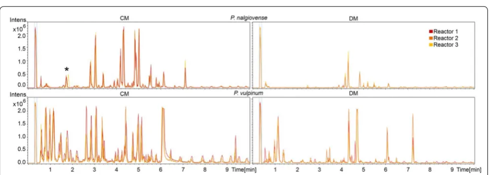
Samples taken during different stages of the submerged cultivations were filtered, extracted, and analyzed for the presence of secondary metabolites in the fermentation medium using reverse phase UHPLC coupled to time-of-flight (QTOF) MS. Analysis of the biological triplicate experiments for each of the tested species in either of the two media were found to be highly reproducible based on the occurrence of base peak chromatogram (BPC) peaks with the same retention times and comparable peak areas in the normalized chromatograms (examples given in Fig. 4).
To allow for identification of the individual compounds found in the samples, the effluent from the UHPLC col-umn was directly analysed by the attached QTOFMS which allowed for the recording of accurate mass. For each sample taken at the end of the cultivation the amount of molecular features (equivalent with the num-ber of different compounds) above an area threshold of 500,000 and an intensity of 10,000 counts in the UHPLC– QTOFMS data was determined. The ten different spe-cies each displayed a unique metabolic profile in the two tested media (Table 2 and Additional file 2). A mini-mum of two compounds was observed in P. flavigenum Table 1 Physiological characteristics of ten Penicillium species cultivated in 1 L bioreactors in DM and CM
Biomass yield on glucose (g DW/g glucose), duration of lag phase (h) and observed morphology at the time point where the CO2 was at its maximum
Species Biomass yield growth phase (Ysx)
DM (g DW/g glucose) Lag phaseDM (h) MorphologyDM Lag phaseCM (h) MorphologyCM
P. flavigenum 0.58 ± 0.05 14.3 ± 0.5 Small clumps/dispersed 10.9 ± 0.3 Dispersed
P. nalgiovense 0.60 ± 0.17 22.5 ± 0.8 Dispersed 14.5 ± 0.1 Dispersed
P. coprophilum 0.37 ± 0.03 23.3 ± 0.6 Pellet 12.2 ± 0.6 Dispersed
P. decumbens 0.55 ± 0.02 17.8 ± 0.9 Dispersed 13.8 ± 0.2 Dispersed
P. vulpinum 0.54 ± 0.14 37.6 ± 1.4 Pellet + clumps + wall 18.3 ± 0.2 Dispersed
P. polonicum 0.25 ± 0.01 30.3 ± 1.7 Wall growth 11.3 ± 0.1 Dispersed
P. antarcticum 0.58 ± 0.03 19.7 ± 0.2 Dispersed 12.5 ± 0.1 Dispersed
P. steckii 0.37 ± 0.02 10.9 ± 2.3 Pellet + wall growth 13.5 ± 0.2 Dispersed
P. solitum 0.29 ± 0.13 19.9 ± 1.2 Pellet + wall growth 9.9 ± 0.2 Dispersed
P. arizonense 0.67 ± 0.04 21.4 ± 0.4 Dispersed 14.0 ± 0.8 Dispersed
Fig. 2 Length of non-exponential growth phase of ten Penicillium
species cultivated in 1 L bioreactors in DM and CM. Non-exponential growth phase was the time in hours between the end of exponential growth phase and the maximum CO2 off gas value. Bars show
aver-age and standard deviation if triplicate submerged fermentations. P.
flavigenum in DM, P. polonicum in CM and P. solitum in CM showed
cultivated in DM, and a maximum of 34 compounds in P. steckii cultivated in CM (last column in Table 2). In eight out of the ten species a greater number of compounds were detected in the cultivations conducted in CM com-pared to DM; exceptions were P. polonicum, where cul-tivation in DM showed a greater number of peaks than in CM, and P. solitum, where the number of compounds was equal in DM and CM.
Additionally, metabolite profiling was performed to identify the known compounds produced during the fer-mentations. In total 34 different metabolites were iden-tified, from which most of them could be confirmed by
comparison of HRMS, UV/Vis and MS/HRMS to a refer-ence standard. Most compounds were unique to one of the ten species, with the exception of chrysogine iden-tified in three species (P. nalgiovense, P. flavigenum and P. antarcticum), fungisporin in three species (P. copro-philum, P. nalgiovense and P. flavigenum) and andrastin A in two species (P. vulpinum and P. decumbens), while andrastin C was only identified in P. decumbens. Addi-tionally, the roquefortine pathway was identified in three species (P. coprophilum, P. vulpinum and P. flavigenum), where depending on the media and species, different intermediates and end products of this pathway could be
Fig. 3 Maximum specific growth rates and maximum CO2 production rates for ten Penicillium species cultivated in 1 L bioreactors in DM and CM.
Dark grey bars: maximum specific growth rates µmax (h−1) and standard deviations calculated by linear regression of minimum three biomass dry
weight data points during the exponential phase. For P. vulpinum, P. polonicum, P. steckii, and P. solitum in DM there were less than three dry weight samples during exponential phase and therefore µmax could not be calculated. Light grey bars: maximum production rate of CO
2 (h−1) and standard
deviations calculated by linear regression of accumulated CO2 values during exponential growth. All values are based on independent triplicate
fermentations, except for the ones with an asterix; there the values are based on duplicate fermentations
Table 2 M etab olit es pr oduc ed in diff er en t gr
owth phases of t
en
Penic
illium
sp
ecies
Total amount of molecular features per sample
(id number of metabolite)
Species Media Idenfied metabolites Sample 1S ample 2 Sample 3 P. coprophilum DM neoxalin e
a (1), meleagri
n
a (2),
oxalin
e
a (4), roqueforne
C
a (5), dechlorogriseofulvin a (6),
griseofulvin
a(7
)
10 (1, 2, 4, 5)
13 (1, 2,
4, 5)
14 (1, 2, 4, 5, 6, 7)
CM
neoxalin
e
a (1), meleagri
n a (2), N6-formyl-roqueforne-c( 3), oxalin e
a (4), fungisporin (6),
dechlorogriseofulvin
a (7), griseofulvin a (8), dehydrogriseofulvi
n
a (9
)
10 (2, 3, 4)
18 (1, 2, 3, 4, 7, 8, 9) 19 (1, 2, 3, 4, 6, 7, 8, 9)
P. nalgiovens
e
DM
chrysogine
a(10), citreo isocumarin a(11), citreo-isocumari
na
nalog-5 (12), diapornol
a(13),
diapornic acid
a (14), 6-methoxy-citreoisocoumari
n
a (15) fungisporin (9
)
0
2 (10, 14
)
10 (10, 11, 12, 13, 14, 15, 9)
CM
chrysogine
a (10), citreo isocumarin a (11), citreo-isocumari
na
nalog-5 (12), diapornol
a (13),
diapornic acid
a (14), 6-methoxy-citreoisocoumari
n
a (15) fungisporin (9
)
5 (14, 15
)
17 (10, 11, 12, 13, 14, 15
,9
18 (10, 11, 12, 13, 14, 15, 9)
P. vulpinum
DM
desoxypatulinic acid (16), patuli
n
a (17), neopatulin (18), meleagrin a (2), oxalin
e a (4), (-)-3-butyl-7-hydroxyphthalide (19), 3-[2'(R)hydroxybutyl]-7-hydroxyphthalide (20), andrasn A a (21)
17 (16, 17
,2
, 4,
20,
21
)
21 (16, 17, 2, 4, 19, 20, 21
)
CM
desoxypatulinic acid (16), patuli
n
a (17), neopatulin (18), meleagrin a (2), oxalin
e a (4), (-)-3-butyl-7-hydroxyphthalide (19), 3-[2'(R)hydroxybutyl]-7-hydroxyphthalide (20), andrasn A a (21)
23 (16, 17, 2, 4, 19, 20, 21
)
27 (16, 17, 2, 4, 19, 20, 21
)
28 (16, 17, 2, 4, 19, 20, 21
) P. polonicu m DM l-tyrosi n
a (22), penicillic acid a (23), leucyltryptophanyl-diketopiperazine a (24), aspterric acid a (25),
verrucoforne
a (26),
10
(22, 24, 25, 26
)
19 (22, 23, 24, 25, 26) 21 (22, 23, 24, 25, 26)
CM
l-tyrosi
n
a (22), leucyltryptophanyl-diketopiperazin
e
a (24), aspterric acid a (25), verrucoforne a (26)
,
2 (26)
4 (25, 26
)
16 (22, 24, 25, 26
) P. decumben s DM andrasn A
a (27), andrasn C (2
8)
3 (27)
4 (27, 28
)
16 (27, 28
)
CM
calbistrin
A
a (26), andrasn
A
a (27), andrasn C (28)
5
24 (26, 27, 28
)
24 (26, 27, 28
)
P. flavigenu
m
DM
chrysogine
a(10), meleagri
n
a(2
)
0
1 (10)
2 (10, 2)
CM
chrysogine
a (10), meleagri
n
a (2), N6-formylroqueforne (3), roqueforne
C
a (5), fungisporin (6
)
0
1 (2
)
5 (10, 2, 3, 5, 6)
P. stecki i DM trichodermamide C a (30) 3 21 25 (30) CM trichodermamide C a (30) 13 29 34 (30) P. antarccum DM chrysogine
a (10), 2-Pyruvoylamino+benzamid
e
a (31), 2-Acetyl-4(3H)-quinazolone a (32)
7 (10, 31, 32
)
8 (10, 31, 32
)
CM
chrysogine
a (10), 2-Pyruvoylamino+benzamid
e
a (31), 2-Acetyl-4(3H)-quinazolone a (32)
6 (10, 31, 32
)
9 (10, 31, 32
) P. arizonense DM pseuron A( 33 ) 4 8( 33 ) CM pseuron A( 33 ) 6( 33 ) 8( 33 ) P. solitu m DM
atlannone A (34)
5 (34)
5(
34
)
CM
(no known compounds)
4
4
Column 3: metabolit
es (and their id numbers) iden
tified with der
eplica
tion, in or
der of r
et
en
tion time
. C
olumn 4, 5 and 6: per sample the t
otal amoun
t of molecular f
ea
tur
es (iden
tified and unk
no wn metabolit es), in bet w een br
ackets the id number of the iden
tified c
ompounds in each sample
. A hea
t map fr
om y ello w t o dar k or ange f ollo w
s the t
otal amoun
t of molecular f
ea
tur
es in the samples
a C
onfir
med with a r
ef
er
enc
e standar
identified; in P. flavigenum only meleagrin and precur-sors were identified while both in P. coprophilum and P. vulpinum the end product of the pathway, oxaline was identified. Three species–metabolite relationships have, to our knowledge, not been described in literature before: P. vulpinum and P. decumbens producing andrastin A and P. flavigenum producing chrysogine.
To identify in which stages of the fermentation the various metabolites were produced, we examined the individual metabolite production profiles covering the three analyzed stages of the fermentation, one during the growth phase, one a few hours after the CO2 val-ues started decreasing and one in the stationary phase (depicted as triangles in Fig. 1 and exact values in Addi-tional file 2). This analysis revealed production of specific metabolites was associated with specific growth phases of the cultivations. For example for P. coprophilum in DM, the metabolites from the roquefortine/oxaline pathway were already present in the first sample, while griseoful-vin and dechlorogriseofulgriseoful-vin could only be detected in the last sample. Another example of this stage depend-ing production was P. steckii grown in CM where the vast majority of the metabolites were identified at the second sample point, while trichodermamide C was only first observed at the third sample point. Many of the metabo-lites in P. steckii were identified as belonging to the family of hynapenes and arohynapenes (based on UV and accu-rate mass) but because of a lack of reference standards could not be assigned to specific peaks and are therefore not shown in Table 2. In some cases, metabolites were produced only in CM or DM; penicillic acid was only produced in P. polonicum in CM, calbistrin A was only detected in P. decumbens in CM, and atlantinone A was only detected in P. solitum in DM. This was also seen for unidentified metabolites (Additional file 2). In 17 out of the 20 cultivations the metabolite levels in the third sam-ple were higher than in the second samsam-ple, showing that part of the metabolites started to be produced only after the growth phase.
Discussion
The fungal kingdom contains a huge reservoir of bioac-tive secondary metabolites; however, the development from discovery of a relevant metabolite to application remains a challenge. The recent genome sequencing of ten secondary metabolite rich Penicillium species facili-tates exploitation of their genome sequences and/or their use as native producing cell factories. The aim of this study was to determine the suitability of these ten Peni-cillia as novel cell factories for native compounds by test-ing their growth performance and secondary metabolite production in submerged cultivations.
Cultivation of the fungal species in controlled sub-merged bioreactors proved that the ten wild type Peni-cillium species all could be cultivated using the same conditions. The cultivations proved to be highly repro-ducible, both in defined medium (DM) and rich medium (CM). In 18 out of the 20 cultivations, replicate values for CO2 off gas and biomass dry weight concentrations were reproducible with maximum CO2 production rates hav-ing standard deviations between 1 and 27%. The cultiva-tions performed in CM showed dispersed growth until the maximum CO2 off gas was reached, while five out of the ten species cultivated in DM showed clump/pelleted growth. The fungal morphology can have an enormous impact on the production of enzymes and primary or secondary metabolites [18]. For example, micro-colonies are required for the production of citric acid by A. niger [19]. However, there is not a defined formula on how morphology affects productivity and it should be tested and optimized for the relevant species and metabolites. The work conducted in this study can be used as a start-ing point in optimization of process parameters for the production of a specific secondary metabolite.
Similar to the growth profiles, the UHPLC–QTOFMS metabolite profiles showed to be reproducible in the bio-logical triplicates of the batch fermentations. The base peak chromatograms revealed that all ten species pro-duced secondary metabolites in submerged cultivations, with most of the metabolites being unique to one of the ten species. The media composition had an influence on secondary metabolite profiles, with eight out of ten spe-cies producing a higher number of compounds in CM than in DM. This is not surprising as the yeast extract containing CYA medium is regarded as a secondary metabolite inducing medium [24]. Some species such as P. solitum and P. flavigenum produced very few metabo-lites in both media; these species could be interesting for the development of secondary metabolite free produc-tion hosts, especially P. flavigenum since it showed to have a high reproducibility and a high growth rate. It has been shown in both P. chrysogenum [25] and in Strepto-myces species [26, 27] that removal of highly producing secondary metabolite pathways can increase the produc-tion of other native, or heterologous secondary metabo-lites. However, the species producing very few secondary metabolites still contain many secondary metabolite bio-synthetic gene clusters (BGCs). In a recent large scale genomic analysis of Penicillium genomes, it was found that 24 Penicillium genomes (including the ten used in this study) contained in total 1317 putative BGCs, cor-responding to an average of 55 clusters per species [15]. This means that a great number of BGCs were inactive in the conditions used in this study, especially when taken into account that one BGC is often responsible for the biosynthesis of a whole family of related metabolites.
Dereplication of the extracts showed that the species produced a broad spectrum of secondary metabolites belonging to several different metabolite classes (polyke-tides, non-ribosomal peptides and terpenes). In total we identified 34 different known compounds, of which many for the first time in submerged fermentations. Here, it has to be taken into account that because of the chemi-cal diversity of the secondary metabolites the extraction could not be optimal for all compounds, and it was only possible to obtain semi-quantitative data. Several of the identified compounds are known to have antibacterial, antifungal and anti-cancer activities, such as the antifun-gal griseofulvin, the antibiotic roquefortines [28] as well as the promising anti-cancer metabolites andrastin A [29, 30] and calbistrin A [31]. At the same time also sev-eral mycotoxins such as patulin and penicillic acid were produced of which elimination need to be considered in a production process. Additionally, we described three new species–metabolite relationships: to our knowledge, this is the first time that P. decumbens and P. vulpinum are described as andrastin A producers and P. flavigenum
as chrysogine producer. Furthermore, it was shown that the species produce many unknown compounds at lev-els above the used detection area threshold; these could be interesting targets for isolation, bioactivity testing and structural elucidation, especially because it is less labori-ous to isolate metabolites from submerged cultures than from agar plates.
In order to utilize metabolic reprogramming tech-niques to optimize production of a secondary metabo-lite, the corresponding BGC of the metabolite must first be identified. The BGCs responsible for production of the detected andrastin A, patulin, griseofulvin, pseuro-tin A, fungisporin and roqueforpseuro-tine/meleagrin pathways have all previously been identified and characterized [32–42]. Although the experimental characterization was done in different species, represents for each of the listed BGCs, except for andrastin A, were recently iden-tified in at least one of the ten analyzed species used in this study [15]. Several other metabolites with predicted BGCs and previously detected in solid cultivations of the corresponding strains, could not be detected in the submerged cultivations of the same strain. This was the case for yanuthones previously detected in extracts of P. flavigenum grown on agar plates [15], and fumagillin, pyripyropenes, austalides, and tryptoquivalines, previ-ously detected in extracts of P. arizonense grown on agar plates [14]. In this context it is interesting that a species like P. arizonense only secreted pseurotin A, despite the large potential for producing a vast number of second-ary metabolites. This would be an advantage if pseurotin A has to be produced in an industrial fermentation, but a disadvantage if any of its other secondary metabolites should be produced [14]. Furthermore we detected small amounts of penicillin G in the CM extracts of the known penicillin G producer P. nalgiovense, but the intensity and area were lower than the threshold values used in this study. It should be considered that in the experiments conducted in this study the biomass was filtered out and only the secreted metabolites were studied, while in plate experiments the intracellular metabolites are also taken into account.
Conclusions
process optimization, classical strain development and/ or with metabolic engineering approaches. Furthermore, all ten species used in this study were recently genome sequenced, which makes the use of rational approaches for improving the product yields or eliminating the pro-duction of other compounds more accessible thanks to the ongoing development of CRISPR/Cas9 genome editing techniques, which has recently been successfully demonstrated in filamentous fungi [43, 44]. The work conducted in this study can assist in deciding the opti-mization strategy for specific secondary metabolites pro-duced by one of the ten Penicillium species.
Authors’ contributions
MW, KFN, and JN conceived the study. SG and JCN performed the cultivations and data analysis on physiological parameters. SG performed the chemical extractions and analysis of metabolites. SG, KFN, JCF and TOL evaluated the data on secondary metabolites. SG and JCN wrote the manuscript. MW and RF supervised the work and co-wrote the manuscript. All authors read and approved the final manuscript.
Author details
1 Department of Biotechnology and Biomedicine, Technical University of Den-mark, 2800 Kgs. Lyngby, Denmark. 2 Department of Biology and Biological Engineering, Chalmers University of Technology, 412 96 Gothenburg, Sweden. 3 Novo Nordisk Foundation Center for Biosustainability, Technical University of Denmark, 2800 Kgs. Lyngby, Denmark.
Acknowledgements
The authors acknowledge Tina Johansen, Martin Nielsen and Milica Ran-delovic for assistance with fermentations; and Christopher Phippen and Aaron John Christian Andersen for assistance with analytics.
Competing interests
The authors declare that they have no competing interests.
Availability of data and materials
All data generated or analysed during this study are included in this published article (and its Additional files 1, 2).
Additional files
Additional file 1. Physiological data. Physiological characteristics for triplicate fermentations of each species in DM and CM. Figures show the CO2 exhaust values and log value of the accumulated CO2. Furthermore a red line through the data points in exponential phase and highlighted in light grey the exponential phase; the time between start of non-exponential growth until the maximum CO2 off gas value. Additionally the R-squared value for the exponential phase, CO2 production rate, the number of data points in exponential phase, the end point (in hours) of the exponential phase and the end point of the non-exponential phase (in hours) are shown.
Additional file 2. Secondary metabolite data. In each separate tab one species with all triplicate samples taken in the DM and CM media. The time points of the samples taken (in hour after inoculation) and the metabolites detected with their maximum m/z value, retention time and intensity. The metabolites were first identified if they had an absolute area higher than 500,000 counts and intensity higher than 10,000 in the time last sample. The presence of these compounds was searched for in all the other samples. Data was generated by UHPLC–DAD–QTOFMS on a maXis HD orthogonal acceleration quadrupole time-of-flight mass spectrometer (Bruker Daltonics).
Consent for publication Not applicable.
Ethics approval and consent to participate Not applicable.
Funding
This work was supported by the European Commission Marie Curie Initial Training Network Quantfung (FP7-People-2013-ITN, Grant 607332).
Publisher’s Note
Springer Nature remains neutral with regard to jurisdictional claims in pub-lished maps and institutional affiliations.
Received: 22 August 2017 Accepted: 26 September 2017
References
1. Visagie CM, Houbraken J, Frisvad JC, Hong S-B, Klaassen CHW, Perrone G, et al. Identification and nomenclature of the genus Penicillium. Stud Mycol. 2014;78:343–71.
2. Frisvad JC, Smedsgaard J, Larsen TO, Samson RA. Mycotoxins, drugs and other extrolites produced by species in Penicillium subgenus Penicillium. Stud Mycol. 2004;49:201–41.
3. Fleming A. On the antibacterial action of cultures of a penicillium, with special reference to their use in the isolation of B. influenzae. Bull World Health Organ. 1929;79:780–90.
4. Oxford AE, Raistrick H, Simonart P. Studies in the biochemistry of micro-organisms: griseofulvin, C(17)H(17)O(6)Cl, a metabolic product of Penicil-lium griseofulvum Dierckx. Biochem J. 1939;33:240–8.
5. Gosio B. Sperimentate su culture pure di bacilli del carbonchio demonst-rarano notevole potere antisettica. GR Accad Med Torino. 1893;61:484. 6. Brown BAG, Srnale TC, Pharmaceuticals B, Park B, King TJ, Hasenkamp
R, et al. Crystal and molecular structure of compactin, a new antifun-gal metabolite from Penicillium brevicompactum. J Chem Soc Perkin. 1976;1:1165–8.
7. Frisvad JC, Filtenborg O. Terverticillate penicillia: chemotaxonomy and mycotoxin production. Mycologia. 1989;81:837–61.
8. Florey HW, Jennings MA, Gilliver K, Sanders AG. Mycophenolic acid—an antibiotic from Penicillium brevicompactum Dierckx. Lancet. 1946;247:46–9.
9. United Nations [Internet]. [Cited 2017 July 9]. http://www.un.org/pga/71/2016/09/21/ press-release-hl-meeting-on-antimicrobial-resistance/.
10. Aminov RI. A brief history of the antibiotic era: lessons learned and chal-lenges for the future. Front Microbiol. 2010;1:1–7.
11. Ling LL, Schneider T, Peoples AJ, Spoering AL, Engels I, Conlon BP, et al. A new antibiotic kills pathogens without detectable resistance. Nature. 2015;517:455–9.
12. O’Connor SE. Engineering of secondary metabolism. Annu Rev Genet. 2015;49:1–24
13. Gibbs PA, Seviour RJ, Schmid F. Growth of filamentous fungi in sub-merged culture: problems and possible solutions. Crit Rev Biotechnol. 2000;20:17–48.
14. Grijseels S, Nielsen JC, Randelovic M, Nielsen J, Nielsen KF, Workman M, et al. Penicillium arizonense, a new, genome sequenced fungal spe-cies, reveals a high chemical diversity in secreted metabolites. Sci Rep. 2016;6:35112.
15. Nielsen JC, Grijseels S, Prigent S, Ji B, Dainat J, Nielsen KF, et al. Global analysis of biosynthetic gene clusters reveals vast potential of secondary metabolite production in Penicillium species. Nat Microbiol. 2017;2:17044. 16. Kildgaard S, Mansson M, Dosen I, Klitgaard A, Frisvad JC, Larsen TO, et al.
Accurate dereplication of bioactive secondary metabolites from marine-derived fungi by UHPLC–DAD–QTOFMS and a MS/HRMS library. Mar Drugs. 2014;12:3681–705.
• We accept pre-submission inquiries
• Our selector tool helps you to find the most relevant journal
• We provide round the clock customer support
• Convenient online submission
• Thorough peer review
• Inclusion in PubMed and all major indexing services
• Maximum visibility for your research
Submit your manuscript at www.biomedcentral.com/submit
Submit your next manuscript to BioMed Central
and we will help you at every step:
18. Krijgsheld P, Bleichrodt R, van Veluw GJ, Wang F, Müller WH, Dijksterhuis J, et al. Development in Aspergillus. Stud Mycol. 2013;74:1–29.
19. Gomez R, Schnabel I, Garrido J. Pellet growth and citric acid yield of
Aspergillus niger 110. Enzyme Microb Technol. 1988;10:188–91. 20. Nielsen J, Johansen CL, Jacobsen M, Krabben P, Villadsen J. Pellet
formation and fragmentation in submerged cultures of Penicillium chrysogenum and its relation to penicillin production. Biotechnol Prog. 1995;11:93–8.
21. Robin J, Jakobsen M, Beyer M, Noorman H, Nielsen J. Physiological char-acterisation of Penicillium chrysogenum strains expressing the expandase gene from Streptomyces clavuligerus during batch cultivations. Growth and adipoyl-7-aminodeacetoxycephalosporanic acid production. Appl Microbiol Biotechnol. 2001;57:357–62.
22. Prosser JI, Tough AJ. Growth mechanisms and growth kinetics of filamen-tous microorganisms. Crit Rev Biotechnol. 1991;10:253–74.
23. McNeil B, Berry DR, Harvey LM, Grant A, White S. Measurement of autoly-sis in submerged batch cultures of Penicillium chrysogenum. Biotechnol Bioeng. 1998;57:297–305.
24. Frisvad JC. Media and growth conditions for induction of secondary metabolite production. Fungal Second Metab Methods Protoc Methods Mol Biol. 2012;944:47–58.
25. Salo OV, Ries M, Medema MH, Lankhorst PP, Vreeken RJ, Bovenberg RAL, et al. Genomic mutational analysis of the impact of the classical strain improvement program on β-lactam producing Penicillium chrysogenum. BMC Genom. 2015;16:937.
26. Komatsu M, Uchiyama T, Omura S, Cane DE, Ikeda H. Genome-minimized
Streptomyces host for the heterologous expression of secondary metabo-lism. Proc Natl Acad Sci. 2010;107:2646–51.
27. Gomez-Escribano JP, Bibb MJ. Engineering Streptomyces coelicolor for heterologous expression of secondary metabolite gene clusters. Microb Biotechnol. 2011;4:207–15.
28. Kopp-Holtwiesche B, Rehm HJ. Antimicrobial action of roquefortine. Eur J Appl Microbiol Biotechnol. 1979;6:397–401.
29. Rho MC, Toyoshima M, Hayashi M, Uchida R, Shiomi K, Komiyama K, et al. Enhancement of drug accumulation by andrastin A produced by Penicil-lium sp. FO-3929 in vincristine-resistant KB cells. J Antibiot. 1998;51:68–72. 30. Shiomi K, Uchida R, Inokoshi J, Tanaka H, Iwai Y, Omura S. Andrastins A-C,
new protein farnesyltransferase inhibitors, produced by Penicillium sp. FO-3929. Tetrahedron Lett. 1996;37:1265–8.
31. Bladt TT, Durr C, Knudsen PB, Kildgaard S, Frisvad JC, Gotfredsen CH, et al. Bio-activity and dereplication-based discovery of ophiobolins and other fungal secondary metabolites targeting leukemia cells. Molecules. 2013;18:14629–50.
32. Matsuda Y, Awakawa T, Abe I. Reconstituted biosynthesis of fungal meroterpenoid andrastin A. Tetrahedron. 2013;69:8199–204.
33. Rojas-Aedo JF, Gil-Durán C, Del-Cid A, Valdés N, Pamela Á, Vaca I, et al. The biosynthetic gene cluster for andrastin A in Penicillium roqueforti. Front Microbiol. 2017;8:1–11.
34. Tannous J, El Khoury R, Snini SP, Lippi Y, El Khoury A, Atoui A, et al. Sequencing, physical organization and kinetic expression of the patulin biosynthetic gene cluster from Penicillium expansum. Int J Food Microbiol. 2014;189:51–60.
35. Chooi YH, Cacho R, Tang Y. Identification of the viridicatumtoxin and griseofulvin gene clusters from Penicillium aethiopicum. Chem Biol. 2010;17:483–94.
36. Maiya S, Grundmann A, Li X, Li SM, Turner G. Identification of a hybrid PKS/NRPS required for pseurotin A biosynthesis in the human pathogen
Aspergillus fumigatus. ChemBioChem. 2007;8:1736–43.
37. Wiemann P, Guo C-J, Palmer JM, Sekonyela R, Wang CCC, Keller NP. Proto-type of an intertwined secondary-metabolite supercluster. Proc Natl Acad Sci USA. 2013;110:17065–70.
38. Tsunematsu Y, Fukutomi M, Saruwatari T, Noguchi H, Hotta K, Tang Y, et al. Elucidation of pseurotin biosynthetic pathway points to trans-acting C-methyltransferase: generation of chemical diversity. Angew Chem Int Ed. 2014;53:8475–9.
39. Ali H, Ries MI, Lankhorst PP, Van Der Hoeven RAM, Schouten OL, Noga M, et al. A non-canonical NRPS is involved in the synthesis of fungisporin and related hydrophobic cyclic tetrapeptides in Penicillium chrysogenum. PLoS ONE. 2014;9:e98212.
40. Ali H, Ries MI, Nijland JG, Lankhorst PP, Hankemeier T, Bovenberg RAL, et al. A branched biosynthetic pathway is involved in production of roquefortine and related compounds in Penicillium chrysogenum. PLoS ONE. 2013;8:1–12.
41. Ries MI, Ali H, Lankhorst PP, Hankemeier T, Bovenberg RAL, Driessen AJM, et al. Novel key metabolites reveal further branching of the roquefortine/ meleagrin biosynthetic pathway. J Biol Chem. 2013;288:37289–95. 42. García-Estrada C, Ullán RV, Albillos SM, Fernández-Bodega MÁ, Durek P,
Von Döhren H, et al. A single cluster of coregulated genes encodes the biosynthesis of the mycotoxins roquefortine C and meleagrin in Penicil-lium chrysogenum. Chem Biol. 2011;18:1499–512.
43. Pohl C, Kiel JAKW, Driessen AJM, Bovenberg RAL, Nygård Y. CRISPR/ Cas9 based genome editing of Penicillium chrysogenum. ACS Synth Biol. 2016;5:754–64.