Original Articles
Differentiation of Human Embryonic Stem Cells into
Embryoid Bodies Comprising the Three Embryonic
Germ Layers
Joseph Itskovitz-Eldor
1, Maya Schuldiner
2, Dorit Karsenti
1, Amir Eden
2, Ofra
Yanuka
2, Michal Amit
1, Hermona Soreq
3, and Nissim Benvenisty
21
Department of Obstetrics and Gynecology, Rambam Medical Center, Faculty of
Medicine, The Technion, Haifa, Israel
Departments of Genetics
2and Biological Chemistry
3, The Institute of Life Sciences,
The Hebrew University, Jerusalem, Israel
Accepted November 25, 1999
rived from totipotent cells of the embryo and therefore, can serve as a putative source of nu-merous types of differentiated cells. Of the vari-ous ES cell lines, murine (mES) cells have been investigated extensively and were shown to be genuinely pluripotent, being able to differentiate into all embryonic cell types (1,2). The multipo-tency of embryonic stem cells is evident from three main features: (A) These undifferentiated cells can be injected into the blastocyst cavity Address correspondence and reprint requests to: Dr.
Nissim Benvenisty, Department of Genetics, The Hebrew University, Jerusalem 91904, Israel. Phone: 972-2-6586774; Fax: 972-2-6586975; E-mail:nissimb@leonardo. huji.ac.il
Abstract
Background: Embryonic stem (ES) cells are lines of cells that are isolated from blastocysts. The murine ES cells were demonstrated to be true pluripotent cells as they differentiate into all embry-onic lineages. Yet, in vitro differentiation of rhesus ES cells was somewhat inconsistent and disorga-nized. The recent isolation of human ES cells calls for exploring their pluripotential nature.
Materials and Methods: Human ES cells were grown in suspension to induce their differentiation into embryoid bodies (EBs). The differentiation sta-tus of the human ES cells and EBs was analyzed by following the expression pattern of several lin-eage-specific molecular markers using reverse tran-scription polymerase chain reaction (RT-PCR) and in situ hybridization.
Results: Here we report the induction in vitro of cystic embryoid bodies from human ES cells. Our findings demonstrate induction of expression of
cell-specific genes during differentiation of the hu-man ES cells into EBs. In the huhu-man EBs, we could show a characteristic regional expression of embry-onic markers specific to different cellular lineages, namely, -globin (mesoderm), neurofilament 68Kd (ectoderm), and -fetoprotein (endoderm). More-over, we present a synchronously pulsing embryoid body that expresses the myocardium marker -car-diac actin. In addition, dissociating the embryoid bodies and plating the cells as monolayers results in multiple morphologies, among them cells with neu-ronal appearance that express neurofilament 68Kd chain.
Conclusion: Human ES cells can reproducibly dif-ferentiate in vitro into EBs comprising the three em-bryonic germ layers. The ability to induce formation of human embryoid bodies that contain cells of neu-ronal, hematopoietic and cardiac origins will be use-ful in studying early human embryonic develop-ment as well as in transplantation medicine.
Introduction
de-J. Itskovitz-Eldor et al.: Characterization of Human Embryoid Bodies 89
and the resultant embryos implanted in pseudo-pregnant mice. The injected ES cells contribute to all cell types in the chimeric progeny mice, in-cluding the germ layer. Thus, in the next gener-ation, mice with the genotype of the ES cells are born (3,4). (B) Subcutaneous injection of embry-onic stem cells into syngeneic mice induces ter-atomas, which may include cells of endodermal, ectodermal or mesodermal origin (5). (C) In vitro aggregation of ES cells results in formation of embryoid bodies (EBs), with regional differenti-ation into embryonically distinct cell types. Dur-ing their in vitro maturation, mES cells undergo massive morphological changes, and acquire various molecular markers of differentiated cell types. These include -globin, a marker of hematopoietic cells (6,7), neurofilament-68Kd, a marker of neuronal cells (8,9), and albumin, which is a marker of hepatic cells (9). Thus, the capacity of mES cells to undergo terminal differ-entiation in vitro to cells of the mesoderm, ecto-derm and endoecto-derm lineages has been well doc-umented. Most recently, ES cell lines, derived from human embryos produced by in vitro fer-tilization, were established (10). The embryos were cultured to the blastocyst stage and inner cell masses were isolated and grown as ES cells. The established cell lines have normal kary-otypes, proliferate in culture with an undifferen-tiated phenotype and differentiate into the vari-ous embryonic lineages in teratomas (10). Clearly, any potential future use of human ES (hES) cells will depend on their capacity to un-dergo embryonic differentiation in vitro. To this end, we initiated a study aimed at inducing hES cell differentiation in vitro into EBs.
Unlike the murine ES cell lines, EBs for-mation from primate ES cells presented con-siderable difficulties. Thus, EBs differentiation from ES cells derived from the common mar-moset (11) were well organized, but appeared inconsistent and asynchronous, and differenti-ation of ES cells from the rhesus monkey (12) was disorganized, so that the resultant EBs failed to form vesicular structures (13). To over-come these obstacles, we explored the poten-tial of the hES cells to aggregate in suspension and form EBs.
Materials and Methods
Cell CulturehES cells (H9 clone; 10) were grown on mouse embryo fibroblasts in culture medium that con-sisted of 80% KnockOut® DMEM (an
opti-mized Dulbeco’s modified Eagle’s medium for ES cells; Gibco-BRL, Gaithersburg, MD), 20% KnockOut® SR (a serum-free formulation; Gibco-BRL), 1 mM glutamine (Gibco-BRL), 0.1 mM -mercaptoethanol (Sigma, St. Louis, MO), 1% nonessential amino acids stock (Gibco-BRL), 103 units/ml leukemia inhibitor
factor (LIF) (Gibco-BRL), and 4 ng/ml basic fi-broblast growth factor (bFGF; Gibco-BRL). Under these conditions, most of the cells were kept in an undifferentiated state. To induce for-mation of EBs, ES cells were transferred using either colagenase (1 mg/ml; Gibco-BRL) or trypsin/EDTA (0.1%/ 1 mM) to plastic Petri dishes (Miniplast, Ein-Shemer, Israel) to allow their aggregation and prevent adherence to the plate. Usually about 106 ES cells were
incu-bated in each 50 mm Petri plate. The hEBs were grown in the same culture medium, ex-cept that it lacked LIF and bFGF.
RNA and RT-PCR
Total RNA was extracted as described (14) and cDNA was synthesized from 1 g total RNA, using random hexamer (pd(N)6) as
primer (Pharmacia Biotech, Uppsala, Swe-den) and Moloney Murine Leukemia Virus (M-MLV) reverse transcriptase (Gibco-BRL). cDNA samples were subjected to polymerase chain reaction (PCR) amplification with DNA primers selective for the human genes. For each gene, the two DNA primers derived from different exons, so as to ensure that the PCR product represented the specific mRNA species and not the genomic DNA. PCR was performed under linear conditions, to reflect the original amount of the specific transcript. The PCR primers used and the reaction con-ditions were:
-fetoprotein; AGAACCTGTCACAAGCT-GTG and GACAGCAAGCTGAGGATGTC; Product: 676 base pair (bp); 20 cycles at 60C in 1 mM MgCl2.
-globin; GACTGAGAGGACCATCATTG and TCAGGACAGAGGATACGACC; Prod-uct: 397 bp. 25 cycles at 60C in 1 mM MgCl2.
PCR products were analyzed by Southern blot hybridization (15). Probes were radiola-belled by random priming (Boehringer Mann-heim, Indianapolis, IN) using [-32P]dCTP (3000 Ci/mM; NEN - Life Science Products, Boston, MA).
In Situ Hybridization
The EBs were fixed in 4% paraformaldehyde and embedded in paraffin. 5 m paraffin–
embedded serial sections of EBs were hybrid-ized to specific 5-biotinilated RNA probes and labeled with a fluorogenic product of streptavidin-conjugated alkaline phosphatase (16). When in situ hybridization was per-formed on cells from dissociated EBs, the cells were grown on glass cover slides coated with fibronectin (50 g/ml in phosphate-buffered saline (PBS; Boehringer Mannheim). The probes used were 50-mer 2-O-methyl 5-biotinylated cRNA of either
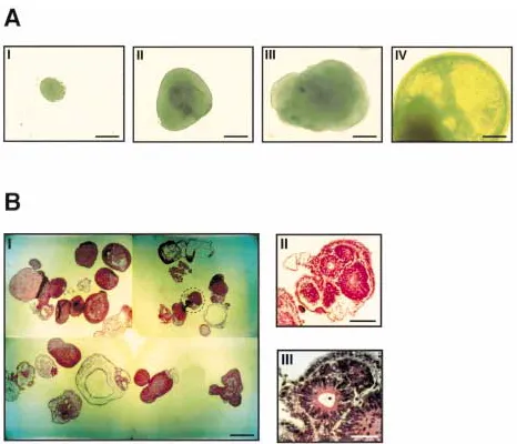
-fetopro-Fig. 1. Formation of human cystic embryoid bodies.(A) Time scale of hEBs development. Shown from left to right is a gradual growth and transformation with time of simple, densely packed hEBs into cystic hEBs. To induce formation of EBs, ES cells were transferred to plastic Petri dishes to allow their aggregation and grown in culture medium lacking leukemia inhibitor factor (LIF) and basic fibroblast growth factor (bFGF). Shown from I to IV are EBs from 3, 7, 10 and 14 days,
J. Itskovitz-Eldor et al.: Characterization of Human Embryoid Bodies 91
tein–TTGTCCCTCTTCAGCAAAGCGAGTTC-CTGGCCTTGGCAGCATT, -globin–TGATG- GTCCTCTCAGTCTTGGTCAGAGACATG-GCGGCAGGGTGGGCAGCT, neurofilament 68Kd–CCTGCGTGCGGATGGACTTGAG-GTCGTTGCTGATGGCGGCTACCTGGCTC, or
-cardiacactin–CGGTGGACAATGGATGGGC-CTGCCTCATCGTACTCTTGCTTGTAATCCA.
Results
To explore the potential of the hES cells to differentiate in vitro, we tried to aggregate the hES in suspension in order to form EBs. In suspension, the hES cells were cultured without LIF and bFGF. In addition, we grew the cells on Petri dishes to prevent their ad-herence to the plate. Under these conditions, hES cells consistently aggregated and formed EBs. Figure 1A shows EBs photographed from 3 to 14 d after initiation of cellular ag-gregation of the hES cells. Initially, these bodies were largely composed of densely packed hES cells, creating simple EBs (Fig. 1A-I). Soon after, the center of the bodies be-came cavitated (Fig. 1A-II and III) and the bodies began to accumulate fluid and turn into cystic EBs (Fig. 1A-IV). Twenty days af-ter initiation of cellular aggregation 20–90% of the structures were cystic (Fig. 1B-I). In parallel, we noted the development of a
vari-ety of cells with epithelial and endodermal morphology (Fig. 1B-II and III).
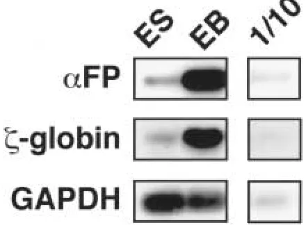
To examine the differentiation status of the cultured hEBs, we extracted RNA from 20-day-old hEBs and from ES cells grown on mouse embryo fibroblasts (as feeder cells). cDNA was reverse-transcribed from this RNA and the expression of several marker genes was tested by PCR under nonsaturating lin-ear conditions using several human-specific DNA primers. For each RNA transcript, the DNA primers derived from separate exons to allow verification that the PCR product repre-sented the cDNA and not the genomic DNA. In addition, in each set of reactions, a sample of RNA (and not cDNA) was used as a tem-plate to control for any DNA contamination. The identity of each of the amplified PCR products was verified by sequence analysis, using an ABI 377 sequencer (Perkin Elmer, Foster City, CA). Robust expression of -feto-protein, an endodermal marker (17) and
-globin, a marker of early hematopoietic cells (18; Fig. 2), demonstrated that the EBs had begun differentiating. The house-keep-ing gene, glyceraldehyde 3-phosphate dehy-drogenase (GAPDH), served as an internal control. Some significant differentiation also occurred in the ES cells grown on feeders, as demonstrated by low levels of -fetoprotein and -globin in the ES samples. By compari-son with the EB samples, the significantly more intense PCR products from the hEB RNA supported and extended the conclusion
Fig. 2. Expression of cell-specific genes in hu-man cystic embryoid bodies.Shown is an RT-PCR analysis of expression of -fetoprotein (FP),
-globin and glyceraldehyde–3–phosphate dehy-drogenase (GAPDH) in human embryonic stem (ES) cells grown on feeder cells, or in 20 day-old
Fig. 3. In situ hybridization analysis of embry-oid bodies.Shown is in situ hybridization detec-tion of expression of neurofilament 68Kd subunit (NF), -globin (Glob), -fetoprotein (FP) and -cardiac actin (cAct) in 20-day-old EBs. Shown are 5 m paraffin–embedded serial sections of three
different EBs stained with hematoxylin and eosin (H&E) or hybridized to specific 5-biotinilated RNA probes and labeled with a fluorogenic product of streptavidin-conjugated alkaline phosphatase (16). Con-hybridization with nonspecific RNA. Scale bar, 100 m.
from microscopic examination that the hEB structures were more differentiated.
To regionally characterize the differentiat-ing cells within the EBs we examined by in situ hybridization the expression of four cell-specific molecular markers, all of which were transcribed very early during embryonic dif-ferentiation. Thus, serial sections of the EBs were hybridized with 5-biotinylated 2 -O-methyl cRNA probes specific to -fetoprotein (17), -globin (18), -cardiac actin (19) or neurofilament 68Kd (20). As shown in Fig. 3, each of these probes reproducibly labeled distinct cell layers in the hEBs. The endoder-mal marker -fetoprotein was primarily ex-pressed in the interior part (Fig. 3) and in some hEBs it was also expressed in the exte-rior layer. A similar pattern of expression of
-fetoprotein was observed by others in EBs derived from human embryonic germ cells (21). In each of the sections, the labeled cells were localized in a specific area, suggesting that they were either clonal, and derived from the same progenitor cell, or that the different
cells were all affected by similar signals and, thus, differentiated to the same specific lin-eage.
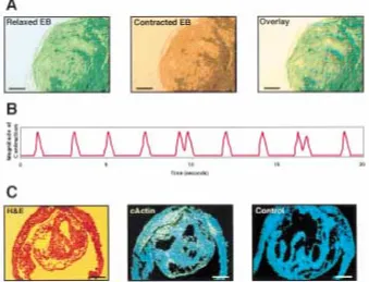
Differentiation into the myocardial lineage was reported to induce development of pulsing muscle in mEBs (22). In a minority of the cys-tic hEBs, rhythmic pulsing was observed. In Fig. 4, we demonstrate a large vacuolated em-bryoid body, including cardiac muscle cell lay-ers that were pulsing in a synchronous rhythm of about 30 pulses/minute (Fig. 4A and B). In situ hybridization of sections from this EB with a probe for -cardiac actin, a marker of embry-onic myocardial cells (19), revealed that the central cavity was indeed surrounded by car-diac muscle cells (Fig. 4C).
J. Itskovitz-Eldor et al.: Characterization of Human Embryoid Bodies 93
Discussion
Our findings demonstrate that hES cells readily differentiate to cystic EBs in a similar manner and time scale to those reported for mES cells. In ES cells derived from the primate rhesus mon-key, it was reported that differentiation was somewhat disorganized and the cells did not de-velop cystic embryoid bodies (13). However, hu-man embryonic germ cells also developed into embryoid bodies similar to our ES cells (21). Un-til now, the hES cells were shown to differenti-ate into the various embryonic lineages only in teratomas. Here, we demonstrate that human ES cells, when differentiating in suspension in vitro, acquire molecular markers specific to the three embryonic germ layers. The different markers used reflect development into hematopoietic cells (-globin), myocardial cells (-cardiac actin), neuronal cells (neurofilament 68Kd) and endodermal cells (-fetoprotein). The differentiating cells also acquired characteristic morphologies, distinct for the hEB regions
ex-pressing different markers. They further devel-oped new functions, as evident from the appear-ance of pulsing muscle cells. As in the mouse, the embryoid bodies displayed the potential to differentiate into various lineages, but no pattern formation or organized organogenesis was ob-served.
Our findings open the field to direct in vitro differentiation of human ES cells into specific lineages. These can serve as a source of mature cells, which may be used in cell trans-plantation and offer an opportunity to study in vitro processes involved in early human em-bryogenesis.
Acknowledgments
We are grateful to Oren Schuldiner for assis-tance and many helpful suggestions. The study was partially supported by the Alon Founda-tion (to N.B.).
Fig. 4. Cardiac muscle differentiation depicted in a pulsing embryoid body.(A) Contracting em-bryoid body. Shown is a video micrograph of a pulsing EB in a relaxed or contracted state. Light absorption in the video micrographs was color coded in either green or red; the two photos were overlaid to demonstrate the changes in opacity, re-flecting contraction during the pulsation of the EB. Scale bar, 200 m. (B) Rhythmic contraction.
References
1. Robertson EJ. (1987) Embryo-derived stem cell lines. In: Robertson EJ (ed). Teratocarcinomas and Embryonic Stem Cells, a Practical Approach. IRL Press, Oxford, pp. 71–112.
2. Dushnik-Levinson M, Benvenisty N. (1995) Embryogenesis in vitro: study of differentiation of embryonic stem cells. Biol. Neonate67:77–83. 3. Capecchi MR. (1989) Altering the genome by homologous recombination. Science 244:
1288–1292.
4. Rossant J, Joyner AL. (1989) Towards a molec-ular-genetic analysis of mammalian develop-ment. Trends Genet.5:277–283.
5. Wobus AM, Holzhausen H, Jakel P, Schoneich J. (1984) Characterization of a pluripotent stem
cell line derived from a mouse embryo. Exp. Cell. Res.152:212–219.
6. Wiles MV, Keller G. (1991) Multiple hematopoi-etic lineages develop from embryonic stem (ES) cells in culture. Development111:259–267. 7. Lindenbaum MH, Grosveld F. (1990) An in vitro
globin gene switching model based on differen-tiated embryonic stem cells. Genes Dev. 4:
2075–2085.
8. Bain G, Kitchens D, Yao M, Huettner JE, Gott-lieb DI. (1995) Embryonic stem cells express neuronal properties in vitro. Dev. Biol. 168:
342–357.
9. Levinson-Dushnik M, Benvenisty N. (1997) In-volvement of hepatocyte nuclear factor 3 in en-doderm differentiation of embryonic stem cells.
Mol. Cell. Biol.17:3817–3822.
Fig. 5. Dispersed cell cultures derived from hEBs.hEBs were grown for 5 d in suspension and then were dissociated with trypsin and plated as a monolayer on cover slides coated with fibronectin. Shown are cells with fibroblast (A) or neuronal (B) appearance. Insert in (B) is a higher magnification
J. Itskovitz-Eldor et al.: Characterization of Human Embryoid Bodies 95
10. Thomson JA, Itskovitz-Eldor J, Shapiro SS, et al. (1998) Embryonic stem cell lines derived from human blastocysts. Science282:1145–1147. 11. Thomson JA, Kalishman J, Golos TG, Durning M, Harris CP, Hearn JP. (1996) Pluripotent cell lines derived from common marmoset (Callithrix jacchus) blastocysts. Biol. Reprod.55:254–259. 12. Thomson JA, Kalishman J, Golos TG, et al. (1995)
Isolation of a primate embryonic stem cell line.
Proc. Natl. Acad. Sci. U.S.A.92:7844–7848.
13. Thomson JA, Marshall VS. (1998) Primate em-bryonic stem cells. Curr. Top. Dev. Biol.38:133–165. 14. Chirgwin JM, Przybyla AE, MacDonald RJ, Rutter WJ. (1979) Isolation of biologically ac-tive ribonucleic acid from sources enriched in ri-bonuclease. Biochemistry18:5294–5299.
15. Southern EM. (1975) Detection of specific se-quences among DNA fragments separated by gel electrophoresis. J. Mol. Biol.98:503–517. 16. Grifman M, Galyam N, Seidman S, Soreq H.
(1998) Functional redundancy of acetyl-cholinesterase and neuroligin in mammalian neuritogenesis. Proc. Natl. Acad. Sci. U.S.A. 95:
13935–13940.
17. Krumlauf R, Hammer RE, Tilghman SM, Brin-ster RL. (1985) Developmental regulation of
-fetoprotein genes in transgenic mice. Mol. Cell. Biol.5:1639–1648.
18. Leder A, Weir L, Leder P. (1985) Characteriza-tion, expression, and evolution of the mouse embryonic -globin gene. Mol. Cell. Biol. 5:
1025–1033.
19. Sassoon DA, Garner I, Buckingham M. (1988) Transcripts of alpha-cardiac and -skeletal actins are early markers for myogenesis in the mouse embryo. Development104:155–164. 20. Julien JP, Meyer D, Flavell D, Hurst J, Grosveld
F. (1986) Cloning and developmental expres-sion of the murine neurofilament gene family.
Br. Res.387:243–250.
21. Shamblott MJ, Axelman J, Wang S, et al. (1998) Derivation of pluripotent stem cells from cul-tured human primordial germ cells. Proc. Natl. Acad. Sci. USA95:13726–13731.
22. Sanchez A, Jones WK, Gulick J, Doetschman T, Robbins J. (1991) Myosin heavy chain gene ex-pression in mouse embryoid bodies. An in vitro developmental study. J. Biol. Chem. 266: