Amplification of neural stem cell
proliferation by intermediate progenitor
cells in
Drosophila
brain development
www.neuraldevelopment.com
NEURAL
DEVELOPMENT
Open Access
Research article
Amplification of neural stem cell proliferation by intermediate
progenitor cells in
Drosophila
brain development
Bruno C Bello*, Natalya Izergina, Emmanuel Caussinus and
Heinrich Reichert
Address: Biozentrum, University of Basel, CH-4056 Basel, Switzerland
Email: Bruno C Bello* - bruno.bello@unibas.ch; Natalya Izergina - natalya.izergina@stud.unibas.ch; Emmanuel Caussinus - emmanuel.caussinus@unibas.ch; Heinrich Reichert - heinrich.reichert@unibas.ch * Corresponding author
Abstract
Background: In the mammalian brain, neural stem cells divide asymmetrically and often amplify the number of progeny they generate via symmetrically dividing intermediate progenitors. Here we investigate whether specific neural stem cell-like neuroblasts in the brain of Drosophila might also amplify neuronal proliferation by generating symmetrically dividing intermediate progenitors.
Results: Cell lineage-tracing and genetic marker analysis show that remarkably large neuroblast lineages exist in the dorsomedial larval brain of Drosophila. These lineages are generated by brain neuroblasts that divide asymmetrically to self renew but, unlike other brain neuroblasts, do not segregate the differentiating cell fate determinant Prospero to their smaller daughter cells. These daughter cells continue to express neuroblast-specific molecular markers and divide repeatedly to produce neural progeny, demonstrating that they are proliferating intermediate progenitors. The proliferative divisions of these intermediate progenitors have novel cellular and molecular features; they are morphologically symmetrical, but molecularly asymmetrical in that key differentiating cell fate determinants are segregated into only one of the two daughter cells.
Conclusion: Our findings provide cellular and molecular evidence for a new mode of neurogenesis in the larval brain of Drosophila that involves the amplification of neuroblast proliferation through intermediate progenitors. This type of neurogenesis bears remarkable similarities to neurogenesis in the mammalian brain, where neural stem cells as primary progenitors amplify the number of progeny they generate through generation of secondary progenitors. This suggests that key aspects of neural stem cell biology might be conserved in brain development of insects and mammals.
Background
Neural stem cells are primary precursors that have the ability to renew themselves at each division such that one of the two daughter cells retains stem cell identity, while the other enters a program of differentiation and contrib-utes to a continuous supply of neural cell types.
Under-standing how neural stem cells maintain their pluripotent state and how their progeny differentiate into distinct neural fates is of central importance for understanding nervous system development (for recent reviews, see [1-3]). Neural stem cells must exert a tight control over pro-liferative divisions so as to generate the appropriate
Published: 19 February 2008
Neural Development 2008, 3:5 doi:10.1186/1749-8104-3-5
Received: 27 November 2007 Accepted: 19 February 2008
This article is available from: http://www.neuraldevelopment.com/content/3/1/5
© 2008 Bello et al.; licensee BioMed Central Ltd.
number of neural progeny necessary to populate the nerv-ous system but not to produce so many self-renewing daughters that neoplastic overgrowth occurs [4]. There-fore, a better comprehension of the mechanisms that con-trol the behavior of neuronal stem cells and their progeny may also be important for understanding brain tumors [5,6].
The Drosophila central nervous system is an excellent sim-ple model system for analyzing the molecular mecha-nisms that control neural stem cell divisions (for recent reviews, see [7,8]). Drosophila neural stem cells, called neuroblasts (NBs), delaminate as single cells from the neuroectoderm and undergo repeated asymmetric cell divisions, each of which self-renew the NB while produc-ing a smaller neural progenitor cell called a ganglion mother cell (GMC). Compared to the NB, the GMC adopts a radically opposite fate and undergoes a single neurogenic division to produce two cells that exit the cell cycle and differentiate (reviewed in [9-12]). During embryogenesis, each NB produces a lineage of 10–20 pri-mary neural cells that contribute to the functional cir-cuitry of the larva. Following a period of quiescence, most NBs resume their asymmetric mode of proliferative divi-sions during post-embryonic development and generate the lineage-related clusters of secondary adult-specific neurons that make up the bulk of the adult central brain and thoracic ganglia [13-16].
Mechanisms involved in NB division and neural prolifer-ation during embryogenesis have been studied in great detail (reviewed in [7,17-19]). NB divisions are known to be molecularly as well as morphologically asymmetric, and a number of key intrinsic and extrinsic factors that control the asymmetrical and self-renewing divisions of these NBs have been identified. Among these, a central role is played by molecular polarity cues that establish the apico-basal polarity of the NB and enable the asymmetric segregation of localized cell-fate determinants from the NB to the GMCs at each asymmetric cell division. Although considerable insight has been attained into the mechanisms by which NB polarity is established and maintained, little is known about the function of the pro-teins that are asymmetrically localized to the GMC. The best characterized of these fate determinants is the home-odomain protein Prospero, which is synthesized in the NB and localized at the cell cortex in a polarized manner. Upon segregation to the GMC, Prospero acts in the nucleus to repress NB-specific gene expression (including genes required for self-renewal) and activate genes for GMC fate specification and terminal differentiation of post-mitotic neurons [20-23]. Asymmetric segregation of Prospero protein is mediated by the adaptor coiled-coil protein Miranda. Once segregated from the NB to the GMC, Miranda is degraded, thereby releasing Prospero
from the cell cortex and allowing it to enter the nucleus [24-26]. Indeed, the nuclear localization of Prospero is one of the first molecular differences between the self-renewing NB and a differentiating cell [27,28].
During the postembryonic period of neurogenesis, the NBs of the central brain and thoracic ganglia are thought to undergo a similar proliferation program and express many of the asymmetric cell fate determinants that char-acterize embryonic neurogenesis [29,30]. Nuclear locali-zation of Prospero is manifest in GMCs and postmitotic neurons of the larval brain, and loss of prospero in somatic clones results in massive overproliferation of cells that express molecular markers of NBs [31-33]. Additionally, numerous other molecular control elements are likely to be required for the continuous mitotic activity of NBs dur-ing postembryonic life (reviewed in [34]).
Controlled neuronal proliferation is especially important for the generation of the adult brain. The mature brain of
Drosophila is an exceedingly complex structure with numerous highly organized neuropil assemblies, such as the mushroom bodies, central complex and antennal lobes, as well as other specialized neuropils and major fiber tracts required for complex behavioral functions [35]. Remarkably, approximately 95% of the neurons that make up the adult brain are post-embryonic in origin, and in the central brain all of these neurons are produced by a set of only about 100 bilaterally symmetrical NBs [36,37]. Given the fact that 100 NB pairs generate the tens of thou-sands of differentiated, spatially heterogeneous neurons in the adult central brain, sophisticated mechanisms for lineage- and region-specific amplification control of NB proliferation are likely to be required during post-embry-onic brain development. However, with the exception of rough estimates, which suggest that each brain NB might undergo between 40 and 60 rounds of post-embryonic mitosis to produce lineages of 100–150 neurons, very lit-tle is known about this process and the underlying molec-ular mechanisms.
pro-The DM brain NBs generate a large number of progeny during larval development Figure 1
duce neural progeny, implying that they are IPs. The pro-liferative divisions of these IPs are morphologically symmetrical, but molecularly asymmetrical in that cell fate determinants such as Prospero and Miranda are segre-gated into only one of the daughter cells. The IPs are gen-erated by a specific set of NBs that do not segregate Prospero to their smaller daughter cell, thereby allowing this cell to retain proliferative capacity instead of
undergo-ing its final neurogenic division. The amplification of NB proliferation through IPs reported here for Drosophila
bears remarkable similarities to mammalian neurogene-sis, where neural stem cells as primary progenitors often amplify the number of progeny they generate via symmet-rically dividing secondary progenitors (reviewed in [2]). This suggests that key aspects of neural stem cell biology
The DM NBs generate an exceptional number of neuronal progenitors Figure 2
might be conserved in brain development of flies and mammals.
Results
Large neuroblast lineages are located in the dorsomedial brain hemispheres
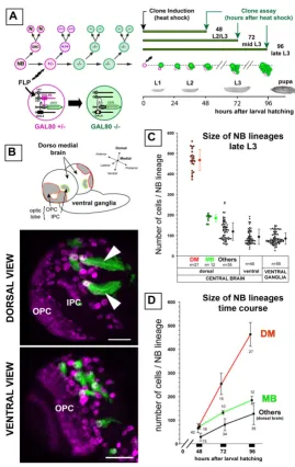
Since most of the secondary, adult-specific neurons of the brain are generated during larval development [38], we used mosaic-based MARCM techniques to label NB line-ages (hereafter referred to as 'NB lineline-ages' or 'NB clones') in the developing larval nervous system [39]. Random mitotic recombination was induced in NBs within a few hours after larval hatching (ALH) in order to achieve pos-itive labeling of their clonal post-mitotic progeny (Figure 1a). Labeled NB clones typically consisted of a single NB, unequivocally recognizable as a large cell of roughly 10
μm in diameter, and an associated cluster of smaller cells representing its larval progeny (Figure 1a,b) [40,41].
Prominent among these were unusually large clones recoverable at the DM margins of the brain hemispheres (Figure 1b). Six NBs located in the most medial position of each hemisphere were found to generate this type of clone, hereafter referred to as 'DM lineages' or 'DM clones'. As detailed below, the parental DM NBs were eas-ily identifiable owing to the signature pattern of Miranda-positive cells that followed the lateral to medial orienta-tion of their progeny in these labeled clones. Morpholog-ically, DM NBs were indistinguishable from other NBs in the central brain or in the ventral ganglia. Thus, cell vol-ume measurements of DM and non-DM NBs in third lar-val instar brains gave comparable lar-values of 344 ± 94 μm3
(n = 12) and 424 ± 110 μm3 (n = 13), respectively.
Prelim-inary analysis of the axonal tracts suggests that the large NB clones in the dorsal brain correspond to the pl and pm subgroups of the Dorsoposterior medial (DPM) lineages previously described (data not shown) [16].
To compare the proliferative capacity of the DM NBs with that of other NBs in the larval central nervous system, we quantified the number of cells in DM NB lineages, in mushroom body NB lineages, and in other NB lineages scored randomly in different brain and ventral ganglion regions of the late third instar larvae shortly before pupa-tion (96 h ALH). The number of cells in the DM lineages had an average value of 450 (range 370–580). Remarka-bly, this was more than twice the average number of cells observed for the larval lineages of the mushroom body NBs (184 ± 17, n = 17) or for other larval NB lineages scored in other areas of the central nervous system (Figure 1c).
To determine the rate of clone size increase during larval central nervous system development, we counted the number of cells in MARCM-labeled DM NB clones,
mush-room body NB clones and other dorsal brain NB clones at various larval stages (Figure 1d). Following a quiescent phase in the early developing larva, most NBs had entered mitosis by the late second larval instar stage [38]. Our observations show that at this stage (48 h ALH), NBs in the dorsal brain had generated only a small number of postembryonic cells and that no pronounced lineage-spe-cific differences in progeny number was apparent (Figure 1d, 48 h ALH). However, at 72 h and 96 h ALH, the DM lineages had increased markedly in size when compared to other dorsal brain NB lineages, indicating an approxi-mate four-fold increase in their rate of proliferation (Fig-ure 1d).
To investigate this further, we cultured MARCM-labeled brain explants in 5-bromodeoxyuridine (BrdU) and then used anti-BrdU immunocytochemistry to determine the number of cells engaged in S-phase in DM clones com-pared to other NB clones of the central brain. Following a 90 minute pulse of BrdU incorporation in L3 brain explants, we found a markedly higher number of BrdU-positive cells in DM clones (38 ± 8 BrdU BrdU-positive cells, n = 8 clones) than in the other NB clones scored at random in dorsal brain regions of the same specimens (4 ± 1.5, n = 27). (This higher rate of BrdU incorporation in DM clones was also observed at earlier stages and in various conditions of incubation; data not shown.)
These data indicate that a significant amplification of pro-liferation occurs in the DM lineages when compared to other NB lineages of the central brain (hereafter collec-tively referred to as 'non-DM' lineages).
DM lineages contain a large population of mitotically active progenitor cells
The large number of cells found in the DM NB clones could, in principle, be due to an unusually high rate of mitotic activity of the DM NBs. However, immunodetec-tion of mitotic DNA in MARCM clones (via the phospho-histone H3 (PH3) epitope) revealed a comparable mitotic frequency in these NBs (22.5%, n = 40) compared to NBs found in dorsal (16.7 %, n = 48) or ventral (21.6 %, n = 97) brain lineages. This prompted us to search for other types of progenitor cells in these lineages. To this aim, we first characterized molecular markers enabling in situ
detection of mitotically active versus post-mitotic cells in labeled NB lineages of the larval brain.
(Figure 2e). DM lineages contained an average of 56.7 ± 11.8 Elav-negative cells (n = 10 clones) closely associated with the Elav-negative NBs. This was over 10 times more than in non-DM NB clones (4.7 ± 1.7 cells, n = 114), sug-gesting that the DM lineages contain a markedly higher number of mitotically active progenitor cells.
Could these smaller Elav-negative cells associated with the NBs be GMCs? To investigate this, we first studied the expression of the coiled-coil protein Miranda. The
miranda gene has been reported to be expressed in larval NBs but not in their GMCs [42]; Miranda expression might, therefore, be a useful marker for differentiating NB-like cells from GMCs. In non-DM lineages, Miranda was strongly expressed in the NBs but only very weakly Molecular characterization of NB-like and GMC-like progenitors in the progeny of DM NBs
Figure 3
Molecular characterization of NB-like and GMC-like progenitors in the progeny of DM NBs. Confocal images of MARCM-labeled NB clones in the dorsal part of larval brains stained for the markers indicated on the top of the columns. Representative views of
(a-f) non-DM lineages are used as a reference for (g-i") the DM lineages. Clones were labeled with CD8::GFP (membrane marker, green in all panels) and CNN::GFP (centrosomes visualized as bright green spots in e, f, i-i"). Proliferative cells are detected by anti-Cyclin (red in e, f, i-i') and anti-PH3 during mitosis (blue in all panels). In a non-DM NB clone, mitosis is restricted to two cell types: the NB and a sin-gle GMC in close proximity (a-f, asterisks and arrowheads, respectively). NBs show a unique pattern of polarized expression of Prospero and Miranda at the cell cortex during mitosis (a, c) and stable expression of Cyclin E throughout the cell cycle (e, mitosis; f, interphase). In contrast, the GMC is uniquely defined when engaged in mitosis (PH3 positive) by nuclear localization of Prospero (b, inset), weak uniform cortical localization of Miranda (d, inset) and lack of Cyclin E (f, inset). (g-i) In DM clones many progenitors other than the NB are identi-fied as PH3-positive nuclei. These cells show patterns of marker expression usually found in mitotic NBs (IP; arrows) or mitotic GMCs (arrowheads). Lower panels show close up views of the areas boxed in (g-i). The two types of mitotic progenitors can be detected simul-taneously in a single DM lineage (images) and are found at a comparable ratio when quantified in multiple clones using the three independ-ent markers (histograms). IP, small NB-associated intermediate progenitor with NB-like marker expression. Scale bars: 10 μm (a-f) or 15
expressed in the set of smaller, Elav-negative cells associ-ated with the NBs, suggesting that these Elav-negative cells were GMCs (Figure 2a,a'). (Their weak expression of Miranda could be due to perdurance of the protein during cell divisions; see also [29,30]). In DM lineages, Miranda was strongly expressed in the NB; however, in contrast to non-DM lineages, distinct Miranda expression was also observed in many of the smaller, Elav-negative cells asso-ciated with the NBs (Figure 2b,b'). This suggests that the smaller Elav-negative/Miranda-positive cells in the DM lineages might not be GMC-like, but might have proper-ties that are more NB-like. To investigate this further, we next attempted to find other markers for progenitor cells and, thus, examined the expression of Cyclin E (CycE) and PH3 as markers of mitotically active cells.
In green fluorescent protein (GFP)-labeled non-DM NB clones, used as control, a small number of GMCs were observed as small NB-associated cells expressing either CycE or PH3 (Figure 2c,c'). At 96 h ALH we found an aver-age of two CycE-positive cells (range one to five) and a maximum of one cell engaged in mitosis as visualized by anti-PH3 (Figure 2e) [40]. This pattern was consistent with live imaging data obtained in experiments on cul-tured nervous systems to monitor asymmetric NB
divi-sions [43]. Thus, as in the embryo, these larval NBs divide by a budding process that generates a set of smaller GMCs, each GMC is born adjacent to the previous one, and the division of the 'oldest' GMC is delayed compared to that of the NB.
Contrasting with this simple pattern, DM lineages con-tained an average of 38 CycE-positive cells located around the NB, and many scattered mitoses, up to 14 per clone, were observed by PH3 immunoreactivity (Figure 2d,d',e). This strikingly high level of ongoing mitotic activity and engagement in the cell cycle in DM lineages compared to other central brain lineages (including mushroom body lineages) was seen at all stages of larval development examined (Figure 2f). These findings indicate that signifi-cantly elevated mitotic activity occurs among the numer-ous small NB-associated cells in larval DM lineages. Moreover, they are in accordance with the idea that these cells do not adopt a GMC fate, but rather remain mitoti-cally active and continue to proliferate. In this case, these cells would have the characteristics of IPs that amplify the proliferation of their parent NBs (primary progenitors) in the DM lineages.
Live imaging of multiple and repeated division of DM NB daughter cells in MARCM-labeled clones Figure 4
Live imaging of multiple and repeated division of DM NB daughter cells in MARCM-labeled clones. Frames from time-lapse recordings of a DM clone labeled with CD8::GFP and tau::GFP in larval brain cultured over 13 hours. The large NB, not visible in these frames, divided twice during this time period (Additional data file 1). The time is indicated in minutes relative to the start of the recording.
Molecular markers reveal two types of non-neuroblast progenitor cells in DM lineages
If some of the mitotically active cells in DM NB clones are amplifying IPs, they might be expected to have cellular and molecular features in common with proliferating NBs. To investigate this, we first examined the expression patterns of Prospero, Miranda, and CycE in NBs of non-DM lineages, used as control, as well as in the small NB-associated progenitors of the DM lineages. For this, MARCM clones induced at larval hatching were scored at 96 h ALH. Importantly, we further restricted our analysis to cells engaged in mitosis (PH3-positive) in order to identify progenitor cells unambiguously and to obtain valid comparisons, since all markers showed cell-cycle dependent expression (see below). (Clones analyzed at 48 h or 72 h ALH gave comparable results; data not shown.)
In non-DM clones, Prospero was specifically detected at the cellular cortex of the NBs, accumulating on one side during mitosis (Figure 3a; n = 57; 100%). All other cells in the clones expressed Prospero in the nucleus or uniformly throughout the cell, thus including both GMCs and post-mitotic cells. Localization of Prospero was more specifi-cally revealed in the GMCs by co-staining with anti-PH3 (Figure 3b; n = 37; 100%) or CycE (not shown). In strik-ing contrast, in DM lineages 31% of PH3-positive small NB-associated cells expressed Prospero at the cortex in a polarized manner. This expression pattern was, thus, sim-ilar to that observed in dividing NBs (Figure 3g,g", arrow). The remaining dividing, NB-associated cells showed uni-form expression of Prospero throughout the cell at mito-sis; their pattern was, thus, GMC-like (Figure 3g,g' arrowheads).
As expected, the adaptor protein Miranda formed promi-nent cortical crescents in dividing NBs of non-DM clones (Figure 3c, asterisks). In the associated GMCs, Miranda was detected at weaker levels with uniform cortical distri-bution both at interphase and during mitosis (Figure 3c, inset, and Figure 3d, arrowheads). Strikingly, in DM line-ages, 36% of the NB-associated cells showed strong and polarized expression of Miranda during mitosis, as described for dividing NBs (Figure 3h,h", arrows). The remaining dividing cells showed weak and uniform corti-cal locorti-calization of Miranda; their Miranda expression pat-tern was, thus, GMC-like (Figure 3h,h' arrowheads).
To confirm the presence of both NB-like and GMC-like progenitors in the DM NB lineages, we searched for mark-ers of cellular identity that did not rely on the conven-tional criteria of cell size and/or cortical polarity. Significantly, we found that in non-DM lineages (taken as reference lineages), CycE was detected in virtually all the self-renewing NBs during mitosis (Figure 3e, asterisks; n = 74), but never during the terminal division of the GMCs
(Figure 3f, arrowheads; n = 48). This distinctive criterion for cell identity was only applicable during mitosis because all progenitor cells expressed CycE at interphase, irrespective of their size (Figure 3e,f; PH3- nuclei; see also
Figure 2c,d). In DM lineages, some of the small itive cells were negative for CycE but other small PH3-pos-itive cells were posPH3-pos-itive for CycE (Figure 3i,i', arrow and arrowhead). Thus, in agreement with the data obtained using markers of cell polarity, both NB-like and GMC-like progenitors could be identified simultaneously in the progeny of a single DM NB (Figure 3g–i). Furthermore these two types of progenitors were observed specifically in these lineages and at all larval stages examined. Thus, the small CycE-positive/PH3-positive progenitors repre-sented 55% (n = 64), 45% (n = 93) and 40% (n = 105) of the mitotic cells found in DM NB clones at 48 h ALH, 72 h ALH and 96 h ALH, respectively. The small CycE-posi-tive/PH3-positive progenitors were never found associ-ated with NBs of the ventral brain or the ventral ganglia at the corresponding stages (114 PH3-positive cells in 297 clones examined).
Taken together, these data indicate that the larval DM lin-eages contain two types of molecularly distinct progenitor cells other than NBs. Although not readily identifiable by their size, approximately two-thirds of these cells have molecular expression patterns of Prospero, Miranda and CycE that are characteristic of GMCs. In contrast, the remaining third have expression patterns of Prospero, Miranda and CycE that are remarkably similar to the pat-terns found in proliferative NBs. These novel NB-like pro-genitors are hereafter referred to as IPs. Our data further show that IPs are generated by DM NBs throughout larval neurogenesis in a quantitatively stable and balanced ratio with GMC-like progenitors and post-mitotic neurons.
Intermediate progenitor cells divide repeatedly and produce multicellular neuronal clones
Clonal expansion of IPs analyzed by MARCM Figure 5
Asymmetrically dividing DM NBs do not express Prospero Figure 6
Asymmetrically dividing DM NBs do not express Prospero. Confocal images of NB divisions in a canonical NB lineage (top pan-els) compared to a DM lineage (bottom panpan-els). Shown are representative CD8::GFP-labeled clones (green), seen around the NB in late larval brains stained for Miranda (MIRA, blue) and Prospero (PROS, red). Single channels are also shown in gray scale for better contrast.
we repeatedly observed small, NB-associated cells that divided more than once. Two subsequent symmetrical divisions of such a progenitor cell are visible in the still images taken from a time-lapse laser confocal movie (Fig-ure 4b).
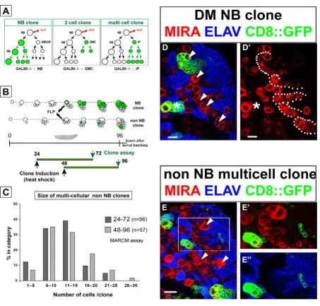
Next, we performed a more detailed analysis of the differ-ent types of MARCM clones that were recoverable in the DM lineages. To date, only two types of multicellular clones have been observed in the central brain following a somatic recombination event in a parental NB and the loss of the GAL80 repressor in one of the post-mitotic sib-lings. Thus, the NB clones described above derive from the proliferation of GAL80-minus NB founders, while two cell clones are obtained from GAL80-minus GMCs (Figure 5a). Other possible recombination events may occur in a GMC, but they result in the labeling of a single post-mitotic daughter cell [39,41]. In DM lineages containing
repeatedly dividing IPs, a third type of non-NB clone con-sisting of more than two labeled cells would be predicted to occur following the loss of the GAL80 repressor (Figure 5a).
Mitotic recombination was randomly induced in progen-itor cells at 24 h and 48 h ALH and progenies were exam-ined in isolated GFP-labeled clones 48 hours later (Figure 5b). As expected, single cell-, two cell-, and NB clones were recovered throughout the central nervous system. Prominent among the latter were the exceptionally large DM NB clones identifiable in the dorsal brain by their medial position and the spatial orientation of the labeled progeny that extend from the typical large cluster of late born Miranda-positive cells (Figure 5d,d'). Consistent with their linear growth rate (Figure 1d), we measured comparable clone sizes for DM NB clones generated dur-ing each of the two overlappdur-ing 48 hour windows (157
Unequal segregation of Prospero/Miranda during symmetric division of IPs Figure 7
cells ± 33, n = 14 clones, and 220 cells ± 43, n = 16 clones, respectively). Likewise, non-DM NBs selected at random in the dorsal brain also generated comparable, albeit smaller, NB clones in the same time periods (63 cells ± 20, n = 40 clones, and 66 cells ± 23, n = 48 clones, respec-tively). Importantly, however, numerous clones lacking a NB and consisting of more than two cells were recovered in these experiments. These multicellular non-NB clones were found only in close spatial association with DM NBs and their progeny (Figure 5e,e'). Cell counts revealed a wide range of clone sizes in these lineages. Most clones, however, comprised 6–25 cells and this class was observed at comparable frequency in the two time win-dows examined (73% and 67%, respectively; Figure 5c). In over 90% of the cases examined, the cells in these mul-ticellular clones expressed Elav, indicating that they were composed exclusively of post-mitotic neurons (Figure 5e,e').
The observed variability in clone size could be due to intrinsic variations in the mitotic capacity of different IPs and/or may result from mitotic recombination occurring in an IP that had already completed a variable number of divisions after its birth. Interestingly, the distribution of clonal cell number appeared remarkably similar when FLP/FRT recombination was induced at 24 h or at 48 h ALH (Figure 5c). This suggests that the mitotic potential of IPs is independent of their birth date from their parental DM NBs during larval development.
These findings imply that IPs in DM lineages can divide several times and produce differentiated progeny in less than 48 hours. Thus, they allow considerable amplifica-tion of the number of neurons produced in comparison to the standard mode of division adopted by other lineages in the central brain.
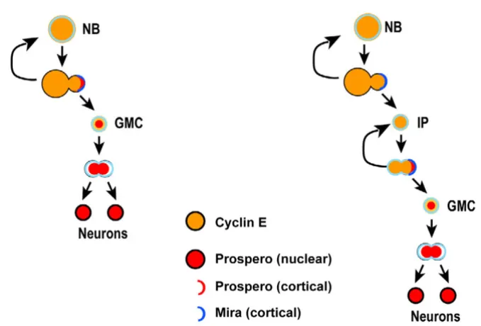
Model for a transient amplifying progenitor cells in DM NB lineages Figure 8
DM neuroblasts do not segregate Prospero protein to their daughter cells
The experiments described above show that DM NBs gen-erate multiply dividing daughter cells that produce neural progeny. Surprisingly, these amplifying IP cells appear to be restricted to the DM lineages. What might explain this restriction? DM and non-DM NBs are not morphologi-cally distinguishable and both divide asymmetrimorphologi-cally to generate smaller progeny cells (Figure 3 and below).
A large amount of evidence indicates that the polarized assembly of multiprotein complexes at the cellular cortex during mitosis is both a characteristic hallmark of NBs and a key determinant in promoting their self-renewing ability. As exemplified in non-DM lineages (Figure 6a,c), Prospero and Miranda are synthesized in the NB and they co-localize on one side of the cortex at metaphase (Figure 6a, asterisk). This asymmetric distribution results in une-qual segregation of these proteins to the budding new GMC as visualized at telophase or soon after cytokinesis (Figure 6c, asterisk). (Older GMCs located in close prox-imity to the newly generated GMC show a much lower level of Miranda and manifest the same type of nuclear localization of Prospero as do all other post-mitotic nuclei of the clone; Figure 6c, n > 50 clones). Importantly, the loss of these fate determinants in mosaic clones leads to unrestricted proliferation of the GMC in situ and the acquisition of neoplastic characters of mutant cells in transplantation assays [31-33,44].
Remarkably, and in contrast to all other Drosophila NBs described to date, Prospero was undetectable in the DM NBs during mitosis (Figure 6b,d). In all DM NB clones examined (n = 25), Miranda, but not Prospero, formed a cortical crescent in the dividing NB at metaphase (Figure 6b, asterisk) and segregated to the smaller daughter cell (Figure 6d). As a result, the IPs that derived directly from the DM NB lacked nuclear Prospero. GFP-labeled DM lin-eages typically contained 28 ± 9 Prospero-negative cells close to the NB (Figure 6b, white dots, n = 14 clones). These are likely to be accumulating IPs in interphase because they showed weak uniform expression of Miranda at the cortex and did not express PH3 (Figure 6b and data not shown). At IP mitosis, however, Prospero was unambiguously detected in these progenitors and showed co-localization with Miranda in a polarized man-ner (Figure 6b, arrows).
These data identify the DM NBs as a unique subset of neu-ral stem cell-like progenitors that do not express and seg-regate Prospero during mitosis, thereby generating daughter cells that are molecularly distinct from GMCs.
Intermediate progenitor cell divisions are morphologically symmetrical but molecularly asymmetrical
Studies on asymmetric neural stem cell division in Dro-sophila have established a simple scheme that links cell size of sibling daughter cells, restriction of mitotic poten-tial and partitioning of fate determinants. Thus, in the canonical scheme exemplified in MARCM-labeled non-DM clones, the only self-renewing cell is the large NB that segregates Miranda/Prospero to its small GMC daughter cell during mitosis (Figure 6a,c). In contrast, the terminal division of the GMC involves the formation of equal-sized daughter cells at telophase and equal partitioning of Miranda/Prospero to both cells (n = 27; Figure 7a,c and data not shown).
The asymmetric division of DM NBs is also associated with the unequal segregation of Miranda to the smaller daughter cell (Figure 6b,d). Moreover, the resulting IP divides symmetrically to generate sibling cells of similar size as examined at telophase (n = 14; Figure 7b,d). Thus, in terms of the morphology of their cell divisions, IP cells are more like GMCs than like NBs. However, in sharp con-trast to GMCs, mitotic IPs show cortical crescents of Miranda and Prospero (Figure 6b) and unequal partition-ing of these two proteins at telophase (Figure 7d; n = 7). Thus, in terms of the segregation of cell fate determinants, dividing IP cells are remarkably more NB-like and differ substantially from GMCs.
Taken together, these findings demonstrate that the prolif-erative divisions of amplifying IPs in DM lineages have novel cellular and molecular features. These divisions are morphologically symmetrical and lead to two daughter cells of similar size, but molecularly asymmetrical in that the differentiating cell fate determinants Prospero and Miranda are segregated into only one cell. The ensuing absence of these differentiating cell fate determinants in the remaining daughter cell is likely to be a significant fac-tor in the mitotic activity of amplifying IP cells.
Discussion
The data presented here are consistent with a novel model for neurogenesis exemplified by the DM NBs, which divide asymmetrically in a stem cell mode to self-renew and generate IP daughter cells (Figure 8b). In this process, they do not segregate the cell fate determinant Prospero into the IP cells, which subsequently repeatedly divide symmetrically (in morphological aspects) yet asymmetri-cally segregate the cell fate determinants Prospero and Miranda during mitosis. The daughter cell that receives the Prospero and Miranda determinants is fated to become a differentiating GMC-like cell, whereas the other daughter cell retains its ability to divide several more times.
This novel model postulates that DM NBs produce exclu-sively IPs and not GMCs. The alternative notion, that the NB sometimes produces an IP and sometimes a GMC, is unlikely given that Prospero is never detected in the NB and, thus, cannot be segregated to one of its daughter cells as would be required for GMC generation. The model also posits that GMCs are produced by IPs through (function-ally) asymmetrical divisions that result in one daughter cell becoming a GMC while the other daughter cell self-renews as an IP. Alternative scenarios, such as one in which IPs first divide symmetrically to expand in numbers and then adopt a GMC fate to generate differentiating neurons, are unlikely given the spatiotemporal pattern of Prospero/Miranda expression and the stable ratio of IPs versus GMCs observed in DM NB clones throughout lar-val development.
The experimental findings that support this novel model have implications for our understanding of neural stem cells and proliferation control. These are discussed in the following.
The NBs of the developing central brain and ventral gan-glia divide asymmetrically in a stem cell mode in which the larger NB self renews and the smaller daughter cell dif-ferentiates into a different cell type, usually a GMC (reviewed by [7,8,10-12,18]). This asymmetric division of the parent NB has been thought to be tightly coupled with the asymmetric segregation of cell fate determinants, and central among these molecular determinants is the tran-scription factor Prospero, which is required in GMCs to inhibit self-renewal and to promote differentiation [20-23,27,28]. Our findings indicate that the asymmetric seg-regation of Prospero does not occur in all dividing brain NBs. Indeed, in the DM NBs the lack of asymmetric segre-gation of Prospero to the IPs may be a key element in imparting (transient) NB-like features to these proliferat-ing cells.
The GMCs of the developing nervous system divide sym-metrically and generate two postmitotic progeny of equal
size. Our findings indicate that IP cells also divide sym-metrically in morphological terms, although Prospero and Miranda are partitioned to only one of their daughter cells. Thus, the morphologically symmetric cell division of a NB-derived daughter cell does not necessarily engen-der equal portioning of differentiation factors into both resulting cells. It has been assumed that only cells of a cer-tain critical size show NB-like proliferative properties. The small size of the GMC would be a key factor promoting cell cycle exit and differentiation of its progeny (see [8]). This simple link between cell size and self renewing/ter-minal division is also called into question by our findings, since IPs are comparable in size to GMCs and yet they pos-sess a very distinct mitotic potential.
The only repeatedly dividing progenitor cell type identi-fied to date in the central nervous system of Drosophila is the NB. Our studies identify the IP cell as a second progen-itor type with the capacity to undergo multiple rounds of divisions. This characteristic is coupled with several cellu-lar and molecucellu-lar features that are shared with NBs. Among these are the specific expression patterns of Pros-pero, Miranda and CycE during mitosis as well as the abil-ity to asymmetrically segregate Prospero and Miranda during cell division. The number of divisions that IPs typ-ically carry out is currently not known with precision. Our observations based on quantification of cell number in multicellular clones suggest an average of three-to five divisions as a conservative estimate. If, as assumed by our model, each IP cell division results in the generation of one GMC-like daughter cell, this estimate would predict a three- to five-fold amplification of the number of neuro-nal progeny in DM lineages compared with other lineages of the central brain and ventral ganglia. This prediction is in reasonable accordance with the amplified cell numbers observed in NB clones of DM versus non-DM lineages. The ultimate fate of the IPs is currently not known. The fact that almost all intermediate precursor-derived multi-cellular clones are composed exclusively of postmitotic neurons suggests that, after multiple divisions, these cells are either eliminated by programmed cell death or that they terminally divide and differentiate.
tumor suppressor genes in the larval brain has been previ-ously reported using somatic cell clones [32].
Conclusion
Here we identify a novel intermediate neural progenitor generated by asymmetric division of a subset of the Dro-sophila brain neuroblasts during the postembryonic phase of neurogenesis. Unlike conventional GMC this interme-diate progenitor express molecular markers of self-renew-ing neuroblasts and undergoes multiple divisions in absence of Prospero. In the dorsomedial brain these pro-genitors amplify the number of neurons that can be gen-erated by the parental neuroblasts at each round of their divisions. The novel IP described here bears remarkable similarities to the IPs that have been identified recently in mammalian brain development (reviewed by [1-3]). In the developing mammalian brain, primary neural stem cells persist in the ventricular zone through asymmetric self-renewing divisions, and IPs, which are thought to derive from these primary neural stem cells, divide sym-metrically in the adjacent subventricular zone [45-47]. The division of IPs in the subventricular zone amplifies the number of cells produced by a given neural stem cell division and may be an important determinant of brain size, since species with larger brains have a larger pool of IPs [48]. The surprising similarities in the patterns of neu-ral stem and IP cell division in Drosophila and mammals suggest that amplification of brain neurogenesis in both groups of animals may rely on evolutionarily conserved cellular and molecular mechanisms.
In mammalian brain development, a two-step model has been proposed where, as a first step, asymmetric divisions of a primary neural stem cell generate a diverse set of molecularly different IPs and, as a second step, multiple symmetric divisions of each of these IPs generate large numbers of neurons of the same subtype [3]. Given this model in mammalian brain development, it will be inter-esting to investigate if different IPs in a given DM lineage of the Drosophila brain are molecularly diverse, and if the neuronal subpopulations they generate during develop-ment acquire distinguishable anatomical and functional characters. If this is the case, the expansion of neurogene-sis and the generation of multiple neuronal subclasses may be intimately related in the brain development of animals as diverse as insects and mammals.
Materials and methods
Fly stocks and MARCM analysis
All Drosophila stocks were reared and maintained on standard yeast-cornmeal-agar medium and all experi-ments were performed at 25°C. Unless otherwise stated, fly stocks carrying transgenes and recombinant chromo-somes were obtained from the Bloomington stock center and assembled using standard genetics. To generate
posi-tively marked MARCM clones y, w, hsFLP; FRT40A, tubP-GAL80LL10/CyO, ActGFPJMR1; tubP-GAL4LL7,
UAS-mCD8::GFPLL6/TM6, Tb, Hu were mated to either w;
FRT40A, UAS-mCD8::GFPLL5 (standard cell lineage
labe-ling with membrane-tethered GFP), or UASp-cnnGFP26.1;
FRT40A, UAS-cLacZBg4-1-2 (for additional labeling of
cen-trosomes), or w; FRT40A, UAS-cLacZBg4-1-2;
UAS-tauGFP12/2/3 (gift of A Brand), for live imaging.
Genera-tion of MARCM clones and larval staging was performed as previously described [31] for this sub-section.
Immunohistochemistry and live imaging
Nervous systems were dissected from larvae, fixed and immunostained as previously described [40]. Primary antibodies were as follows: rabbit anti-PH3 (1:400; Upstate, Charlottesville, Virginia, USA), mouse anti-MIRA (1:50; gift of P Overton), rabbit anti-MIRA (1:200; gift of YN Jan), mouse anti-PROS (1:10; Developmental Study Hybridoma Bank (DSHB), Iowa City, Iowa, USA); mouse anti-ELAV (1:30; DSHB) rat anti-ELAV (1:30; DSHB), mouse anti-CYCE (1:50; gift of H Richardson). Alexa Fluor-conjugated secondary antibodies (Molecular Probes, Invitrogen, Paisley, Renfrewshire, UK) were used at 1:200.
For live imaging, larval brains were dissected in Schnei-der's Drosophila Medium with 10% fetal bovine serum and mounted in 400-5 mineral oil (Sigma Diagnostic, Inc. St Louis, MO, USA) between a glass coverslip and a gas-per-meable plastic foil (bioFOLIE 25, In Vitro System and Services, GmbH, Gottingen, Germany).
Microscopy and image processing
Fluorescently stained nervous systems were imaged using a Leica TCS SP scanning confocal microscope. Z stacks were collected with optical sections at 1–1.5 μm intervals. Pictures in this paper are presented as 'thick-section' merges projected as a flat image using ImageJ [49]. Figures were assembled using Adobe Illustrator and Photoshop. Clone/lineage sizes were determined from confocal Z stacks of sections, spaced by 1 μm. Using ImageJ, cells were marked section-by-section and counted. Typically, 20–50 nervous systems per staining/genotypes/larval stages were examined using 63× oil-immersion objective. Only well isolated clones were recorded from the surface-located NB to the earliest born neurons close to the neuropil. Sample sizes, means and standard deviations for all histograms are indicated in the text and figure legends.
dimension drifting correction and the follow up of the most interesting cells within the sample.
Competing interests
The author(s) declare that they have no competing inter-ests.
Authors' contributions
BB designed the study, wrote the manuscript and per-formed or participated in all experiments. NI perper-formed the cell number quantification and BrdU labeling ments. EC performed the time-lapse cell imaging experi-ments. HR participated in the overall design and coordination of the study and helped write the manu-script. All authors have read and approved the final man-uscript.
Additional material
Acknowledgements
We thank A Brand, P Overton, F Matzuzaki, YN Jan, H Richardson, the Bloomington stock center and the DSHB for strains and/or antibodies. We also thank all members of the Reichert lab for discussions and assistance. This work was supported by a grant from the Swiss NSF to HR.
References
1. Gotz M, Huttner WB: The cell biology of neurogenesis. Nat Rev Mol Cell Biol 2005, 6(10):777-788.
2. Merkle FT, Alvarez-Buylla A: Neural stem cells in mammalian development. Curr Opin Cell Biol 2006, 18(6):704-709.
3. Kriegstein A, Noctor S, Martinez-Cerdeno V: Patterns of neural stem and progenitor cell division may underlie evolutionary cortical expansion. Nat Rev Neurosci 2006, 7(11):883-890. 4. Morrison SJ, Kimble J: Asymmetric and symmetric stem-cell
divisions in development and cancer. Nature 2006,
441(7097):1068-1074.
5. Al-Hajj M, Clarke MF: Self-renewal and solid tumor stem cells.
Oncogene 2004, 23(43):7274-7282.
6. Oliver TG, Wechsler-Reya RJ: Getting at the root and stem of brain tumors. Neuron 2004, 42(6):885-888.
7. Yu F, Kuo CT, Jan YN: Drosophila neuroblast asymmetric cell division: recent advances and implications for stem cell biol-ogy. Neuron 2006, 51(1):13-20.
8. Egger B, Chell JM, Brand AH: Insights into neural stem cell biol-ogy from flies. Philos Trans R Soc Lond B Biol Sci 2008,
363(1489):39-59.
9. Karcavich RE: Generating neuronal diversity in the Drosophila central nervous system: a view from the ganglion mother cells. Dev Dyn 2005, 232(3):609-616.
10. Skeath JB, Thor S: Genetic control of Drosophila nerve cord development. Curr Opin Neurobiol 2003, 13(1):8-15.
11. Pearson BJ, Doe CQ: Specification of temporal identity in the developing nervous system. Annu Rev Cell Dev Biol 2004,
20:619-647.
12. Technau GM, Berger C, Urbach R: Generation of cell diversity and segmental pattern in the embryonic central nervous sys-tem of Drosophila. Dev Dyn 2006, 235(4):861-869.
13. Truman JW, Bate M: Spatial and temporal patterns of neuro-genesis in the central nervous system of Drosophila mela-nogaster. Dev Biol 1988, 125(1):145-157.
14. Prokop A, Technau GM: The origin of postembryonic neurob-lasts in the ventral nerve cord of Drosophila melanogaster.
Development 1991, 111(1):79-88.
15. Truman JW, Schuppe H, Shepherd D, Williams DW: Developmen-tal architecture of adult-specific lineages in the ventral CNS of Drosophila. Development 2004, 131(20):5167-5184.
16. Pereanu W, Hartenstein V: Neural lineages of the Drosophila brain: a three-dimensional digital atlas of the pattern of line-age location and projection at the late larval stline-age. J Neurosci 2006, 26(20):5534-5553.
17. Bardin AJ, Le Borgne R, Schweisguth F: Asymmetric localization and function of cell-fate determinants: a fly's view. Curr Opin Neurobiol 2004, 14(1):6-14.
18. Betschinger J, Knoblich JA: Dare to be different: asymmetric cell division in Drosophila, C. elegans and vertebrates. Curr Biol 2004, 14(16):R674-85.
19. Wang H, Chia W: Drosophila neural progenitor polarity and asymmetric division. Biol Cell 2005, 97(1):63-74.
20. Chu-Lagraff Q, Wright DM, McNeil LK, Doe CQ: The prospero gene encodes a divergent homeodomain protein that con-trols neuronal identity in Drosophila. Development 1991, Suppl 2:79-85.
21. Vaessin H, Grell E, Wolff E, Bier E, Jan LY, Jan YN: prospero is expressed in neuronal precursors and encodes a nuclear pro-tein that is involved in the control of axonal outgrowth in Drosophila. Cell 1991, 67(5):941-953.
22. Li L, Vaessin H: Pan-neural Prospero terminates cell prolifera-tion during Drosophila neurogenesis. Genes Dev 2000,
14(2):147-151.
23. Choksi SP, Southall TD, Bossing T, Edoff K, de Wit E, Fischer BE, van Steensel B, Micklem G, Brand AH: Prospero acts as a binary switch between self-renewal and differentiation in Dro-sophila neural stem cells. Dev Cell 2006, 11(6):775-789. 24. Ikeshima-Kataoka H, Skeath JB, Nabeshima Y, Doe CQ, Matsuzaki F:
Miranda directs Prospero to a daughter cell during Dro-sophila asymmetric divisions. Nature 1997, 390(6660):625-629. 25. Shen CP, Jan LY, Jan YN: Miranda is required for the asymmet-ric localization of Prospero during mitosis in Drosophila. Cell 1997, 90(3):449-458.
26. Schuldt AJ, Adams JH, Davidson CM, Micklem DR, Haseloff J, St John-ston D, Brand AH: Miranda mediates asymmetric protein and RNA localization in the developing nervous system. Genes Dev 1998, 12(12):1847-1857.
27. Knoblich JA, Jan LY, Jan YN: Asymmetric segregation of Numb and Prospero during cell division. Nature 1995,
377(6550):624-627.
28. Spana EP, Doe CQ: The prospero transcription factor is asym-metrically localized to the cell cortex during neuroblast mitosis in Drosophila. Development 1995, 121(10):3187-3195. 29. Akong K, McCartney BM, Peifer M: Drosophila APC2 and APC1
have overlapping roles in the larval brain despite their dis-tinct intracellular localizations. Dev Biol 2002, 250(1):71-90. 30. Ceron J, Gonzalez C, Tejedor FJ: Patterns of cell division and
expression of asymmetric cell fate determinants in postem-bryonic neuroblast lineages of Drosophila. Dev Biol 2001,
230(2):125-138.
31. Bello B, Reichert H, Hirth F: The brain tumor gene negatively regulates neural progenitor cell proliferation in the larval central brain of Drosophila. Development 2006,
133(14):2639-2648.
Additional file 1
Time-lapse analysis of a proliferating dorsomedial NB clone. Cultured explant of larval brain. CD8::GFP and tau::GFP are used to mark cell membranes and mitotic spindles simultaneously. Each frame is a projec-tion of a thick stack of confocal secprojec-tions taken at 1 μm z-intervals. Frames were taken at 1-min intervals over an 810-min period. Two divisions of the large NB cell were observed during this period (red arrowheads) while many associated cells underwent mitosis giving rise to daughter cells of similar sizes (yellow arrowheads).
Click here for file
Publish with BioMed Central and every scientist can read your work free of charge "BioMed Central will be the most significant development for disseminating the results of biomedical researc h in our lifetime."
Sir Paul Nurse, Cancer Research UK
Your research papers will be:
available free of charge to the entire biomedical community
peer reviewed and published immediately upon acceptance
cited in PubMed and archived on PubMed Central
yours — you keep the copyright
Submit your manuscript here:
http://www.biomedcentral.com/info/publishing_adv.asp
BioMedcentral
32. Betschinger J, Mechtler K, Knoblich JA: Asymmetric segregation of the tumor suppressor brat regulates self-renewal in Dro-sophila neural stem cells. Cell 2006, 124(6):1241-1253. 33. Lee CY, Wilkinson BD, Siegrist SE, Wharton RP, Doe CQ: Brat is a
Miranda cargo protein that promotes neuronal differentia-tion and inhibits neuroblast self-renewal. Dev Cell 2006,
10(4):441-449.
34. Maurange C, Gould AP: Brainy but not too brainy: starting and stopping neuroblast divisions in Drosophila. Trends Neurosci 2005, 28(1):30-36.
35. Strausfeld: Atlas of an insect brain. 1976.
36. Younossi-Hartenstein A, Nassif C, Green P, Hartenstein V: Early neurogenesis of the Drosophila brain. J Comp Neurol 1996,
370(3):313-329.
37. Urbach R, Schnabel R, Technau GM: The pattern of neuroblast formation, mitotic domains and proneural gene expression during early brain development in Drosophila. Development 2003, 130(16):3589-3606.
38. Ito K, Hotta Y: Proliferation pattern of postembryonic neu-roblasts in the brain of Drosophila melanogaster. Dev Biol 1992, 149(1):134-148.
39. Lee T, Luo L: Mosaic analysis with a repressible cell marker (MARCM) for Drosophila neural development. Trends Neuro-sci 2001, 24(5):251-254.
40. Bello B, Holbro N, Reichert H: Polycomb group genes are required for neural stem cell survival in postembryonic neu-rogenesis of Drosophila. Development 2007, 134(6):1091-1099. 41. Wu JS, Luo L: A protocol for mosaic analysis with a repressible
cell marker (MARCM) in Drosophila. Nat Protoc 2006,
1(6):2583-2589.
42. Lee CY, Robinson KJ, Doe CQ: Lgl, Pins and aPKC regulate neu-roblast self-renewal versus differentiation. Nature 2006,
439(7076):594-598.
43. Rusan NM, Peifer M: A role for a novel centrosome cycle in asymmetric cell division. J Cell Biol 2007, 177(1):13-20. 44. Caussinus E, Gonzalez C: Induction of tumor growth by altered
stem-cell asymmetric division in Drosophila melanogaster.
Nat Genet 2005, 37(10):1125-1129.
45. Haubensak W, Attardo A, Denk W, Huttner WB: Neurons arise in the basal neuroepithelium of the early mammalian telen-cephalon: a major site of neurogenesis. Proc Natl Acad Sci U S A 2004, 101(9):3196-3201.
46. Noctor SC, Martinez-Cerdeno V, Ivic L, Kriegstein AR: Cortical neurons arise in symmetric and asymmetric division zones and migrate through specific phases. Nat Neurosci 2004,
7(2):136-144.
47. Miyata T, Kawaguchi A, Saito K, Kawano M, Muto T, Ogawa M:
Asymmetric production of surface-dividing and non-surface-dividing cortical progenitor cells. Development 2004,
131(13):3133-3145.
48. Martinez-Cerdeno V, Noctor SC, Kriegstein AR: The role of inter-mediate progenitor cells in the evolutionary expansion of the cerebral cortex. Cereb Cortex 2006, 16 Suppl 1:i152-61. 49. ImageJ: A public domain Java image processing program .