Ivan Enghian Gladwyn-Ng
1†, Shan Shan Li
1†, Zhengdong Qu
1†, John Michael Davis
1, Linh Ngo
1,3, Matilda Haas
1,
Jeffrey Singer
2and Julian Ik-Tsen Heng
1,3,4,5*Abstract
Background:During fetal brain development in mammals, newborn neurons undergo cell migration to reach their appropriate positions and form functional circuits. We previously reported that the atypical RhoA GTPase Rnd2 promotes the radial migration of mouse cerebral cortical neurons (Nature 455(7209):114–8, 2008; Neuron 69(6):1069–84, 2011), but its downstream signalling pathway is not well understood.
Results:We have identified BTB-domain containing adaptor for Cul3-mediated RhoA degradation 2 (Bacurd2) as a novel interacting partner to Rnd2, which promotes radial migration within the developing cerebral cortex. We find that Bacurd2 binds Rnd2 at its C-terminus, and this interaction is critical to its cell migration function. We show that forced expression or knockdown ofBacurd2impairs neuronal migration within the embryonic cortex and alters the morphology of immature neurons. Ourin vivocellular analysis reveals thatBacurd2influences the multipolar-to-bipolar transition of radially migrating neurons in a cell autonomous fashion. When we addressed the potential signalling relationship between Bacurd2 and Rnd2 using a Bacurd2-Rnd2 chimeric construct, our results suggest that Bacurd2 and Rnd2 could interact to promote radial migration within the embryonic cortex.
Conclusions:Our studies demonstrate thatBacurd2is a novel player in neuronal development and influences radial migration within the embryonic cerebral cortex.
Keywords:Neuronal migration, Cerebral cortex, Rho GTPase, Bacurd2, Tnfaip1, Rnd2
Background
During mammalian brain development, newborn neu-rons undergo a well-defined migratory journey in order to arrive at their final location within the developing nervous system and form functional connections with other neural cells [1-3]. Following their birth within the germinal zone of the ventricular neuroepithelium (known as the ventricular zone (VZ)), they migrate through a tran-sitional intermediate zone (IZ) before arriving at their appropriate positions within the cortical plate (CP) and undergo terminal differentiation. Failure in the proper po-sitioning of neurons during brain development can result
in the formation of abnormal neural circuits, leading to in-tellectual impairment and epilepsy in humans [4,5].
While the molecular mechanisms which govern cell migration during brain development are not fully under-stood, recent work has revealed that neuronal migration is intrinsically regulated by the activity of DNA binding transcription factors on a RhoA-like GTPase gene known asRnd2[6,7]. It was discovered that members of the basic helix-loop-helix (bHLH) family of transcriptional activa-tors (such as Neurog2, NeuroD1 and NeuroD2) stimulate Rnd2expression to promote the migration of newborn ex-citatory neurons of the cerebral cortex [6,8]. Furthermore, transcriptional repressors such as COUP-TFI and RP58 negatively regulateRnd2expression in the course of their radial migration and control their multipolar-to-bipolar conversion within the IZ as they enter the CP to complete their migration [9-11]. Together, these multiple regulatory * Correspondence:[email protected]
†Equal contributors
1EMBL Australia, The Australian Regenerative Medicine Institute, Monash
University, Clayton, Victoria 3800, Australia
3The Harry Perkins Institute of Medical Research, Perth, Australia
Full list of author information is available at the end of the article
pathways control appropriate levels of Rnd2gene dosage in neurons to shape their development during cortical neurogenesis.
Despite a deep understanding of the regulation ofRnd2 expression for the positioning of neurons within the nas-cent cortex, the intracellular signalling pathways through which Rnd2 controls cell migration remain less well understood. Nevertheless, Rnd2 and its related family member Rnd3 are both known to control radial migration and neurite outgrowth through their actions on the actin cytoskeleton [6,7,12]. However, while recent studies dem-onstrate that both Rnd proteins commonly suppress RhoA signalling and modulate the filamentous-actin (F-actin) cytoskeleton within cortical neurons as they differentiate within the embryonic cortex [7], the underlying signalling mechanisms for Rnd2 and Rnd3 are known to be different. Notably, Rnd3 mediates actin depolymerisation and promotes cell migration within the embryonic cortex through its downstream effector molecule p190Rho-GAP, while Rnd2 does not signal through this pathway [7]. In addition, Rnd proteins are known to interact with different protein partners in order to elicit their effects on fibroblast cell shape and motility (reviewed in [13,14]), thus the challenge remains to better understand the com-plexity of the downstream signalling pathways through which Rnds function in neural cells as well.
In this study, we wanted to clarify the signalling path-way through which Rnd2 mediates cell migration during neuronal development in mice. We have identified a member of the BTB-domain containing adaptor for Cul3-mediated RhoA degradation (Bacurd2) as a novel binding partner to Rnd2 within the mouse embryonic cerebral cortex. We report that knockdown or forced expression of Bacurd2 disrupts radial cell migration in vivo and that Bacurd2 promotes the multipolar-to-bipolar transition of neurons as they transit from the intermediate zone into the cortical plate. In our exploration of the functions for Bacurd2 and Rnd2, we find both to be crucial to the migration of newborn neurons within the embryonic cerebral cortex.
Results
Bacurd2 interacts with Rnd2 and mediates cell migration within the embryonic cerebral cortex
To identify binding partners to Rnd2, we performed a yeast two-hybrid screen of an embryonic mouse (E15.5) cortex library [15] using an Rnd2 bait construct lacking the C-terminal membrane-binding (CAAX) motif. A sur-vey of 2 × 10⁷independent clones resulted in the isolation of multiple interacting prey clones encoding polypeptides corresponding to full-length Bacurd2, as well as a smaller fragment comprising the C-terminal aa242-316 fragment. Following prey plasmid recovery, complementation tests confirm specificity of interaction between Bacurd2 preys
and the Rnd2 bait, but not pLaminC or with p53 (Additional file 1: Figure S1). To confirm protein-protein interaction between Bacurd2 and Rnd2, we performed immunoprecipitation experiments with epitope-tagged constructs and found that FLAG-tagged Rnd2 binds to EGFP-Bacurd2 fusion protein, but not to EGFP alone (Figure 1A). We also performed immunoprecipitation experiments with mouse embryonic (E14.5) brain lysate using a Bacurd2 antibody (Additional file 2: Figure S2A) to confirm their interactionin vivo(Figure 1B). Bacurd2 and Rnd2 are detected throughout the course of brain development (Additional file 2: Figure S2C). Immuno-staining of embryonic E14.5 cerebral cortex tissue revealed Bacurd2 signal in the VZ, sVZ and IZ, while parallel ex-periments performed with pre-immune serum did not elicit a signal (Additional file 2: Figure S2D-E).
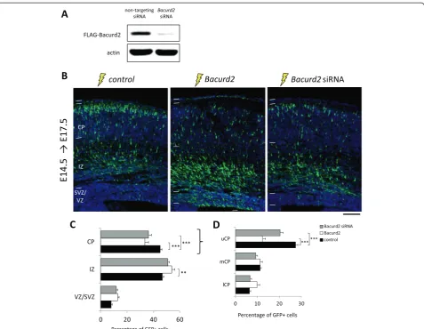
Next, we performed a series ofin uteroelectroporation experiments on E14.5 mouse embryonic cortex to deter-mine whether perturbations to Bacurd2 might disrupt cortical development. To do this, we forced expressed Bacurd2by delivering a bicistronic expression construct encoding Bacurd2 and GFP into embryonic cortical cells and examined the distribution of GFP-labelled cells 3 days later at E17.5. In a reciprocal approach, we suppressed Bacurd2 expression in cells using targeting siRNAs to-gether with an empty (GFP only) vector (Figure 2A). In each condition, the amounts of siRNA (control or target-ing) and expression vector (GFP only, or GFP + Bacurd2 bicistronic vector) were normalised to enable comparisons across conditions. In Figure 2B, we show that while a sig-nificant proportion of GFP-labelled cells had migrated into the CP of control-treated brains, forced expression of Bacurd2or knockdown with siRNAs disrupted their migration within the embryonic cortex, observed as an accumulation of cells within the IZ and a concomitant decrease in cells located within the CP (Figure 2C). Within the CP, a significant proportion of Bacurd2 -overexpressing cells and Bacurd2 siRNA-treated cells failed to reach the upper cortical plate, suggesting that changes to Bacurd2 levels disrupt their ‘intracortical’ positioning (Figure 2D). To account for the possibility that disruptions to Bacurd2 might influence cortical neurogenesis, we performed quantification studies and found no significant differences in the proportions of GFP+/Tuj1+ cells or their distribution within the subcom-partments of the embryonic E17.5 cortex (Additional file 3: Figure S3).
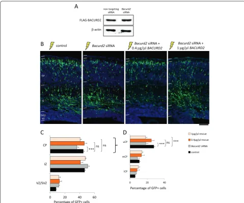
To confirm the specificity of the siRNA-mediated mi-gration defect, we performed rescue experiments whereby cells were co-treated with an expression construct encod-ing human BACURD2 which was refractory to RNAi (Figure 3). Our results show that the defective migration of siRNA-treated cells could be significantly restored to levels resembling control condition when 0.4 μg/μl of
BACURD2 construct was co-delivered with Bacurd2 siRNA (Figure 3C). Interestingly, while co-treatment with either concentrations of BACURD2 enhanced mi-gration into the CP (Additional file 4: Figure S4), we show in Figure 3C,D that the migration profile of siRNA-treated cells was corrected to levels resembling control when co-treated with 0.4μg/μl of BACURD2 (Figure 3C), while co-treatment with a higher concentration (1 μg/μl) of BACURD2 construct disrupted intracortical positioning (Figure 3D). Thus, Bacurd2 cell autonomously controls ra-dial migration, with concentration-sensitive effects.
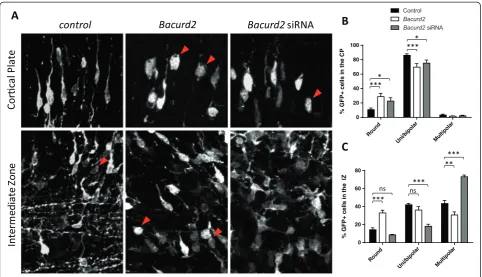
In the course of their radial migration, embryonic cor-tical cells adopt different modes of migration from the germinal VZ, through to the IZ and the CP [16,17]. Hence, we analysed the morphology of GFP-labelled neurons to describe the cellular basis for the defective migration of cells as a result of perturbations toBacurd2. Within the IZ, we found that forced expression of Bacurd2 resulted in a significant increase in the propor-tion of round-shaped cells which have very short pro-cesses (or no detectable propro-cesses at all), together with a corresponding decrease in multipolar-shaped neurons; while the proportion of uni/bipolar-shaped neurons was not significantly different (Figure 4). On the other hand, knockdown of Bacurd2 resulted in a significant increase in the proportion of multipolar-shaped neurons and a concomitant decrease in uni/bipolar neurons, while the proportion of round-shaped neurons was not significantly different. Within the CP, we found that forced expression as well as knockdown ofBacurd2resulted in an increase in the proportion of round-shaped cells, together with a decrease in the proportions of uni/bipolar-shaped cells. These documented changes in cell morphology upon siRNA-mediated knockdown were corrected by co-delivery of 0.4 μg/μl BACURD2 construct (Additional file 5: Figure S5). Together, these results demonstrate that disruptions to Bacurd2 alter the morphologies of
embryonic neurons, and this effect could underlie their defective migration within the embryonic E17.5 cortex.
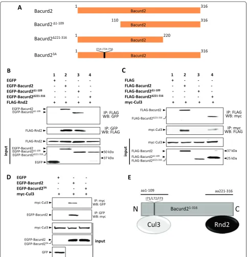
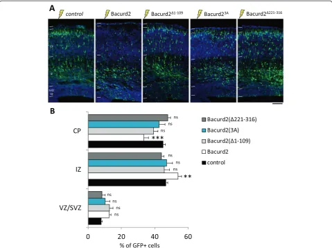
In the following experiments, we wanted to define the interaction domains on Bacurd2 which govern its bind-ing to Rnd2. We cloned truncation mutants of Bacurd2 based on the minimal interaction regions identified in our yeast two-hybrid assay (Additional file 1: Figure S1) and assessed their interaction in co-immunoprecipitation assays using epitope-tagged proteins in heterologous cells (Figure 5). Our results show that while a C-terminal trun-cation mutant Bacurd2(Δ221-316) fails to immunoprecipi-tate Rnd2, an N-terminal mutant Bacurd2(Δ1-109) still interacts with Rnd2 (Figure 5B, lanes 3 to 4). Recently, Bacurd2 was demonstrated to interact with the E3 ubiqui-tin ligase Cul3 at its N-terminus and signal together to promote fibroblast cell migrationin vitro[18]. Given that Bacurd2, Rnd2 and Cul3 proteins are all present during mouse brain development (Additional file 2: Figure S2C), we wanted to confirm their protein-protein interaction. As shown, our co-immunoprecipitation experiments re-veal that while Cul3 interacts with full-length Bacurd2, as well as a C-terminal truncation mutant, the N-terminal mutant Bacurd2(Δ1-109) fails to immunoprecipitate Cul3 (Figure 5C). In addition, we engineered missense muta-tions I71A/L72A/I73A to Bacurd2 (named as Bacurd2(3A), the location of these amino acids are indicated in bold text on Figure 5A) which are reported to disrupt its BTB domain [18], and we found that this variant did not interact with Cul3 (Figure 5D). Therefore, these studies demonstrate that Bacurd2 interacts with Rnd2 as well as Cul3 via the C- and N-termini, respectively (summarised in Figure 5E).
Next, we investigated how the Bacurd2 polypeptide influences neuronal migration by performing in utero electroporation assays. Specifically, we asked if forced
expression of each of the mutated variants of
Bacurd2(Δ221-316)) might affect the migration of E14.5 embryonic cortical cells within the E17.5 cortex in a similar manner to wildtype Bacurd2. As shown in Figure 6, we found that while forced expression of full-length Bacurd2 disrupted the migration of embryonic cortical cells into the CP, forced expression of the N-terminal truncation mutants Bacurd2(Δ1-109) or Bacurd2(3A) (both of which fail to interact with Cul3) did not significantly disrupt the migration profile of treated cells when compared with control. Similarly, forced expression of the Rnd2-binding defective mutant Bacurd2(Δ221-316) mutant did not sig-nificantly disrupt the migration profile of GFP-labelled cells. Therefore, overexpression of all three mutants did
not disturb migration and this suggests that an intact, full-length Bacurd2 polypeptide is important for its cell migra-tion funcmigra-tions within the embryonic cortex.
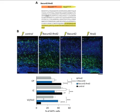
A Bacurd2:Rnd2 chimeric construct influences radial migration within the embryonic cortex
Based on our analysis of Bacurd2 and its mutants in migration (Figure 6), we reasoned that the Bacurd2 polypeptide must coordinate cell migration through its protein-protein interactions at its N- and C-termini. To explore the possibility that Bacurd2 might signal cell migration in concert with Rnd2, we designed a polypep-tide expression construct comprising a fusion between Figure 2Bacurd2 influences cell migration within the embryonic mouse cerebral cortex. (A)Western blotting with HEK293T cell lysates confirms that FLAG-Bacurd2 expression is suppressed by targeting siRNAs, but not by control (non-targeting) siRNAs. Actin was used as loading control.(B)In uteroelectroporation was performed on embryonic mouse E14.5 embryos and analysed 3 days later at E17.5. Cortical cells were electroporated with control vector (GFP only), a bicistronic GFP expression construct which also encodes Bacurd2, orBacurd2siRNA co-electroporated with GFP vector.(C)Quantification reveals that forced expression of Bacurd2, or treatment withBacurd2siRNAs, alters the distribution of cells within the embryonic cortex (N> 4,500 cells from four to six brains per condition;F4,72= 14.97;P< 0.0001; two-way ANOVA followed by Bonferroni’spost
hoctest; ****P< 0.0001.(D)Quantification of GFP+ cells within the CP (divided into the lower, medial and upper CP) reveals that forced expression of Bacurd2 or knockdown of Bacurd2 disrupts the intracortical distribution of GFP+ cells compared with control (N> 1,500 cells from four to six brains per condition;F4,72= 27.89;P< 0.0001; two-way ANOVA followed by Bonferroni’spost hoctest). uCP, mCP and lCP indicate upper, medial and
lower cortical plates, respectively. Scale bar represents 100μm.
the N-terminal Bacurd2(aa1-220) sequence together with the C-terminal sequence of Rnd2(aa181-227) (Figure 7A). It was recently discovered that the C-terminal (aa181-227) region of Rnd2 is important for signalling cell migration in vivo[7], and so we cloned this region of Rnd2 in place of Bacurd2(aa221-316) to generate a chimeric molecule. When we introduced this construct into E14.5 born cortical cells, we found that forced expression of the Bacurd2:Rnd2 disrupts radial migration in a manner which was distinct to Rnd2 or Bacurd2 overexpression
alone (Figure 7B). Notably, we found that forced expres-sion of Bacurd2 led to a significant accumulation of cells in the IZ and a failure of cells to reach the CP, while forced expression of Rnd2 resulted in a significant accu-mulation of cells in the VZ but not the IZ. In contrast, forced expression of Bacurd2:Rnd2 led to a significant accumulation of cells in the VZ and IZ. Consistent with these distinct effects on cell migration, we found that each different treatment altered the morphology of IZ and CP cells in different ways (Additional file 6: Figure S6). Figure 3The defective migration ofBacurd2siRNA-treated cells is augmented by co-delivery of human BACURD2. (A)Western blotting of lysates from P19 embryocarcinoma cells transiently transfected with control siRNA orBacurd2siRNAs, together with an expression construct encoding human BACURD2 as an epitopte-tagged (FLAG) protein. FLAG-BACURD2 protein expression is refractory to Bacurd2 siRNA-mediated knockdown.(B)In uteroelectroporation studies with E14 mouse brains electroporated with GFP vector and control siRNA (‘control’), GFP vector andBacurd2siRNA, andBacurd2siRNA with the indicated concentrations of BACURD2 expression construct are indicated.(C)Quantitation reveals that while Bacurd2 siRNA treatment impairs radial migration, co-delivery of 0.4μg/μl BACURD2 construct restores their migration to control levels, while co-delivery of 1.0μg/μl BACURD2 construct only partially restores their migration within the embryonic cortex (N> 1,450 cells counted per condition;F6,45= 15;P< 0.0001; two-way ANOVA followed by Bonferroni’spost hoctest).(D)An analysis of their intracortical distribution reveals
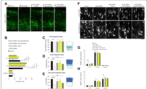
It was reported that suppression ofRnd2by RNAi sig-nificantly disrupted cell migration within the embryonic E17.5 cortex, including their multipolar-to-bipolar tran-sition from the IZ to the CP [6,7,10]. Hence, we wanted to determine if the migration defect of Rnd2-deficient cells could be restored by modulating Bacurd2 signal-ling. We began with control experiments to confirm that the defective migration of Rnd2 shRNA-treated cells could be corrected by co-delivering an expression con-struct encodingRnd2which is not targeted by the shRNA vector (Figure 8A,B) [6,7]. Next, we asked whether forced expression of full-length Bacurd2 could compensate for the defective migration ofRnd2shRNA-treated cells, but we did not observe a restoration of cell migration in our assay (n= 6 brains per condition, data not shown). In con-trast, co-delivery of Bacurd2:Rnd2 significantly improved the migration of Rnd2 shRNA-treated cells (Figure 8A), with cells reaching the cortical plate at levels not signifi-cantly different to control treatment (Figure 8B; 39.37% ± 2.57% of cells within the CP of control samples versus
33.45% ± 2.4% of Rnd2 shRNA+ Bacurd2:Rnd2 treated cortices;F8,39= 17.36;P< 0.0001; two-way ANOVA;post hoc t-testP> 0.05). In addition to this result, we were also interested to determine whether I71A/L72A/I73A substi-tution mutations to the BTB domain of Bacurd2 which disrupt its binding to Cul3 were relevant to its cell migra-tion funcmigra-tions. Thus, we performed parallel rescue experi-ments to co-deliver Bacurd2(3A):Rnd2 (which is defective in Cul3 binding; see Additional file 7: Figure S7) together withRnd2shRNA in embryonic E14.5 cortical cells. Our results show that while treatment with Bacurd2(3A):Rnd2 improved the migration ofRnd2shRNA-treated cells, the proportion of GFP-labelled cells within the CP remained significantly decreased compared with control condition (Figure 8A,B; 13.43% ± 2.76% of cells within the CP of Rnd2shRNA-treated cortices versus 26.38% ± 2.06% in Rnd2shRNA+ Bacurd2(3A):Rnd2 treated cortices versus 39.37% ± 2.57% of cells within the CP of control sam-ples; F8,39 = 17.36; P < 0.0001; two-way ANOVA; post hoc t-test ***P< 0.0001).
Figure 4The effect of forced expression or knockdown of Bacurd2 on the morphology of GFP-labelled neurons within the IZ and CP of the E17.5 embryonic cortex. (A)The morphology of neurons within the CP and the IZ in representative brain sections electroporated with control (GFP only) vector,Bacurd2expression vector orBacurd2siRNAs. Arrowheads point to round-shaped cells.(B)Within the CP, overexpression or knockdown of Bacurd2 leads to a significant increase in the proportion of round cells, and a decrease in uni/bipolar-shaped cells (N> 300 cells counted from three brains per condition;F4,45= 9.63;P< 0.0001, two-way ANOVA followed by Bonferroni’spost hoctest; *P< 0.05, ***P< 0.001). (C)Within the IZ, overexpression ofBacurd2leads to a significant increase in the proportion of round cells and a decrease in multipolar-shaped cells (N> 500 cells counted from three brains per condition;F4,33= 48.56;P< 0.0001; two-way ANOVA followed by Bonferroni’spost hoctest;
*P< 0.05, ***P< 0.001), but the proportion of uni/bipolar-shaped cells remains unchanged. On the other hand, treatment withBacurd2siRNAs leads to a significant decrease in the proportion of uni/bipolar-shaped cells and multipolar-shaped cells, with no significant difference in the proportion of round-shaped cells. Scale bar represents 20μm.
We previously demonstrated that Rnd2 controls the mor-phological transitions undertaken by migrating neurons as they reach the CP, including their multipolar-to-bipolar transition as they leave the IZ and enter the CP [6,7]. Thus, we analysed the migration index of GFP-labelled cells in our current rescue experiments to understand how neurons
enter the IZ (Figure 8C) and the CP (Figure 8D,E). As a control experiment, we first confirmed thatRnd2-deficient cells are defective in their migration from the VZ to the IZ and CP in a cell autonomous fashion, as previously re-ported [6,7] (Figure 8C,D,E). We then observed that the IZ migration defect of Rnd2 shRNA-treated cells is restored Figure 5Bacurd2 interacts with its binding partners through distinct regions of the polypeptide. (A)Schematic representation of Bacurd2 polypeptide and its mutant variants used in our analysis.(B)Reciprocal co-immunoprecipitation assays show that Rnd2 interacts with an N-terminal Bacurd2 truncation mutant but is unable to bind the C-terminal truncation mutant Bacurd2(Δ221-316).(C)Cul3 binds Bacurd2(Δ221-316) but not the N-terminal truncation mutant Bacurd2(Δ1-109).(D)Mutations to I71/L72/I73 in the Bacurd2(3A) polypeptide sequence disrupt its interaction with Cul3.
with either the Bacurd2:Rnd2 or Bacurd2(3A):Rnd2 to levels which are not significantly different to control pro-file (Figure 8C). In contrast, the defective CP-entry of Rnd2-deficient cells was efficiently restored only when Bacurd2:Rnd2 was co-delivered, but not with Bacurd2 (3A):Rnd2 (Figure 8D). Furthermore, we found that the defective intracortical distribution of Rnd2-deficient cells was only corrected by co-delivery of Bacurd2:Rnd2, but not with Bacurd2(3A):Rnd2 (Figure 8E).
Finally, we analysed GFP-labelled neurons within the IZ and CP to determine whether the abnormal morpholo-gies of Rnd2 shRNA-treated neurons could be corrected by co-delivery of Bacurd2:Rnd2. We first investigated the morphologies of neurons within the IZ of Rnd2 shRNA electroporated brains and found a significant increase in the proportion of multipolar-shaped neurons compared
with control treatment, a result which is consistent with our previous reports describing failed multipolar-to-bipolar transition of Rnd2-deficient cells [6-8,10,11] (Figure 8F). Also, we observed that co-delivery ofRnd2 restores the morphologies ofRnd2shRNA-treated neu-rons to a distribution which is not significantly different to control treatment. In contrast, we found that the morphological profiles ofRnd2shRNA + Bacurd2:Rnd2 treated cells within the IZ and CP were restored to a profile resembling control condition, as wereRnd2shRNA cells co-treated with Bacurd2(3A):Rnd2 (Figure 8G,H). Taken together, our results collectively demonstrate that Bacurd2 coordinates cell migration within the embryonic cortex and influences the morphological transitions of im-mature neurons as they transit through the IZ, as well as when their radial distribution within the CP. Despite the Figure 6The effect of forced expression of Bacurd2 and its mutated variants on cell migration within the embryonic E17.5 cortex. (A)Coronal sections of E17.5 embryonic cortex following E14.5in uteroelectroporation with a bicistronic construct encoding GFP vector only (control), or together with Bacurd2 or its mutant variants.(B)Forced expression of Bacurd2 disrupts the migration of GFP-labelled cells in the embryonic cortex, when compared with control treatment. On the other hand, forced expression of the N-terminal mutants Bacurd2(Δ1-109), Bacurd2(3A) or the C-terminal Bacurd2(Δ221-316) variant did not significantly disrupt the migration of cells (N> 4,000 cells counted from four to six brains per condition. Distribution of GFP-labelled cells within the VZ/SVZ, IZ and CP of the E17.5 cortex;F8,96= 5.38; two-way ANOVA followed
by Bonferroni’spost hoctest which compares each column to control; *P< 0.05; ***P< 0.001). Scale bar, 100μm.
caveat that our Bacurd2:Rnd2 chimeric construct repre-sents an artificial model of a Bacurd2-Rnd2 signal trans-ducer, our results suggest that Bacurd2 and Rnd2 promote cell migration within the embryonic cortex.
Discussion
We previously reported that Rnd2 regulates the migra-tion of newborn embryonic cortical neurons [6,7]; hence, we wanted to clarify the downstream signalling pathway through which Rnd2 modulates this activity. In this study, we have identified Bacurd2 as an interacting
partner to Rnd2 which influences radial migration dur-ing cerebral cortex development. Notably, both Rnd2 and RhoA signalling are crucial to radial migration [6,7,19], and Bacurd2 suppresses RhoA to influence cell migrationin vitro[18]. Therefore, we were motivated to characterise the neuronal functions for Bacurd2 within the embryonic cortex. We find that disruptions to Bacurd2impair cell migration and alter the multipolar-to-bipolar transition of embryonic cortical neurons. Thus, our study introduces Bacurd2 as a new player in neuronal development.
Figure 7Forced expression of Bacurd2:Rnd2 impairs radial migrationin vivo. (A)Illustration of the protein resulting comprising the N-terminal region of Bacurd2(1-220) together with the C-terminal region of Rnd2 which mediates the migration of embryonic cortical neurons in vivo[7].(B)Forced expression of Bacurd2:Rnd2 impairs cell migration within the embryonic E17.5 cortex, as do cells which overexpress either Bacurd2 or Rnd2 (N> 2,400 cells counted from three to four brains per condition;F6,63= 55.34;P< 0.0001; two-way ANOVA followed by Bonferroni’s
The ability for immature neurons to migrate is a func-tion which is sensitive to Rnd2 levels, with too much or too little disrupting this process [6,7,10]. Given the role for Bacurd2 in targeting RhoA for degradation by the Cul3 ubiquitin ligase complex, it is possible that Bacurd2 may act as a substrate adaptor for the degradation of Rnd proteins. As such, Bacurd2 could target Rnd2 for degrad-ation via the Cul3 ubiquitin ligase complex so as to pro-mote radial migration. However, the role for Bacurd2 in radial migration is also likely to be mediated through RhoA regulation as well. In the future, it will be important to determine the relative contributions of both of these postulated signalling mechanisms for Bacurd2 which in-fluence the development of cerebral cortical neurons.
In our functional studies, we found that truncation of the C-terminal region of Bacurd2 abolishes its binding to Rnd2 and disrupts its cell migration functions in em-bryonic cortical cells. Furthermore, a truncation of the N-terminal region of Bacurd2 or the introduction of mis-sense mutations I71A/L72A/I73A within its BTB-domain (both of which disrupts its binding to Cul3) similarly abolishes its effects on cell migration in vivo. From these findings, we surmise that the ability for the Bacurd2 poly-peptide to control cell migration relies on its N- and C-terminal domains. With the knowledge that Cul3 and Rnd2 interact with the C- and N- termini of Bacurd2, re-spectively, together with our evidence that these proteins are detected throughout the course of brain development,
A
F
G
H
B
C
D
E
Figure 8The defective migration ofRnd2-deficient neurons is restored by co-delivery of Bacurd2:Rnd2. (A,B)The defective migration ofRnd2 shRNA is corrected by co-delivery of Rnd2, Bacurd2:Rnd2 and, to a lesser extent, Bacurd2(3A):Rnd2 (N> 2,000 cells per condition;F8,39= 17.36;
P< 0.0001; two-way ANOVA by Bonferroni’spost hoctest).(C-E)Cell entry index calculated as the proportion of cells within each subcompartment. Rnd2shRNA treatment impairs VZ-to-IZ entry, but is corrected by co-delivery of Rnd2, Bacurd2:Rnd2 and Bacurd2(3A):Rnd2 (F4,13= 4.210,P= 0.021;
one-way ANOVA) (C). Co-delivery of Rnd2 or Bacurd2:Rnd2 restores IZ-to-CP entry ofRnd2deficient neurons (F4,13= 16.31,P< 0.0001; one-way ANOVA) (D), as well as their migration to the upper CP (N> 295 cells per condition;F4,13= 18.01,P< 0.0001; one-way ANOVA)(E). Bacurd2(3A):Rnd2 does not
significantly improve IZ-to-CP entry(D), nor the intracortical migration ofRnd2-deficient cells(E). (F)The morphology of IZ and CP neurons.(G)Rnd2 shRNA-treated CP neurons show an increase in the proportion of round cells and a concomitant reduction in uni/bipolar shaped cells (N> 250 cells per condition;F8,57= 8.64,P< 0.0001; two-way ANOVA followed by Bonferroni’spost hoctest). Co-delivery of Rnd2, Bacurd2:Rnd2 or Bacurd2
(3A):Rnd2 restores the morphological profile ofRnd2shRNA-treated neurons within the CP.(H)Rnd2shRNA-treated IZ neurons show a significant increase in the proportion of multipolar cells and a concomitant reduction in uni/bipolar shaped cells (N> 640 cells per condition; F8,51= 3.497,P= 0.0027; two-way ANOVA followed by Bonferroni’spost hoctest). Co-delivery of Rnd2, Bacurd2:Rnd2 and Bacurd2(3A):Rnd2
restored the morphologies ofRnd2shRNA-treated IZ neurons to control profile. Data was collected from three to four independent brains per condition. Scale bar represents 100μm(A)and 20μm(F).
plain the basis for this rescue? One possible explanation relates to the migration-promoting functions of Rnd2 and Bacurd2. In the case of Rnd2, it was reported that its aa181-227 C-terminal region is important for subcellular localisation and promotion of activity within the peri-nuclear region of immature neurons to signal migration [7,20]. On the other hand, studies with a triple I71A/ L72A/I73A point mutation variant of Bacurd2(3A) protein show an importance for Cul3 binding to signal cell migra-tion in a wound-healing assay [18] as well as embryonic cell migration (this study). When we attempted to recon-cile these findings through our own experimentation, our results show that Bacurd2:Rnd2 restores the migration of Rnd2 shRNA-treated neurons. While the roles for both Bacurd2 and Rnd2 in neuronal development remain to be better understood, it will be important to determine if these molecules are important for the terminal differenti-ation of cortical neurons, including their dendritic arbor-isation and synaptic connectivity. More broadly, given the roles for Bacurd and Rnd proteins in the regulation of the early steps of neurogenesis [7,12,21,22], a better under-standing of their combined signalling activities will reveal their specific contributions to the development and func-tion of cerebral cortical neurons in health and disease.
Conclusions
The molecular regulation of radial migration is complex and involves multiple signalling factors which promote directional movement as well as neurite outgrowth as immature neurons position themselves within the devel-oping cerebral cortex. We have identified Bacurd2 as a new player which promotes the migration of immature cortical projection neurons in a concentration-sensitive manner. In addition, we have characterised Bacurd2 as a novel interacting partner to Rnd2, a known regulator of radial migration within the embryonic cortex. Our discov-ery supports the notion that Rnd2 interacts with specific binding partners (such as Bacurd2) to control neuronal migration during cortical development.
Methods
Animals - Mice were housed, bred and treated within the animal facilities at Monash University. All animal procedures are approved by the Animal Ethics Commit-tee within Monash University (Licenses MARP/08-104
tants were constructed by the standard PCR cloning strategy or DNA synthesis of the entire cDNA (Life Technologies, Carlsbad, USA). Complementary DNA (cDNA) fragments were cloned into the EcoRI site of pEGFP-C2 (Clontech, Mountain View, USA) and pCIG2-Flag. Expression constructs for Rnd2 and Rnd3 were pre-viously described [2]. The Bacurd2:Rnd2 cDNA encodes the following polypeptide which is engineered as a fusion between aa1-220 of Bacurd2 and aa181-227 of Rnd2: MSGDTCLCPASGAKPKISGFKGGGLGNKYVQLN VGG SLYYTTVRALTRHDTMLKAMFS GRMEVLTDKEGWIL IDRCGKHFGTILNYLRDDTITLPQSRQEIQELMAEAKY YLIQGLVSTCQTALQDKKDSYQPVCNIPIITSLREEDRLI ESSTKPVVKL LYNRSNNKYSYT SNSDDHLL KNIELFDK
LSLRFNGRVLFIKDVIGDEICCWS FYGQGRKLSLGRGH
RQL RRT DSRR GLQRSTQLSGRPDR GNEGEMHKDRAKS CNLM(the Rnd2 C-terminal polypeptide sequence is rep-resented in italics, while the amino acids in bold are targeted for alanine substitution in the Bacurd2(3A):Rnd2 construct). All constructs were sequenced verified and plasmids produced using PureYield™ Midi-prep kits (Promega, Madison, USA).
In utero electroporation - In utero electroporation ex-periments are performed as described [23]. High-quality, low endotoxin plasmid preparations (Qiagen, Limburg, The Netherlands) of DNA vectors were injected at 1μg/μl for each plasmid, together with Fast Green (0.05%, Sigma, St. Louis, USA). For RNAi experiments, Dharmacon siRNA targeting pools for Bacurd2 were injected at 10μM concentration together with GFP expression plasmid at 1
μg/μl concentration. Following recovery from in utero electroporation, the mice were sacrificed by cervical location, and the embryonic brains were harvested by dis-section in cold PBS and preserved for tissue processing, cryosectioning (16μm) and fluorescence immunostaining. Images of brain sections were captured on an epifluores-cence microscope (Olympus, Tokyo, Japan) equipped with a CCD camera (SPOT, Sterling Heights, USA). Subdivi-sions of the embryonic cortex (VZ/SVZ, IZ and CP) were identified based on cell density as visualised with 4′6-dia-midino-2-phenylindole (DAPI) staining. Cell counting was performed blind to the condition on representative fields of sections of electroporated brains using ImageJ software.
Additional files
Additional file 1: Figure S1.Identification of Bacurd2 as a binding partner to Rnd2 in a yeast two-hybrid assay. Representation of results of a yeast two-hybrid interaction screen using an Rnd2 bait which lacks its C-terminal (CAAX) motif.(A)Identification of multiple cDNA prey encoding Bacurd2 polypeptide. Complementation assays confirm association of Bacurd2 preys with Rnd2 bait.(B)Growth of yeast transfected with Rnd2 bait and Bacurd2 prey construct on nutritional selection medium (lacking histidine and adenine). Double-transfected yeast harbouring Bacurd2 prey and pLaminC bait, or Bacurd2 prey with p53 bait do not grow on this selection medium.
Additional file 2: Figure S2.Immunodetection of Bacurd2 and Rnd2.
(A)Western blotting of HEK293T cell lysates transfected with FLAG-Bacurd2 construct and immunoblotted with our mouse monoclonal antibody. A specific signal of approximately 37 kDa is detected, which corresponds to the FLAG-Bacurd2 signal in an immunoblot with FLAG antibody.
(B)Western blotting of HEK293T cell lysates transfected with FLAG-Rnd2 construct and immunoblotted with a rabbit polyclonal antibody (Santa Cruz Biotechnologies, Santa Cruz, USA). A specific signal of approximately 25 kDa is detected and which corresponds to the FLAG-Rnd2 signal in an immunoblot with FLAG antibody.(C)Western blotting using antibodies to Bacurd2, Rnd2 and Cul3 on mouse brain lysates harvested from the indicated timepoints during mouse brain development. The signal for
β-actin was used as a loading control.(D-E)Fluorescence immunostaining of E14.5 embryonic cortex using preimmune serum (D) or Bacurd2 antibody (E, green signal). Nuclei were counterstained with DAPI. Scale bar, 50μm.
Additional file 3: Figure S3.Perturbations toBacurd2do not disturb neuronal differentiation.(A)GFP-labelled cells which co-express Tuj1 (arrow) or do not co-express Tuj1 (arrowheads) are identified in a representative section of E17.5 embryonic cortex.(B)Quantification studies of the proportion of GFP-labelled cells which co-express the neuronal marker Tuj1 (N> 1,700 cells counted from at least three brains per condition; one-way ANOVA followed by Bonferroni’spost hoc t-test) reveal no significant difference in GFP+/Tuj1+ cells following overexpression or knockdown of Bacurd2 (F2,13= 1.6;P= 0.24;P> 0.5).(C)Two-way ANOVA
analysis of GFP+/Tuj1−cells in each cortical subcompartment reveals no significant difference in their distribution following overexpression or knockdown of Bacurd2 (F4,39= 0.44;P= 0.77;P> 0.5). Graph plots
mean ± SEM. Scale bar, 50μm.
Additional file 4: Figure S4.The defective migration ofBacurd2 siRNA-treated cells is augmented by co-delivery of human BACURD2.
(A)To ask if co-treatment with 0.4μg/μl or 1.0μg/μl of BACURD2 construct restores their migration of siRNA-treated cells, we performed two-way ANOVA followed by Bonferroni’s multiple comparisons test. We found that treatment with either concentrations of BACURD2 improved the migration of cortical cells (N> 1,450 cells counted per condition, F6,45= 15,P< 0.0001).(B)An analysis of their intracortical distribution
(that is within the lower, medial and upper CP) reveals that the defective migration of Bacurd2 siRNA-treated cells is restored with co-delivery of 0.4μg/μl of BACURD2 construct, but treatment with 1.0μg/μl of BACURD2 did not restore their positioning within the upper CP (N> 500 cells per condition,F6,42= 15.27,P< 0.0001; two-way ANOVA followed by
Bonferroni’spost hoctest). Graph plots mean ± SEM. Scale bar represents 100μm.
Additional file 5: Figure S5.Bacurd2controls cell shapein vivo.(A)
The morphology of neurons within the CP and the IZ in representative brain sections electroporated with control (GFP only) vector,Bacurd2 siRNAs orBacurd2siRNAs with the indicated concentrations of BACURD2 expression vector. Arrowheads point to round-shaped cells.(B)Within the CP, knockdown ofBacurd2leads to a significant increase in the proportion of round cells and a decrease in uni/bipolar-shaped cells (N> 300 cells counted from three brains per condition;F6,60= 24.60;
P< 0.0001; two-way ANOVA followed by Bonferroni’spost hoctest; *P< 0.05, ***P< 0.001). These effects on cell shape are corrected with co-delivery of 0.4μg/μl of BACURD2 construct.(C)Within the IZ, treatment withBacurd2siRNAs leads to a significant decrease in the proportion of uni/bipolar-shaped cells and multipolar-shaped cells, with no significant difference in the proportion of round-shaped cells when compared with control. (N> 500 cells counted from three brains per condition;F6,48=
22.52;P< 0.0001; two-way ANOVA followed by Bonferroni’spost hoc test; *P< 0.05, ***P< 0.001). Co-delivery of either 0.4μg/μl of BACURD2 construct or 1.0μg/μl of BACURD2 construct corrects theseBacurd2 siRNA-induced changes to cell shape within the IZ. Scale bar represents 20μm.
Additional file 6: Figure S6.Cell shape profiles following treatment withBacurd2:Rnd2,Bacurd2orRnd2.Analysis of the morphologies of IZ and CP neurons following forced expression of Bacurd2:Rnd2, Bacurd2 or Rnd2. There was an interaction between treatment and distribution of cell shapes in the CP (N> 190 cells from three to four brains per condition; F6,60= 8.332,P< 0.0001; two-way ANOVA followed by Bonferroni’spost
hoctest)(A)and cells within the IZ (N> 390 cells from three to four brains per condition;F6,51= 7.537,P< 0.0001; two-way ANOVA followed by
Bonferroni’spost hoctest)(B). Graph plots mean ± SEM.
Additional file 7: Figure S7.Point mutations I71A/L72A/I73A alter binding to Cul3.(A), Diagrammatic representation of Bacurd2 full-length polypeptide sequence, along with Bacurd2:Rnd2 contruct, and a Bacurd2 (3A):Rnd2. Yellow indicates the sequence of Rnd2 polypeptide fused to the C-terminal fragment of Bacurd21-220polypeptide (see the‘Methods’
section for polypeptide sequence). Bold letters identify substitution mutations I71A/L72A/I73A within the BTB-domain of the Bacurd2 polypeptide fragment which mediates Cul3 binding.(B)Reciprocal co-imunoprecipitation experiments were performed to confirm that the Bacurd2:Rnd2 interacts with Cul3, but not with Bacurd2(3A):Rnd2. Input lanes confirm the presence of all proteins evaluated in this experiment.
Abbreviations
bHLH:basic helix-loop-helix; CP: cortical plate; E: embryonic day; EGFP: enhanced green fluorescent protein; IZ: intermediate zone; lCP: lower cortical plate; mCP: medial cortical plate; siRNA: small inhibitory RNA; uCP: upper cortical plate; VZ: ventricular zone.
Competing interests
The authors declare that they have no competing interests.
Authors’contributions
JIH conceived the study and performed Y2H experiments with JMD and ZDQ. Biochemical assays, cell culture experiments and protein-interaction assays were performed by SSL, IEG-N and ZDQ, while mouse electroporation
Fellowship (NHMRC Australia, ID:1011505), a CJ Martin Training Fellowship (ID:310616) and a project grant 566835 (NHMRC, Australia). This work was also supported by NIH grant R01GM082940 to JDS. We thank Drs Laurent Nguyen and Emilie Pacary and members of the Heng lab for critical reading of the manuscript.
Author details
1EMBL Australia, The Australian Regenerative Medicine Institute, Monash
University, Clayton, Victoria 3800, Australia.2Department of Biology, Portland
State University, Portland, Oregon 96207, USA.3The Harry Perkins Institute of
Medical Research, Perth, Australia.4Centre for Medical Research, The
University of Western Australia, Perth, Australia.5Present address: The Harry
Perkins Institute of Medical Research, Perth, Australia.
Received: 24 October 2014 Accepted: 23 February 2015
References
1. Gupta A, Tsai LH, Wynshaw-Boris A. Life is a journey: a genetic look at neocortical development. Nat Rev Genet. 2002;3(5):342–55.
2. Marin O, Rubenstein JL. A long, remarkable journey: tangential migration in the telencephalon. Nat Rev Neurosci. 2001;2(11):780–90.
3. Marin O, Rubenstein JL. Cell migration in the forebrain. Annu Rev Neurosci. 2003;26:441–83. doi:10.1146/annurev.neuro.26.041002.131058041002.131058. 4. Heng JI, Chariot A, Nguyen L. Molecular layers underlying cytoskeletal
remodelling during cortical development. Trends Neurosci. 2010;33(1):38–47. doi:10.1016/j.tins.2009.09.003.
5. Leventer RJ, Guerrini R, Dobyns WB. Malformations of cortical development and epilepsy. Dialogues Clin Neurosci. 2008;10(1):47–62.
6. Heng JI, Nguyen L, Castro DS, Zimmer C, Wildner H, Armant O, et al. Neurogenin 2 controls cortical neuron migration through regulation of Rnd2. Nature. 2008;455(7209):114–8. doi:10.1038/nature07198. 7. Pacary E, Heng J, Azzarelli R, Riou P, Castro D, Lebel-Potter M, et al.
Proneural transcription factors regulate different steps of cortical neuron migration through Rnd-mediated inhibition of RhoA signaling. Neuron. 2011;69(6):1069–84. doi:S0896-6273(11)00116-4 10.1016/j.neuron.2011.02.018. 8. Heng J, Guillemot F. Proneural proteins and the development of the
cerebral cortex. In: Kageyama R, editor. Cortical Development: Neural Diversity and Neocortical Organization. Springer Global; 2013. p. In press. 9. Alfano C, Viola L, Heng JI, Pirozzi M, Clarkson M, Flore G, et al. COUP-TFI
promotes radial migration and proper morphology of callosal projection neurons by repressing Rnd2 expression. Development. 2011;138(21):4685–97. doi:dev.068031 10.1242/dev.068031.
10. Heng JI, Qu Z, Ohtaka-Maruyama C, Okado H, Kasai M, Castro D et al. The zinc finger transcription factor RP58 negatively regulates Rnd2 for the control of neuronal migration during cortical development. Cereb Cortex. 2013. doi:bht277 10.1093/cercor/bht277
11. Ohtaka-Maruyama C, Hirai S, Miwa A, Heng JI, Shitara H, Ishii R, et al. RP58 regulates the multipolar-bipolar transition of newborn neurons in the developing cerebral cortex. Cell Rep. 2013;3(2):458–71. doi:S2211-1247(13)00019-3 10.1016/j.celrep.2013.01.012.
12. Pacary E, Azzarelli R, Guillemot F. Rnd3 coordinates early steps of cortical neurogenesis through actin-dependent and -independent mechanisms. Nat Commun. 2013;4:1635. doi:ncomms2614 10.1038/ncomms2614.
13. Chardin P. Function and regulation of Rnd proteins. Nat Rev Mol Cell Biol. 2006;7(1):54–62. doi:nrm1788 10.1038/nrm1788.
14. Riou P, Villalonga P, Ridley AJ. Rnd proteins: multifunctional regulators of the cytoskeleton and cell cycle progression. Bioessays. 2010;32(11):986–92. doi:10.1002/bies.201000060.
2009;6:841–55. doi:S1097-2765(09)00632-7 10.1016/j.molcel.2009.09.004. 19. Nguyen L, Besson A, Heng JI, Schuurmans C, Teboul L, Parras C, et al.
p27kip1 independently promotes neuronal differentiation and migration in the cerebral cortex. Genes Dev. 2006;20(11):1511–24. doi:gad.377106 10.1101/gad.377106.
20. Riou P, Kjaer S, Garg R, Purkiss A, George R, Cain RJ, et al. 14-3-3 proteins interact with a hybrid prenyl-phosphorylation motif to inhibit G proteins. Cell. 2013;153(3):640–53. doi: 10.1016/j.cell.2013.03.044.
21. Azzarelli R, Pacary E, Garg R, Garcez P, van den Berg D, Riou P, et al. An antagonistic interaction between PlexinB2 and Rnd3 controls RhoA activity and cortical neuron migration. Nat Commun. 2014;5:3405. doi:10.1038/ncomms4405.
22. Golzio C, Willer J, Talkowski ME, Oh EC, Taniguchi Y, Jacquemont S, et al. KCTD13 is a major driver of mirrored neuroanatomical phenotypes of the 16p11.2 copy number variant. Nature. 2012;485(7398):363–7. doi:nature11091 10.1038/nature11091.
23. Breuss M, Heng JI, Poirier K, Tian G, Jaglin XH, Qu Z, et al. Mutations in the beta-tubulin gene TUBB5 cause microcephaly with structural brain abnormalities. Cell Rep. 2012;62(6):1554. doi:S2211-1247(12)00414-7 10.1016/ j.celrep.2012.11.017.
Submit your next manuscript to BioMed Central and take full advantage of:
• Convenient online submission
• Thorough peer review
• No space constraints or color figure charges
• Immediate publication on acceptance
• Inclusion in PubMed, CAS, Scopus and Google Scholar
• Research which is freely available for redistribution