Article
Simulating Low-Level Laser Propagation From Skin
Surface To Lumbar Disc, Knee, Femur and Prostate
Gland By Monte Carlo Method
Trinh Tran Hong Duyen 1†*, Tran Anh Tu 2†*
1 Laser Technology Laboratory, Faculty of Applied Science, Ho Chi Minh City University of Technology,
Vietnam National Ho Chi Minh City; [email protected]
2 General Physics Laboratory, Faculty of Applied Science, Ho Chi Minh City University of Technology,
Vietnam National Ho Chi Minh City; [email protected]
† These authors contributed equally to this work.
* Correspondence: [email protected]; [email protected] Tel.: +84283.8635.776
Abstract: Nowadays, the uses of laser and optics in the medical areas are extremely vivid, especially low-level laser therapy. The light with the wavelength of 633 nm to 1200 nm could penetrate and propagate deep in biological tissue. To develop the low-level laser therapy device, optimizing light delivery is critical to accurately stimulate the biological effects inside the biological tissue. Nevertheless, each form of the tissues at each zone on the body had various refractive optic, absorption, scattering, and anisotropy coefficients. This paper describes the simulation results of low-level laser propagation from skin surface at the lower spine, the knee, the femur and the prostate gland with four wavelengths (633 nm, 780 nm, 850 nm, and 940 nm) by the Monte Carlo method. These simulation results are the base for developing the low-level laser therapy device, that could be used in clinical for treating the fracture, knee osteoarthritis, spinal degeneration, and benign prostatic hypertrophy.
Keywords: Monte Carlo simulation, propagation, wavelength, low-level laser therapy LLLT
1. Introduction
The low-level laser therapy (LLLT) is getting more consideration and applying widely in the medical arena. This application promotes tissue regeneration, reduces inflammation and relieves pain. Unlike other medical laser applications, LLLT does not have an ablative or thermal mechanism, but rather a photochemical effect which means the light is absorbed and causes the photochemical reactions inside the biological tissue. The reason why the technique is termed low level is that the optimum levels of energy density delivered are low and it is not comparable to other forms of laser therapy as practiced for ablation, cutting, and thermal tissue coagulation. The degree of photochemical reactions and the biological responses depend on the absorbed dose of light in biological tissue. The estimated absorbed dose within tissue requires understanding and studying thoroughly. The radiative transport equation (RTE)1 is the popular equation for describing particle propagation in the complex structure of tissues [1]. One of the effective methods for solving the RTE equation is the simulation of propagation light in a scattering environment by the Monte Carlo method. This spread method has improved and applied in simulating the propagation of light in
1 Radiative transport equation (RTE) :
) sˆ t, ε(r, π
4 p(sˆ,sˆ')d2sˆ' s μ ) sˆ t, I(r, ) t μ a (μ ) sˆ t, L(r, . sˆ ) sˆ t, I(r, t υ 1 + + + − = +
biological tissue, especially in the calculation and optimizing light delivery for low-level laser treatment [2-6].
This paper presents the establishment of the model and the propagation of low-level laser from the skin surface to the lumbar disc, knee, femur and prostate gland by Monte Carlo method. This simulation is based on the popular and prestigious Monte Carlo method for simulating light propagation in multi-layered tissue, the anatomical structure parameters in the human body, as well as the optical parameters of the biological tissues. These simulation results show the distribution of power density2 in the range (1.0 - 10-4 W/cm2) of specific wavelengths (633 nm, 780 nm, 850 nm, and 940 nm), respectively. These results show the "effective operating area" of the low-level laser beam on the tissue being treated. Thereby allowing selected wavelength, optical power and the appropriate dosage for treatment.
2. Materials and Methods
Monte Carlo method
The main base of modeling the light propagation simulation in biological tissues in this paper is the Monte Carlo method [7]. The Monte Carlo method is applied to simulate the propagation of light in biological tissue based on the RTE equation and the simulation calculation, computation of photon propagation in absorption and scattering medium [4-6,8-11]. A photon is a unit of light. The wave properties of a photon are ignored, this paper only deals with particle properties. Therefore, the phase parameter and the polarization of light are not mentioned. In the simple case, the photon is put into an independent environment and its movement is recorded until it is absorbed or scattered out of the observation area. Although the results are highly accurate, this method requires repeated calculations many times to achieve the desired accuracy, resulting in a lot of time to perform the simulation. For example, to achieve an accuracy of 1%, the movement of 10,000 photons must be recorded.
A classic simulation method described by Prahl [3] and programmed by Wang and Jacques [11]. This method uses the technique of capturing hidden photons, known as "existing weights". Without stopping for each photon that will scatter until it is finished by an absorption process, these photons are given an "initial weight" and gradually decrease with each move at each scattering position. This improves the efficiency of the statistical method, which avoids the computational process of the photons moves many steps to make it just ended in an absorption process.
Parameters
Parameters are structures of tissues, optical properties and scattering functions of tissues3. These parameters are assumed to be homogeneous in the infinitesimal volume of biological tissue. The laser beam is assumed to be an extremely narrow beam, Gaussian distribution, exposure perpendicular to a laminated biological tissue sample as Figure 1.
Figure 1. Model of class structure and photon scattering[11,12]
2 Power density at which the laser beam can cause biological effects on tissue stimulation.
3 Thickness d (cm), absorption coefficient µa (cm-1), scatteringcoefficient µs (cm-1), total attenuation coefficient
These layers of tissue are considered parallel to each other, infinitely broad and characterized by structural and optical parameters. Although tissue never really wide to infinity, it can be regarded as extremely broad in terms of comparing the spatial distribution of the photons.
Simulation models
The structure of the human skin consists of stratum-corneum, epidermis, dermis with an approximate thickness (0.06– 0.1 cm), (0.0006– 0.015 cm), (0.06 –0.3 cm) [13,14]. In this paper, we use the modeling of general skin with an approximate thickness of 0.3 cm.
The first modeling structure of lumbar disc from skin consists of skin ~ 0.3 cm; fat ~ 1.0 cm; muscle ~ 1.0 cm; the lumbar spine. The second modeling structure of the knee from the skin at lateral patellar facets consists of skin ~ 0.2 cm; fat ~ 0.9 cm; muscle ~ 0.7 cm; synovial fluid. The third modeling structure of the femur from skin consists of skin: ~ 0.3 cm; fat ~ 1.2 cm; muscle ~ 2.0 cm; femur. The fourth modeling structure of the prostate gland of the skin: skin ~ 0.2 cm; fat ~ 0.2 cm; muscle ~ 1.6 cm; prostate gland.
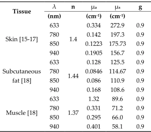
The absorption coefficient µa and the scattering coefficient µs are probability density functions, their inverse can be explained as the average mean distance for absorption and scattering. The total attenuation coefficient µtis the sum of the absorption coefficient µa and scattering coefficient µs, characterize the interaction of the photon average per unit of path length. Anisotropy coefficient g, the value of the cosine of the angle average deviation scattering θ - the angle between the direction of the photons being scattered and incident photons, characterize isotropic properties of the medium. All of these parameters characterize the properties of each tissue layer and are referenced from reputable international publications and are presented in Tables 1.
Table 1. The optical parameters of the tissues
Tissue λ n µa µs g
(nm) (cm-1) (cm-1)
Skin [15-17] 633 780 850 940 1.4
0.334 272.9 0.9
0.142 197.3 0.9
0.1223 175.73 0.9
0.1905 156.7 0.9
Subcutaneous fat [18] 633 780 850 940 1.44
0.128 125.5 0.9
0.0846 114.67 0.9
0.086 110.9 0.9
0.168 108.6 0.9
Muscle [18] 633 780 850 940 1.37
1.32 89.6 0.9
0.331 71.2 0.9
0.295 66.0 0.9
0.401 58.1 0.9
The simulation results presented below are 0.1% accurate and calculated with 1,000,000 photons based on the improved Monte Carlo Multi-Layered (MCML) program [11] and the companion
program called CONV [19] to enhance the performance and shorten simulation. Laser power will change to suit the position needed to achieve desired results with the continuous mode,
Gaussian-shaped spatial beam profile (radius 1/e2 is 0.15 cm).
These simulation results show the distribution of power density in 10-4 W/cm2 - at which the laser beam can cause the stimulation biological effect on tissue. The simulation results of the
spreading of 633 nm wavelength reach a depth smaller than the remaining wavelengths. It can be
3. Results
Figure 2 shows the simulation result from the skin surface to lumbar disc at the lower-back with
the total energy of the Gaussian laser beam is 20 J. The penetrating ability into the tissue of 780 nm and 850 nm is deeper (~ 6.0 cm) compared to 940 nm (~ 5.0 cm). The 633 nm wavelength is less penetrating into tissue (~ 3.5 cm).
Figure 2. The penetrating ability of wavelengths (633 nm, 780 nm, 850 nm, and 940 nm) from the skin to the lumbar disc at the lower back.
Figure 3 shows the simulation result from the skin surface at the knee with the total energy of
the Gaussian laser beam is 10 J. The penetrating ability into the tissue of 780 nm, 850 nm and 940 nm is quite similar (~ 4.5 cm). The 633 nm wavelength is less penetrating into tissue (~ 2.0 cm).
Figure 3. The penetrating ability of wavelengths (633 nm, 780 nm, 850 nm, and 940 nm) from the skin surface at the position of the knee.
Figure 4. The penetrating ability of wavelengths (633 nm, 780 nm, 850 nm, and 940 nm) from the skin surface to the femur.
Figure 5 shows the simulation result from the skin surface to the prostate gland with the total
energy of the Gaussian laser beam is 30 J. The penetrating ability into the tissue of 780 nm, 850 nm is quite similar (~ 5.5 cm) compared to 940 nm (~ 4.8 cm). The 633 nm wavelength is less penetrating into tissue (~ 3.0 cm).
Figure 5. The penetrating ability of wavelengths (633 nm, 780 nm, 850 nm, and 940 nm) from the skin surface to the prostate gland.
On the other hand, when the irradiation time increases, the "depth of penetration" and "impact radius" of the beam also increase significantly. For the pelvic area (lumbar disc, knee, thigh), it does not require as much energy of the beam as when it is exposed to the deep (prostate gland). This is not entirely true for all cases of the body, because tissue structures are different in each region, thickness
and each tissue has different characterizes.
4. Conclusions
skin surface to the lumbar spine, knee, femur and prostate gland. These wavelengths are useful and suitable for developing the low-level laser therapy device. As the irradiation time increases, the "depth of penetration" and "impact radius" of the beam also increase.
These simulation results allow further studies to apply low-level laser of 780 nm, 850 nm, and 940 nm wavelengths in the low-level laser therapy non-invasively from the skin surface in the treatment of fracture, knee osteoarthritis, spinal degeneration, and benign prostatic hypertrophy.
Author Contributions: All authors contributed equally to this work. All authors have read and agreed to the published version of the manuscript.
Funding: This research received no external funding.
Acknowledgments: All authors would like to thank the support of Dr.Eng. Tran Trung Nghia, Associate Dean of Faculty of Applied Science, Ho Chi Minh City University of Technology, Vietnam National Ho Chi Minh City for his supervision, suggestions, contributions, support as well as improve the content of this article.
Conflicts of Interest: The authors declare no conflict of interest. The funders had no role in the design of the study; in the collection, analyses, or interpretation of data; in the writing of the manuscript, or in the decision to publish the results.
References
1. Chandrasekhar, S. Radiative transfer. Oxford University Press: London, 1950.
2. Wilson, B.C.; Adam, G. A Monte Carlo model for the absorption and flux distributions of light in tissue.
Medical Physics 1983, 10, 824-830, doi:10.1118/1.595361.
3. Prahl, S.; Keijzer, M.; Jacques, S.; Welch, A. A Monte Carlo Model of Light Propagation in Tissue. SPIE Inst. Ser. IS 1989, 5.
4. Flock, S.T.; Patterson, M.S.; Wilson, B.C.; Wyman, D.R. Monte Carlo modeling of light propagation in highly scattering tissues. I. Model predictions and comparison with diffusion theory. IEEE Transactions
on Biomedical Engineering 1989, 36, 1162-1168, doi:10.1109/TBME.1989.1173624.
5. Flock, S.T.; Wilson, B.C.; Patterson, M.S. Monte Carlo modeling of light propagation in highly scattering tissues. II. Comparison with measurements in phantoms. IEEE Transactions on Biomedical Engineering
1989, 36, 1169-1173, doi:10.1109/10.42107.
6. Keijzer, M.; Jacques, S.L.; Prahl, S.A.; Welch, A.J. Light distributions in artery tissue: Monte Carlo simulations for finite-diameter laser beams. Lasers in Surgery and Medicine 1989, 9, 148-154, doi:10.1002/lsm.1900090210.
7. Metropolis, N.; Ulam, S. The Monte Carlo Method. Journal of the American Statistical Association 1949, 44, 335-341, doi:10.1080/01621459.1949.10483310.
8. Keijzer, M.; Pickering, J.W.; van Gemert, M.J.C. Laser beam diameter for port wine stain treatment.
Lasers in Surgery and Medicine 1991, 11, 601-605, doi:10.1002/lsm.1900110616.
9. Jacques, S.L.; Wang, L. Monte Carlo Modeling of Light Transport in Tissues. In Optical-Thermal Response
of Laser-Irradiated Tissue, Welch, A.J., Van Gemert, M.J.C., Eds. Springer US: Boston, MA, 1995;
10.1007/978-1-4757-6092-7_4pp. 73-100.
10. Wang, L.; Jacques, S.L. Hybrid model of Monte Carlo simulation and diffusion theory for light reflectance by turbid media. J. Opt. Soc. Am. A 1993, 10, 1746-1752, doi:10.1364/JOSAA.10.001746. 11. Wang, L.; Jacques, S.L.; Zheng, L. MCML—Monte Carlo modeling of light transport in multi-layered
tissues. Computer Methods and Programs in Biomedicine 1995, 47, 131-146, doi:https://doi.org/10.1016/0169-2607(95)01640-F.
12. Abdo, A.; Ersen, A.; Sahin, M. Near-infrared light penetration profile in the rodent brain. Journal of
biomedical optics 2013, 18, 75001, doi:10.1117/1.JBO.18.7.075001.
13. Chaurasia, B.D. Human anatomy; CBS Publishers & Distributors Pvt Ltd: India, 1991; Vol. 1.
14. Odland, G.F.S.o.s.I.G.A.E.P.B.a.m.b.o.t.s.O.U.P.O., 3- 62. Structure of skin. In Physiology. Biochemistry
and molecular biology of the skin, Goldsmith, L.A., Ed. Oxford University Press: New York, 1991; pp. 3-62.
15. Mobley, J. Optical Properties of Tissue. In Biomedical Photonics Handbook, Vo-Dinh, T., Ed. CRC Press: 2003.
16. Anderson, R.R.; Parrish, J.A. The Optics of Human Skin. Journal of Investigative Dermatology 1981, 77, 13-19, doi:https://doi.org/10.1111/1523-1747.ep12479191.
17. Schmitt, J.M.; Zhou, G.X.; Walker, E.C.; Wall, R.T. Multilayer model of photon diffusion in skin. J. Opt.
18. Bashkatov, A.N.; Genina, E.A.; Kochubey, V.I.; Tuchin, V.V. Optical properties of human skin,
subcutaneous and mucous tissues in the wavelength range from 400 to 2000 nm. Journal of Physics D:
Applied Physics 2005, 38, 2543-2555, doi:10.1088/0022-3727/38/15/004.
![Figure 1. Model of class structure and photon scattering [11,12]](https://thumb-us.123doks.com/thumbv2/123dok_us/983118.1597921/2.595.109.479.565.662/figure-model-class-structure-photon-scattering.webp)