e-ISSN: 2278-067X, p-ISSN: 2278-800X, www.ijerd.com
Volume 12, Issue
2
(
Febr
uary 2016), PP.09-15
DNA Microrray: A Miniaturized High Throughput
Technology
Imtiyaz Murtaza
1⃰, Omi Laila
2, Shah Ubaid-ullah
31Biochemistry and Molecular Biotechnology Laboratory, Biochemistry Division, S.K University of Agricultural
Sciences and Technology,
Shalimar Campus, Srinagar, India – 191121
2
Biochemistry and Molecular Biotechnology Laboratory, Biochemistry Section, Division of Post Harvest Technology, S.K University of Agricultural Sciences and Technology,
Shalimar Campus, Srinagar, India – 191121.
3
Department of Biotechnology, School of Life Sciences, Central University of Kashmir (CUK), Transit Campus, Sonwar, Srinagar, India – 190004.
⃰ Correspondence to:[email protected]
Abstract:-
Since last decade or so, new types of experiments are changing the way to analyze complex research problems in biological systems at mini levels and with much simplicity. Such type of experiments called as high throughput experiments use automated technology along with conventional methods. The main advantage of such technology is ‗parallelization‘ i.e. several thousands of tests can be run simultaneously rather than doing them one by one. One of such miniaturized technology known as DNA microarray enables us to study simultaneously the behavior of all the genes of an organism under different conditions in a single go. Over the period of time, different types of microarrays have been developed to answer different biological questions. In fact, microarray technology is a fast approach to study thousands of DNA and protein molecules simultaneously. A DNA microarray (also commonly known as gene chip, DNA chip, or biochip) is a collection of microscopic DNA spots attached to a solid support such as glass chip. A DNA microarray works by exploiting the ability of a given mRNA/cDNA molecule to bind specifically to, or hybridize to, the DNA template from which it originated. By this technology, scientists determine the expression levels of hundreds or thousands of genes within a cell by simply measuring the amount of mRNA/cDNA bound to each site on the array in a single experiment rather than going by gene-by-gene analysis. Using DNA Microarray analysis coupled with computational methods, the nature and the function or molecular mechanism of action of different genes of an organism can be investigated in one go. The present paper provides a basic overview on various types of DNA microarrays used in various biological endeavors.Keywords:- Microarray, surface chemistry, cDNA, hybridization, .gene expression
I.
INTRODUCTION
A far as DNA microarray is concerned it consists of a solid support usually a microscope slide, onto which DNA molecules have been chemically attached. The actual purpose of microarray is to detect the presence and abundance of labeled nucleic acids in a biological sample, which hybridize to the DNA on the array via Watson-crick duplex formation and can be detached via the label (10). The set of mRNA messages that are transcribed in a given cell type under a certain set of conditions is referred to as the transcriptome (11). In the majority of microarray experiments, the labeled nucleic acids are derived from the mRNA of a sample or tissue, and so the microarray measures gene expression (12). The DNA Microarray technology surveys many thousands of genes to investigate gene expression, transcription factor binding profiles, DNA methylation profiles, DNA copy numbers and genomic sequences (9,13). Microarray have been found to have great potential to be used as powerful tool in diagnostics (14). They can equally play a vital role in toxicogenomics (15)
II.
DNA MICROARRAY AND SURFACE CHEMISTRY
DNA microarray is a high-throughput technology that allows study of thousands of genes like mutation and single nucleotide polymorphism (SNP), drug discovery or development of diagnostic kits (16, 17, 18). They consist simply of small, solid supports onto which the sequences from thousands of different genes are immobilized, or attached, at fixed locations. The supports themselves are usually glass microscope slides, the size of two side-by-side pinky fingers, but can also be silicon chips or nylon membranes (19). The DNA is printed, spotted, or actually synthesized directly onto the support. A microarray consists of a solid surface on which strands of polynucleotide called probes have been attached or synthesized in fixed positions. There exist two types of expression microarrays that are most popular among users. One of the main differences among them relies on how probes are put on the slide. Among these spotted or cDNA microarrays take their name because probes are synthesized apart and printed mechanically on the slide (20). The term cDNA refers to the complimentary copy of the original sequence and each probe represents the sequence of one gene, while as in oligonucleotide chips, supplied or prepared by Genechip or Affymetrix (c), the probes are directly synthesized on the surface (21). The oligonecleotide to be immobilized on surface needs a chemical functionalization of the surface, so that it provides stable conditions for attachments and an easy presentation of the molecules for proper binding to occur (22). A large number of methods and strategies are in use for the modification of surface. The chemical surface coating can generally be divided into three main building blocks. The binding moiety can either consist of a silane for glass surface, of thiols for immobilization on gold surfaces, or poly-l-lysine which bind to glass and dielectric materials such as the oxides of titanium, tantalum and niobium (23-26). The linker moiety can be simple such as a simple constructions as a propyl chain in case of amino propyl silane (aminosilane slide), or an extended polyethylene glycol and a complex 3-D polymeric network of a hydrogen. The molecular structure of the linker mainly determines the properties of the slide with respect to the behavior of the immobilized compounds such as specific and unspecific binding (adsorption), structure changes, accessibility and chemical stability (27). Moreover, spot morphology, i.e., the distribution of spotted substance on the surface, depends on the combination of surface energy (wettability) and surface tension of spotting solution. A stable attachment of the probe molecules is achieved by covalent bond formation to the chemical coating (28). For this, the linker moiety is activated with reactive groups, which are attached at the end of or distributed over the molecular scaffold . Functional groups used on commercially available slides can be electrophillic, such as aldehyde, epoxy, iso (thio) cyanate, N-hydroxy succinamide ester (NHS). Nickel, nitrilo-tri acetic acid or nucleophillic, such as silicarbazide and amino. The electrophillic groups are highly reactive to amino, thiol, and hydroxyl groups and can be used for a covalent immobilization of peptides, proteins, and oligonucleotides under mild coupling conditions. However, due to the reactivity of these functional groups, slide surfaces are susceptible to decay processes caused by humidity and oxygen. The amount of amino-modified oligo nucleotides which bind and epoxy saline slide is thereby reduced by a factor of about 2, when the slide is kept at room temperature and 50% humidity for 24 hrs (22). NHS ester activated surfaces are even more susceptible to humidity leading to a considerable deactivation even by storing under optimal conditions (29). The deactivation of surface functional groups with time is crucial when some thousand samples are to be printed, especially for covalent immobilization of small molecules and proteins on microarray surfaces, new reactive groups were introduced including a protected isocyanate group, diazobenzylidene, azido benzoate attached to poly-l-lysine, or diazirine derivatives using well known photo affinity labeling reactions (22).
widely used and work reliable especially for DNA applications. Standard protocols often recommend the immobilization of c-DNA by UV crosslinking or baking on both types of slides. However, these procedures lead to the uncontrolled binding of DNA via radical reactions initiated by the UV light and various side reactions at elevated temperatures. The shorter length of immobilized DNA, the more these effects and thus need to be considered since they may have immense consequence for the hybridization (31,22).
III.
TYPES OF DNA MICROARRAY
There are three basic types of samples that can be used to construct DNA microarrays, two are genomic; oligonucleotides and cDNA while third one is "transcriptomic", that is, it measures mRNA levels (32,10,12). The main difference lies in the kind of immobilized DNA and hence the information that is derived from each type of chip. The target DNA used will also determine the type of control and sample DNA that is used in the hybridization solution (33). In this review we will discuss spotted microarray, In-situ synthesized oligonucleotide Arrays, Affymetrix Technology, Maskless Photoprotection Technology and Inkjet array synthesis.
Spotted microarrays
The first microarrays to be designed were manufactured while using this technology. There are three main types of spotted arrays which can be subdivided in two ways: by the type of DNA probe, or by the attachment chemistry of the probe to the glass. The DNA probes used on the spotted array can either be cDNA, PCR products or oligonucleotides. In the first case, highly parallel PCR is used to amplify DNA from a clone library and the amplified DNA is purified. However, in the second case, DNA oligo-nucleotides are pre synthesized for use on the array (34-36). The probe can either be attached covalently or non-covalently. In covalent attachment, a primary aliphatic amine (NH2) group is added to the DNA probe and this group is linked covalently to chemical linker on the glass slide. In case of oligonucleoties, amino group can be either added to 5‘ or 3‘ end, but in most of the cases it is added to 5‘ end. For cDNA, the amino group is added to the 5‘end of the primer, used for amplification of target gene. Electrostatic interactions between phosphate backbone of probe and NH2 groups attached to glass slide hold the probe in case of non-covalent attachment. Thus, the probe is attached to glass slide at many points. Since oligonucleoties are shorter than cDNA, so this type of attachment is usually for cDNA microarrays (37).
In-situ synthesized oligonucleotide Arrays
Here, the oligonucleotides are built up base by base on the surface as compared to presynthesized oligonucleotides in spotted arrays (38). The nucleotides are attached by phosphodiester linkages between the 5‘ hydroxyl group of the last nucleotide and the phosphate of the next nucleotide. Each nucleotide on the glass has protective group on its 5‘ position to prevent the addition of more than one base during each round of synthesis. This protective group has to be converted into –OH group either with acid or with light for next round of synthesis. Currently, three main technologies are in use for making in-situ synthesized arrays: deprotection using light (Affymetrix ® technology, deprotection without masks (Nimblegen and Febit) and chemical deprotection coupled with inkjet technology (39).
Affymetrix Technology
Fig. 1: A typical Affymetrix® Gene Chip (www.affymetrix.com).
Maskless Photoprotection Technology
This technology is similar to Affymetrix technology in that light is used to convert the protective group at each step of synthesis to OH. But here light is directed via micomirror arrays instead of masks used in Affymetrix technology (7). These are solid state silicon devices that are at the core of some data projectors in which an array of computer controlled mirrors can be used to direct light to appropriate parts of the glass slide at each step of oligonucleotide synthesis (4).
Inkjet array synthesis
In inkjet array synthesis, deprotection is done chemically. At each step of synthesis, droplets of appropriate base are applied on desired glass slide by the same nozzles which are used in inkjet printers which fire A, C, G and T nucleotides instead of firing cyan, magenta, yellow and black ink (37). One of the main advantages of micromirror and inkjet technologies over both Affymetrix technology and spotted Arrays is that the oligonucleotide being synthesized on each feature is entirely controlled by the computer input given to the array-maker at the time of array production. Therefore this technology is highly flexible, with each array able to contain any oligonucleotide the operator wishes. However such technologies are less efficient for making large number of identical arrays (4; 44)
IV.
CONDUCTING MICROARRAY EXPERIMENTS
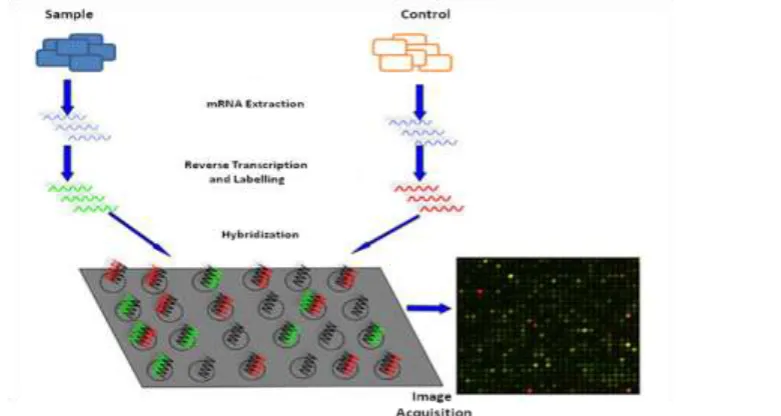
The power of a DNA microarray lies in the fact that is that there may be many thousands of different DNA molecules bonded to an array that makes possible to measure the expression of thousands of genes in sample simultaneously at one time. The DNA microarray measures the gene expression levels in sample by following difficult steps as shown in figure 2
Sample preparation and labelling
There are number of different ways in which a sample can be prepared and labelled for microarray experiments. In all the cases, the first step is to extract the RNA from the tissue of interest along with its control tissue and its conversion into cDNA. The cDNA probes used for array hybridization can be labelled with radioisotopes like P32 or by directly labelling with fluorescent markers. Nowadays, most of the laboratories use fluorescent labelling, with the two dyes Cy3 (excited by green laser) and Cy5 (excited by a red laser). In most of the experiments, the two samples each labelled with different dye are hybridized to the arrays, that finally allows the simultaneous measurement of both the samples (3). However, indirect labelling, instead of using fluorescently labelled dC, amino-allyl-modified dC is used in the reverse transcription reaction for making cDNA. Following this reaction, the cDNA is reacted with an ester of the dye, and in this way hence dye becomes attached to dC of cDNA. The third and least common method for labeling is by random primed labeling using the klenow fragment of DNA polymerase I (7,8).
Hybridization & Washing
Hybridization involves the duplex formation between the probe on the glass and labeled DNA (or RNA) via Watson-Crick base pairing. Hybridization is affected by many factors like, temperature, humidity, salt concentrations, form-amide concentration, volume of target solution and operator. To avoid non-specific binding, slides need to be treated before hybridization. This procedure depends on the slide type and spotting chemistry. The hybridization solution containing the probe is placed onto an array, covered with a coverslip and then placed in a humidified chamber over a period of 12 – 24 hours. Hybridizations can be set-up manually or robotically, however the latter provides better control and reduce the handling variations. The temperature employed depends on the GC content and type of array used. The stringency of the hybridization depends on the concentration of salts. Most of the hybridizations take place in approximately 1M Na+ (45, 37,3) . The next step involving the washing, removes the excess of hybridization solution to ensure that only labelled target is bound to probes on the array for measurement. The washing also reduces the chances of cross hybridization. Washing is either done with low salt buffers or by using higher temperatures ( 4).
Image acquisition
After hybridization and washing, the signal intensities of all the spots on a microarray slide are captured by scanners. The scanners have one or two lasers that are focused on the array, and the scanners which are used for two-color arrays use two lasers. In two-color arrays, the output image is usually two monochrome images, one for each of the two lasers in the scanner. These are combined to create the familiar red-green false colors images of the microarray (46).
V.
CONCLUSIONS
ACKNOWLEDGMENT
The Authors are highly thankful to Prof Girish Sharma, Amity University, Noida, UP for his inputs.
REFERENCES
[1]. T. Victor , F. Francesco, A. B. Hugo, ―DNA Microarrays: a Powerful Genomic Tool for Biomedical
and Clinical Research‖ Molecular Medicine, Vol. 13(9-10),: pp.527–541, 2007.
[2]. D. K. Garner, A. Weissman, C. M. Howells, Z. Shoham ―Text book of assisted reproductive Techniques‖, Informa Healthcare, 4th edition, pp358, 2012
[3]. C. A. Nsofor, "DNA microarrays and their applications in medical microbiology". Biotechnology and Molecular Biology Reviews, Vol. 9(1), pp. 1-11, 2014
[4]. K. Dill, R. Liu, P. Grodzinski, ―Integrated analytical systems. Microarrays preparation, microfluidics, detection methods and biological application‖, Springer, 2009, pp.276
[5]. N.M. M. Pires, T. Dong, U. Hanke, N. Hoivik, ―Recent developments in optical detection technologies in Lab-on-a-Chip Devices for biosensing applications‖. Sensors, Vol.14, pp. 15458-15479, 2014 [6]. A. Schulze and J. Downward, "Navigating gene expression using microarrays [mdash] a technology
review". Nature Cell Biology, Vol. 3, pp. E190-E195, 2001.
[7]. A. Singh, N. Kumar, "A review on DNA microarray technology". International Journal of Current Research and Review, Vol. 5(22), pp.01-05, 2013.
[8]. X.Teng, and H. Xiao, "Perspectives of DNA microarray and next-generation DNA sequencing technologies". Science in China Series C: Life Sciences,Vol. 52(1), pp. 7-16, 2009.
[9]. S.P.A. Fodor, J.L. Read, M.C. Pirrung, L. Stryer, A.T. Lu, D. Solas, "Light-directed, spatially addressable parallel chemical synthesis." Science, Vol. 251(4995),pp. 767-773,1991.
[10]. R. Bumgarner,"Overview of DNA microarrays: types, applications, and their future". Current protocols in molecular biology, Chapter 22(Unit 22.1), 2013.
[11]. G. MacBeath,"Protein microarrays and proteomics." Nature Genetics, Vol. 32, pp. 526 - 532, 2002 [12]. R. Bumgarner, "DNA microarrays: Types, Applications and their future". Current protocols in
molecular biology,Vol. 101, 22.1.1–22.1.11, 2011.
[13]. X-Q. Xia, Z. Jia, S. Porwollik, F. Long, C. Hoemme, K. Ye, C. Muller-Tidow, M McClelland, and Y
Wang, "Evaluating oligonucleotide properties for DNA microarray probe design". Nucleic Acids Research, Vol 38(11), pp. e121, 2010.
[14]. S. Schumacher, S. Muekusch, H. Seitz,"Up-to-Date applications of microarrays and their way to commercialization," Microarrays, Vol. 4(2), pp.196, 2015.
[15]. L. Geue, B. Stieber ,S. Monecke, I.Engelmann, F. Gunzer, P. Slickers, S.D. Braun, R.
Ehricht ,"Development of a Rapid Microarray-Based DNA Subtyping Assay for the Alleles of Shiga Toxins 1 and 2 of Escherichia coli". Journal of Clinical Microbiology, 52(8), pp.2898-2904, 2014 [16]. D.G. Wang, J.B. Fan, C.J. Siao, A. Berno, P.Young, R. Sapolsky, G. Ghandour, N. Perkins, E.
Winchester, J. Spencer, L. Kruglyak, L. Stein, L. Hsie L, T. Topaloglou, E. Hubbell, E. Robinson, M. Mittmann, M.S. Morris, N. Shen, D. Kilburn, J. Rioux, C. Nusbaum, S. Rozen, T.J. Hudson, R. Lipshutz, M. Chee, E,S. Lander, "Large-scale identification, mapping, and genotyping of single-nucleotide polymorphisms in the human genome." Science, Vol. 280(5366), pp.1077-1082, 1998. [17]. C. Debouck and P. N. Goodfellow, "DNA microarrays in drug discovery and development." Nature
Genetics,Vol. 21(1 Suppl),pp. 48-50,1999.
[18]. J-C. Cho, and J. M. Tiedje, "Bacterial Species Determination from DNA-DNA Hybridization by Using Genome Fragments and DNA Microarrays". Applied and Environmental Microbiology, Vol.67(8),pp. 3677-3682,2001.
[19]. S. N. Kumar, "A Proper Approach on DNA Based Computer." American Journal of Nanomaterials, Vol. 3(1), pp.1-14, 2015.
[20]. A. Sassolas, B. D. Leca-Bouvier,L.J. Blum . "DNA Biosensors and Microarrays". Chemical Reviews,Vol. 108(1), pp. 109-139, 2008.
[21]. D. Gershon,"Microarrays go mainstream". Nature Methods, Vol.1, pp. 263-270, 2004.
[22]. J. Sobek, K. Bartscherer, A. Jacob, J.D. Hoheisel, P. Angenendt, "Microarray technology as a universal tool for high-throughput analysis of biological systems". Jens Combinatorial Chemistry and High Throughput Screening, Vol. 9(5), pp. 365-380, 2006.
[23]. G.Hardiaman ―Microarray Innovations:Technology and Experimentation.. CRC Press Newyork USA,
2009.
[25]. C.D. Hodneland, Y.S.Lee , D-H. Min , M. Mrksich, "Selective immobilization of proteins to self-assembled monolayers presenting active site-directed capture ligands" in Proc. National Academy of Sciences of the United States of America,Vol 99(8), pp.5048-5052, 2002.
[26]. D. Neuschäfer, W. Budach, C. Wanke , S.D. Chibout, "Evanescent resonator chips: a universal platform with superior sensitivity for fluorescence-based microarrays." Biosensors and Bioelectronics, Vol. 18(4), pp. 489-497, 2003.
[27]. T. Kaufmann and B. J. Ravoo, "Stamps, inks and substrates: polymers in microcontact printing". Polymer Chemistry, Vol. 1(4),pp. 371-387, 2010.
[28]. Q. Xu and K. S. Lam, "Protein and Chemical Microarrays—Powerful Tools for Proteomics." Journal of Biomedicine and Biotechnology, 2003(5), pp. 257-266, 2003.
[29]. W. Gong, K. He, M. Covington, S.P. Dinesh-Kumar, M. Snyder, S.L. Harmer, Y-X. Zhu, XW Deng,"The Development of Protein Microarrays and Their Applications in DNA-Protein and Protein-Protein Interaction Analyses of Arabidopsis Transcription Factors." Molecular Plant, Vol. 1(1), pp. 27-41, 2008.
[30]. S. Seetharaman, M. Zivarts , N. Sudarsan, R.R. Breaker,Immobilized RNA switches for the analysis of complex chemical and biological mixtures". Nature Biotechnology,Vol. 19(4), pp. 336-341, 2001.
[31]. H.Y. Wang , R.L. Malek , A.E. Kwitek , A.S. Greene , T.V. Luu , B. Behbahani ,
B. Frank , J.Quackenbush, N.H. Lee, "Assessing unmodified 70-mer oligonucleotide probe performance on glass-slide microarrays". Genome Biology, Vol. 4(1), pp. R5, 2003.
[32]. N.C. Roy, E. Altermann, Z.A. Park, W.C.McNabb, "A comparison of analog and Next-Generation transcriptomic tools for mammalian studies." Briefings in Functional Genomics Vol. 10(3), pp.135-150, 2011.
[33]. T. Maier, M. Güell, L. Serrano, "Correlation of mRNA and protein in complex biological samples". FEBS Letters, Vol. 583(24), pp.3966-3973, 2009.
[34]. L.Smith and A. Greenfield, "DNA microarrays and development". Human Molecular Genetics, Vol. 12(suppl 1), pp. R1-R8, 2003.
[35]. M. Dufva, "Fabrication of high quality microarrays". Biomolecular Engineering, 22(5–6),pp.173-184, 2005.
[36]. V. Trevino, F. Falciani, H.A. Barrera-Saldaña, "DNA Microarrays: a Powerful Genomic Tool for Biomedical and Clinical Research". Molecular Medicine, Vol. 13(9-10), pp. 527-541, 2007.
[37]. D. Stekel, "Microarray bioinformatics'. Annals of Botany, Vol. 93, pp. 615-617, 2004.
[38]. Roasetta, Agilent and Oxford Gene Technology E.J.Masaro,S.N Austad.‖handbook of biology of aging‖ 6th Edition. pp.504, 2006.
[39]. A. H. Broderick, M. R. Lockett, M. E. Buck, Y. Yuan, L. M. Smith, D. M. Lynn, ―In situ Synthesis of Oligonucleotide Arrays on Surfaces Coated with Crosslinked Polymer Multilayers‖ Chemical Material, Vol. 24(5),pp. 939–945, 2012
[40]. A.C. Pease, D. Solas , E.J. Sullivan, M.T. Cronin, C.P.Holmes, S.P., "Light-generated oligonucleotide arrays for rapid DNA sequence analysis"in Proc. National Academy of Sciences of the United States of America, vol. 91(11), pp. 5022-5026. 1994.
[41]. R.J. Lipshutz, S.P. Fodor, T.R. Gingeras, D.J. Lockhart, "High density synthetic oligonucleotide arrays." Nature Genetics, Vol. 21(1 Suppl), pp. 20-24, 1999.
[42]. D.J. Lockhart, H. Dong, M.C. Byrne, M.T. Follettie, M.V. Gallo, M.S. Chee,M. Mittmann, C. Wang,M. Kobayashi, H. Horton, E.L. Brown. "Expression monitoring by hybridization to high-density oligonucleotide arrays". Nature Biotechnology,Vol. 14(13), pp.1675-1680, 1996.
[43]. M. J. Heller "DNA Microarray Technology: Devices, Systems, and Applications". Annual Review of Biomedical Engineering,Vol. 4(1), pp. 129-153, 2002.
[44]. S.Dasari, "Microarray Based Genotyping: A Review". Journal of Cancer Sciences, Vol.1,pp.2377-9292, 2014.
[45]. K. Mirnics,"Microarrays in brain research: the good, the bad and the ugly". Nature Reviews Neuroscience, Vol. 2(6), pp. 444-447, 2001.