muscle tone and their relationship to
models of subluxation / joint dysfunction
Part II
Gary A. Knutson,
DC*
Edward F. Owens, Jr.,
MS, DC**
The relationship of muscles to the causes and effects of the pathophysiologic entity referred to as chiropractic subluxation or joint dysfunction is critical. Part I of this paper reviewed the complexities of skeletal muscle in regards to anatomy, active and passive tone, detection of muscle tone, neurophysiology, and how muscle function fits into a variety of subluxation/joint dysfunction models. The concluding part of the review culminates in a
hypothesis to describe and explain varying degrees of muscle tone that may be encountered clinically. It is hoped that knowledge of the differing levels of muscle tone and their causes will help the clinician to better determine the underlying cause of a neuro-musculoskeletal problem allowing application of necessary and proper intervention. .
(JCCA 2003; 47(4):269–283)
K E Y W O R D S : skeletal muscle, muscle tone, subluxation,
joint dysfunction.
Les muscles jouent un rôle essentiel dans les causes et effets de l’entité physiopathologique couramment appelée « subluxation » ou « dysfonctionnement des articulations ». Dans la première partie du présent article, on a examiné la complexité des muscles squelettiques quant à l’anatomie, à la tonicité active et passive, à la présence de tonicité, à la neurophysiologie et au fonctionnement des muscles dans une variété de modèles de subluxation ou de dysfonctionnement des articulations. La conclusion de cet examen aboutit à une hypothèse pour décrire et expliquer les divers degrés de tonicité musculaire pouvant être reconnus sur le plan clinique. Nous espérons qu’une connaissance approfondie des divers degrés de tonicité musculaire et de leurs causes aidera le clinicien à mieux
cerner les causes sous-jacentes des troubles neuromusculosquelettiques afin de permettre l’intervention nécessaire et adéquate. (JACC 2003; 47(4):269–283)
M O T S C L É S : muscle squelettique, tonicité musculaire,
subluxation, dysfonctionnement des articulations.
* 840 W/17th, Suite 5, Bloomington, IN 47404. E-mail [email protected] ** Director of Research, Sherman College of Straight Chiropractic,
P.O. Box 1452, Spartanburg, SC 29304. © JCCA 2003.
ground in muscle physiology is overlaid on a variety of mechanisms that have been proposed to explain joint dysfunction. Finally, a guide to various types and causes of altered muscle tone that may be experienced in clinical practice is presented.
Introduction
back-Functional characteristics of muscle tone in models of joint dysfunction
The following section examines how the varying proper-ties of muscle physiology may be related to myopathology in differing models of joint dysfunction, which include: facilitation, central sensitization, flexor/nociceptive re-flex, pain-spasm-pain, gamma loop, sub-maximal contrac-tion, thixotropy and post contraction sensory discharge.
Facilitation/Korr-Denslow hypothesis
This is a neurological reflex based theory whose salient points are based on the premise that unbalanced, abnormal afferent signals enter the spinal cord and cause an increase in segmental neural activity, including active a -motoneu-ron firing, and which result in reflexive muscle hyperto-nicity. Input from the now activated muscles then reflexes into the cord, exacerbating the feedback loop.1 Korr
pro-posed that the segmental spinal motoneuron excitability was the result of the afferent output from muscle spindles.2
Central sensitization
In this theory, an injury that causes nociceptive signals can produce increases in muscle tone that are maintained long after the nociception ceases. These findings led to a theory of spinal cord plasticity now called central sensitization,3
or long-term potentiation.4 The effect of central
sensit-ization is to reduce the associated segmental neural thresh-olds prompting increased excitability and summation of the a-motoneuron(s) to threshold. Such a spinal learning mechanism has been suggested as a potential basis for osteopathic somatic dysfunction5 and chiropractic
sub-luxation.6,7 It has been theorized that chronic, repetitive
joint dysfunction is caused by some injury long ago whose afferent stimulation led to plastic changes in the spinal cord leading to a “neural scar”.5 Stimulation of this
sensi-tized area leads to reflexive muscle contraction and signs of dysfunction.
The source of afferent input leading to central sensit-ization has not been adequately defined. In a recent paper, the authors write: “It is not clear how central hyperexcit-ability is maintained and eventually causes chronicity, but most likely an outgoing nociceptive afferent barrage is needed”.8 Slosberg provides a review of central sensitization
and how this phenomenon may relate to joint dysfunction.6
Flexor or nociceptive reflex response to injury
Wyke, as reported in a review by Slosberg,9 found that
activation of nociceptors by irritation of the joint capsule generated an intense, non-adapting muscle hypertonicity to guard the joint. The musculoskeletal reflexes triggered by nociceptive input are termed “nocifensive reflexes”.10
The prototypical nocifensive reflex is the flexion reflex, in which stimulation of nociceptors causes muscular contrac-tion – less than a spasm – of the flexor muscle and inhibi-tion of the extensor muscle about the associated joint.11
Woolf and McMahon found that the prolonged nocicep-tive stimulation changed the reflexive muscular response such that, “Essentially, the high-threshold phasic flexor withdrawal response has become a low-threshold tonic one”. This change in muscular response was thought to be due to “a remarkable degree of functional plasticity of the system”.12 A group of interneurons in lamina 4–6 of the
dorsal horn of the spinal cord has been shown to encode the muscular contractions in nociceptive reflexes,13 and
this may be where plasticity of the nervous system occurs. Muscular reactions to nociceptive input may also be learned and stored in the cerebellum. Kottke has suggested that movement is based upon the development of cerebel-lar engrams that are acquired through learning.14 The
nocifensive reflex can be a conditioned reflex,15 and the
muscular reactions to a nociceptive stimulus have been shown to be learned by, and stored in, the cerebellum.16
Engrams occurring at the reflex level contain the codes for exciting and inhibiting the appropriate muscles. Such find-ings give support to a hypothesis that the cerebellum learns nocifensively induced muscle reactions to acute joint dys-function, and could account for a relatively constant mis-alignment pattern.
There is much evidence to show that the effect of injury to the mechanoreceptors of a joint can cause loss of aware-ness of joint position (loss of kinesthesia).17–19 McLain, for
example, found few mechanoreceptors in the facet capsule of the joints in the cervical spine, and commented that the loss of any of these receptors could lead to denervation of the joint with the potential for loss of protective muscular reflexes.20 This loss of kinesthetic sense allows the joint to
inter-neurons or cerebellar engram reacts with the pattern of muscle contraction learned from the prior nociceptive event. Hence, a pattern of joint dysfunction or postural distortion could be established.
Whether flexor or nociceptive reflex muscle contrac-tions can be maintained for long periods of time is contro-versial. Animal research indicates that a prolonged pathological nociceptive input from deep tissues abolishes the capability of muscle to facilitate the flexor reflex for extended periods of time.21 A further problem with the
flexor reflex model is that, in back pain, hypertonicity is often in the extensor muscles, not flexor muscles. Mense suggests that the activation of the g-motor system by muscle nociceptors must be postulated to account for extensor hypertonicity.22
Pain-spasm-pain positive feedback loop
The “vicious cycle” hypothesis of pain and muscle tone was proposed by Travell in 1942 who wrote, “According to this view, limitation of motion is primarily a reaction to pain rather than the result of structural lesion. If muscle spasm causes pain, and pain reflexly produces muscle spasm, a self-perpetuating cycle might be estab-lished ...”.23
Roland provides a review of evidence for the pain-spasm-pain cycle, arguing very generally that pain can cause muscle spasm and that muscle spasm can cause pain.24 However, subsequent to that review there have
been several studies examining what happens when a painful stimulus is applied to a muscle. While a linkage between muscle pain and increased muscle spindle sensi-tivity has been found, there was no effect of muscle pain on
a-motoneuron excitability.25 In their review, Matre et al.
found that “… a large number of well-controlled studies have shown no statistical significant difference in resting EMG activity between painful and non-painful muscles”.26
Indeed, a painful, inflamed muscle has been found to have lower than normal tone.27
If a muscle becomes hypertonic – spasmed – as a reflex, that spasm, while it may or may not be painful, does not seem to cause a positive feedback loop that results in further spasm. Roland points out that if the pain-spasm-pain theory is correct, eliminating pain-spasm-pain with analgesics or muscle spasm via biofeedback, physical therapy, or mus-cle relaxant drugs should interrupt the putative feedback loop.24 Although this type of positive feedback loop may
be active in some cases of musculoskeletal pain, many patients seeking chiropractic/manipulative evaluation have already been treated using methods that should abolish this hypothetical cycle, presumably without effective relief of their complaint.
Gamma loop
Recently, another type of feedback loop has been proposed and investigated with a series of experiments.25,28–31
Ac-tive muscle contraction generates metabolites of fatigue, including bradykinin, which stimulates group III and IV chemoreceptive nociceptors, or metaboreceptors.32 Group
III and IV metaboreceptors synapse with and excite g -motoneuron cells. Depolarization of g-motoneurons ex-cites homonymous muscle spindles which causes an increase in Ia and II output. The Ia and II input to the cord then stimulates a-motoneurons to summation causing fur-ther muscle contraction generating more metabolites, completing the positive feedback loop. Any increased ac-tivity in secondary (II) spindle afferents, which project back to the gamma system, constitutes a second positive feedback loop which may perpetuate the muscle contrac-tion in absence of the group III and IV input.29
Interest-ingly, as mentioned previously, there is incomplete cortical control of the static bag2 and chain spindle
fibers,33 which give rise to the secondary (II) spindle
afferents. Once a static gamma®spindle II® static gamma positive feedback loop has been created, cortical inhibitory signals may not be able to break the cycle.
Considerable research has demonstrated evidence for this mechanism28,29,34–41 however, some has not.42,43 It
may be worth noting that the two studies with negative results were done on the lumbar spine muscles of the cat, whereas most of the other studies were conducted on muscles of the head and neck. Perhaps the concentration of b2c spindles in the cervical spine and their predominant association with type II afferents is a factor in the conflict-ing findconflict-ings. Further information regardconflict-ing the gamma positive feedback loop hypothesis as proposed by Johansson/Sojka can be found in a recent review.44
Muscle stiffness associated with ischemia and fatigue – the establishment of a gamma positive feedback loop – would result in lowered nociceptive thresholds to me-chanical stimulation.45 This has been confirmed in studies
thresholds occur with spinal joint dysfunction the associ-ated paraspinal muscles would be painful to touch. Palpa-tion for abnormal pain thresholds has been shown to be a reliable characteristic of joint dysfunction.48–50
The gamma positive feedback loop itself has spinal cord connections by which a central sensitization phenomenon may occur. There are neurons connecting to the group II/g -motoneurons synapses which release noradrenaline (NA) and serotonin (5–HT). NA strongly depresses synaptic actions of group II afferents on gamma motoneurons thus effectively depressing the positive feedback from group II afferents to the gamma motoneuron. 5–HT has the oppo-site effect – to enhance activity of g-motoneurons.51
Jankowska and Gladden conclude, “Changes in the bal-ance between opposite effects of NA and 5–HT releasing neurons on g-motoneurons may be used to adjust the effec-tiveness of positive feedback to these neurons to the needs of different movements”.51
According to Pedersen et al., “The [gamma loop] hy-pothesis implies that sustained muscle contractions, in-flammation and/or ischaemia may lead to activation of chemosensitive group III and IV muscle afferents which increases the stretch sensitivity and the discharge rate of muscle spindle afferents. If the excitatory load on the g -muscle-spindle system is high enough, as a result of a massive input from, for instance, group III and IV muscle afferents due to reduced muscular circulation in combina-tion with inflammacombina-tion and/or muscle contraccombina-tions, this may turn the system into a ‘vicious circle’”.28 This positive
feedback loop could then become the source of the “afferent barrage” required for central sensitization.8
Over time, the spinal cord neural influence on the group II/
g-motoneuron synapse is altered to favor the excitatory action of serotonin (5–HT). This creates a central excitatory state able to enhance the influence of any signal from spindle group II afferents. This central excitatory state would remain in effect even if the initiating event and the ensuing positive feedback loop has been eliminated. Such a central, lowered threshold to re-establishing the positive feedback loop (causing muscle hypertonicity) could ac-count for recurrence of joint dysfunction at the same joint. If this hypothesis proves to be true, a complete correction of spinal joint dysfunction would involve breaking the segmental positive feedback loop and keeping it from recurring during the time it takes for the spinal cord serot-onin neurons to shift back to their normal – pre-positive
feedback loop – state. Speculatively, these goals may be facilitated by having the patient rest post manipulation and through the use of rehabilitation techniques.
Along with these potential positive feedback loops, mus-cle fatigue has also been shown to dramatically decrease the inhibitory afferent input to the a-motoneuron pool from Golgi tendon organ receptors.52 This cycle of fatigue,
gamma stimulation, alpha stimulation and Golgi inhibition has been forwarded as a hypothesis to explain exercise associated muscle cramps.53
While nociception from a muscle due to tissue injury or inflammation leads to inhibition of that muscle,27
noci-ception from muscle ischemia and the products of metabo-lism may indeed lead to positive feedback loop-induced hypertonicity. This positive feedback loop would stabilize with the muscle being hypertonic yet in dynamic equilib-rium with its vascular supply. This dynamic equilibequilib-rium would likely be at or about the critical force of the muscle, a concept discussed next.
Sub-maximal contraction
In most cases of active muscle contraction and hypertonic-ity, the decisive factor for the occurrence of fatigue and pain seems to be the vascular environment of the muscle. A muscle will compress its own blood vessels if it con-tracts with a force above approximately 30% maximal contractile force,54 40% in back extensor muscles.55 In
lower contraction forces, fatigue is not as certain.
a-motoneurons appear to fire in order of increasing size. Input to the motoneuron pool first excites to threshold the smallest, most excitable a-motoneurons connected to slow-twitch, fatigue-resistant type I muscle fibers.56
Mus-cles, particularly postural musMus-cles, are composed predomi-nantly of type I fibers, which, as we have seen, are suited for sustained contraction. One study suggested that during sub-maximal contraction – less than 40% maximum voluntary contraction (MVC) – of the erector spinae, the metabolic effects of fatigue may be counteracted or de-layed by the rotational recruitment of additional motor units. Such a compensatory mechanism may reflect a functional requirement of the back muscles to maintain static “postural” contractions for long periods of time.57
The phenomenon of rotation of slow twitch (type I) motor unit contraction may help explain the ability to sustain low-level active contraction.58
called the critical force of a muscle. Critical force is around 15 to 20% of a maximum voluntary contraction (MVC), and is that point below which an isometric con-traction can be maintained for a very long time without fatigue.58 To get a feel for this level of muscle contraction,
a 10% MVC is approximately the force used to maintain the arms in a horizontal working position.59 The critical
force point of a muscle is a controversial concept. Others have found that despite adequate blood flow, in long term contractions, a muscle can become fatigued although not exhausted.60,61 Figure 1 provides a graphic representation
of active muscle contraction relative to intramuscular blood flow.
Related to submaximal contraction is the so-called “Cinderella Hypothesis” (referring to Cinderella who worked continuously) proposed by Hagg in 1991.62 This
hypothesis states that type I fiber motor units are at risk of overload in conditions of low level, prolonged and sus-tained activation which causes fiber damage and pain.63–65
The hypothesis is based on the neurophysiological finding of ordered recruitment of motor units (Henneman princi-ple) with increasing force and the finding of abnormal morphological characteristics in trapezius myalgia.64 There
has been some research supportive of this hypothesis.63
Kadefors et al. suggests the possibility that sustained, monotonous contraction may result in a reduction of the neurophysiological activation threshold, making eventual
relaxation of the muscle difficult to achieve.63 If muscle
activation is kept below the critical force level, there is no metabolic (lactic acid, bradykinin) build-up to activate nociceptors to cause immediate pain despite the sustained contraction. Repetitive long-term contraction, however, may lead to physical damage of the muscle fibers resulting in pain.
Mense and Stahnke found that while contractions of moderate force are an effective stimulus for some muscle mechanoreceptors (group III and IV) another sub-popula-tion of these slowly conducting afferents is activated dur-ing ischemic work.66 This second population of group III
and IV receptors (metaboreceptors), “… is not excited or is only weakly excited by contractions without ischemia.”66
Pushing a muscle to a level of contraction past the critical force and into an ischemic contraction would generate enough metabolites to depolarize group III and IV metaboreceptors and be experienced cortically as pain.
Impaired muscle circulation has been found in cases of chronic neck and shoulder pain,67,68 is suspect in low back
pain,69 and has been shown to occur when spinal
biome-chanics change from lordosis to kyphosis.69 Ashton-Miller
et al. quote a study which showed that muscle contraction at a level of as little as 4% MVC combined with work loading caused pain and significant increases in sick leave due to musculoskeletal complaints.70
Sub-maximal contraction on the border of ischemia
may also lead to decreased performance of muscle fatigue characteristics.71–74 Mannion, in reviewing various causes
of back pain writes, “… the evidence implicating highly fatigable back muscles in the development of low back pain is somewhat more substantial”.75 While the
underly-ing cause(s) of the abnormal fatigability in back pain have not been adequately described, chronic sub-maximal con-traction resulting in limited blood flow affecting muscle performance is seen as a possibility. McGill notes that, “… complete relaxation of the low-back muscles is neces-sary to avoid compromising performance and an increased risk of musculoskeletal disorders”.72
Additional indirect evidence for the hypothesis of long term, sub-maximal contraction is the finding by Roland that, on balance, chronic back pain patients have increased spinal muscular activity at rest or following exercise, but reduced muscle activity during spinal movement.24 When
at rest, muscle contraction at or just above the critical force level would not be opposed by stretching or movement that would promote intra-muscular blood flow. Inactivity would allow for build-up of metabolites, activation of metabore-ceptors, and be experienced cortically as pain. Exercise would tend to quickly fatigue these already contracted muscles, also leading to ischemia and pain.76
A critical force contraction phenomenon may also be responsible for the long-term muscular hypertonicity asso-ciated with the asymmetric tonic neck reflex.77 This postural
reflex causes changes in muscle tone that do not attenuate
as long as the reflex is left alone.77–79
Finally, the stimulation of type III and IV metabore-ceptors with the by-products of muscle metabolism has been shown to cause changes in skin blood flow pat-terns,80–83 increased heart rate,84–86 blood pressure,85,86
rate of breathing,80 and renal86 and splenic86 sympathetic
nerve activity. All of these reflexive effects in response to metaboreceptor stimulation work to increase blood flow to the ischemic muscle(s), and are elements of what is known as the pressor reflex.
Given the intimate connection of muscles to joint dys-function, one might then expect that manipulation would be found to have an effect on some of the noted physiologi-cal effects of metaboreceptors. Although no direct link between manipulation and a decrease in metaboreceptor activity has been studied, there have been many studies, some controlled and blinded, showing a correlation be-tween manipulation and a significant decrease in blood pressure.87–90 Study of other physiological effects of
metaboreceptors and manipulation for joint dysfunction may be fruitful areas of investigation.
Thixotropy
As was noted previously, there is a tendency for the actin and myosin filaments to stick together when inactive for a period of time. If a muscle has been lengthened or short-ened for a period of time, cross-bridges form at that length and/or tone, changing the resting properties of that
mus-Figure 2 Point A is normal resting muscle tone. As the muscle actively contracts (alpha excitation) muscle tone increases. From point B to C the muscle is being held in an actively contracted state. At point C, alpha excitation stops, and the muscle fibers relax. At point D, the muscle tone stops decreasing due to cross-bridges in the muscle fibers remaining in their prior contracted position. This is muscle – intra- and extrafusal – thixotropy. Note the muscle tone is increased despite no active alpha excitation. A rapid stretch of the muscle, point E to F, brings the muscle tone back to its normal resting value.
Muscle tone (resistance to stretch)
A
B C
D E
F
Thixotropy
cle91–95 (Figure 2). Because muscle thixotropy depends on
passive muscle physiology – the resting positions of actin-myosin cross-bridges – a thixotropic muscle may be hypertonic without EMG activity. In the standing human, the center of gravity of the upper body is anterior of the ankle joint, suggesting that some constant force in the gas-tocnemius or soleus muscle is required to counter the for-ward moment and prevent toppling. Basmajian and Deluca, however, noted that the posterior calf muscles are electromyographically silent on quiet standing, except for occasional bursts of corrective activity.96 Simon and
Mense used the passive thixotropic property of muscle to account for the non-contractile forces that must be gener-ated by the calf and other postural muscles. They estimate that the thixotropic bonds formed when a muscle is ac-tively contracted, then relaxed, can stiffen the muscle up to ten times its normal resting tone.54
How muscle thixotropy could be related to the patho-physiology of joint dysfunction is not difficult to imagine. Nociception may cause a slowly fatiguing reflexive mus-cle contraction.10,97 When the nociceptive input and active
contraction cease, the reflexively contracted muscle may not return to a normal relaxed length and tone, but remain in a shortened, hypertonic state due to the cross-bridge bonds formed during the previous contraction.
Clinically the effect of thixotropy on the resting length and/or tone of muscles is largely unexplored. One report has presented evidence that the cause of lagophthalmos (incomplete closure of the eyelid) in facial nerve palsy (e.g. Bell’s Palsy) is due to thixotropy of the levator palpebrae muscle.98 Treatment consisted of passive
clo-sure of the affected eyelid followed by manual stretching of the upper eyelid in a downward direction as far as possible, stretching the muscle to break down the “stuck” cross-bridges. Other researchers speculate that “limber-ing-up manoeuvres commonly employed by athletes, danc-ers and physiotherapists”, involve the loosening of the thixotropic muscle (intra and extrafusal) bonds that have been re-set due to prior contraction.99 Others find that
passive and active stretching does not seem capable of re-setting the “hung up” intrafusal fibers.100 In these cases,
sudden stretching,95,100 of sufficient velocity94 and
magni-tude95 break down the re-formed intrafusal fiber
cross-bridges. Adjustment may provide the sudden stretch of sufficient velocity and magnitude to re-set thixotropic muscles.
Post contraction sensory discharge
During a contraction of extrafusal muscle fibers, related muscle spindles are temporarily unloaded, and Ia and II discharge decreases.101,102 With Ia and II input to the
spi-nal cord inhibited, the gamma efferent sigspi-nal to the muscle spindles is increased;100,103 Matthews calls this “automatic
gain compensation”.104 This gain compensation is used to
establish a new “normal” muscle length and/or tension or “fusimotor set”.105
Greater g-motoneuron activity causes increased (over the normal, resting muscle) Ia and II spindle output. Experi-ments in cats have shown that in sampled Ia fibers, dis-charge rates after muscle contraction had increased by 60%, and a number of these receptors had been silent prior to the contraction.106 This increased Ia and II muscle spindle
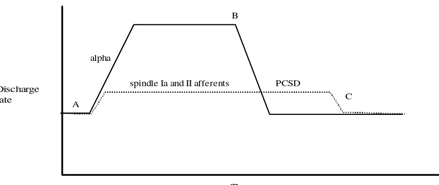
sen-sory output is known as post contraction sensen-sory discharge (PCSD)93,105–107 (Figure 3). The PCSD phenomenon allows
for continued muscle tension, shortened length,91,99,106 and
mostly Ia (with some II) spindle output.91,93,99,100,106
Eldred, Hutton and Smith provide a concise summary of the effects of PCSD:108
1 A persisting increase in discharge at a maintained mus-cle length and in response to stretch appears after a muscle has undergone contraction, under the condition that the efferent background is otherwise quiet.
2 Fusimotor activation alone, i.e., contraction of intra-fusal fibers, can produce this effect, though extraintra-fusal contraction also seems to contribute.
3 The enhanced discharge, as it was recorded, arises to a major extent in Ia afferent fibers.
4 The cause of the effect is probably mechanical in nature. The mechanical nature of PCSD is thought to be due to re-setting of the intrafusal actin-myosin cross-bridges in the contracted/lengthened muscle. Given that kinesthetic sense is almost exclusively from muscle spindles109
A similar mechanism of spindle dyregulation, where the intrafusal representation of the extrafusal muscle length no longer matches the actual length, was proposed by Donaldson111 to explain the rapid recovery of patients with
chronic back pain.112 Rapid response of patients with
chronic back pain to manipulation has also been reported.113
Because the recovery in these chronic cases was so rapid, degenerative muscle changes are not likely to be involved and the mechanism appears to be neurological rather than muscular in nature. The post contraction sensory discharge phenomenon involving “hung up” intrafusal fibers after long term contraction or stretch is a similar “dysregulation” leading to chronic but reversible increases in muscle tone. We have noted that neural commands and feedback depend on muscle spindle signals,109,114 and muscle
hyper-tonicity has been associated with evidence of increased spindle output.115,116 The phenomenon of PCSD could
explain what Panjabi had noted regarding spinal stability. “The active musculoskeletal subsystem may develop
dete-rioration of its ability to receive and/or carry out the neural commands, to provide accurate feedback of muscle ten-sion information to the neural control unit, or to produce coordinated and adequate muscle tensions”.117 This loss in
the integrity of the stabilizing capacity of the spinal sup-port system could be a precursor to back pain.117,118 Panjabi
writes, “One example of the kind of error that might occur is that one or more muscles may fire in a manner that is undesirable; too small or too large forces and/or too early or too late firing. This may happen either due to the faulty information transmitted from the spinal system transduc-ers or due to the fault of the control unit itself. Such an error may cause excessive muscle tension, resulting in soft tissue injury and pain. This may explain some of the instances of acute low back pain initiations where negligi-ble or marginal loads are involved (e.g., while picking up a piece of paper from the floor)”.117
No one particular model of joint dysfunction and its muscular component has reached consensus. One, some, Figure 3 At point A, the muscle is resting. Contraction begins as the a-motoneurons fire, gamma signal to the muscle spindles causes contraction of the intrafusal fibers to match the extrafusal contraction thereby retaining coordination between muscle length and spindle signal. Muscle spindle afferent firing is increased. At point B, alpha firing stops. The discharge from muscle spindle Ia and II afferents, however, remains higher than resting levels. This is post contraction sensory discharge (PCSD). The elevated levels of spindle afferent firing keeps the muscle tone – the resistance to stretch – higher than resting tone. A quick stretch with sufficient magnitude of the muscle, at point C, resets the intrafusal fibers to extrafusal length, bringing the muscle spindle afferent discharge, and muscle tone, back to the resting normal.
A
B
alpha
spindle Ia and II afferents Discharge
rate
Time
PCSD
C
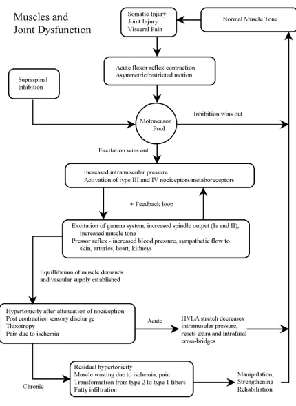
or none of the above models may be correct. It is possible, if not likely, that more than one model involving active and passive aspects of muscle tone will characterize joint dys-function. Perhaps, for example, nociceptive muscular re-flexes predominate in the acute phase of dysfunction with ischemia, mechanoreceptor, thixotropic and sub-maximal contraction mechanisms in chronic phases. Figure 4 shows a flow chart outlining the varying characteristics of muscle function and how they may relate to joint dysfunction.
Altered muscle tone – clinical application
Given the complex inter-relationship of muscle physiol-ogy and joint dysfunction, we would propose the following hypothetical assignment of abnormal clinical muscle tone variations and associated findings. It is important to note, especially in acute presentations, that hypertonicity of a muscle is due to some factor other than an injury/inflam-mation in that muscle; injured muscles are inhibited.27 As
Mense noted, teleologically, inhibition induced by myosi-tis would be an advantage, because it could reduce the forces acting on the damaged muscle.119 Involuntary
mus-cle hypertonicity is more often a secondary reflex reaction to other injury, or, potentially a sign of primary pathologic dysfunction. Also, each of the below variations of muscle tone might lie on a continuum from mild to severe.
• Full muscle spasm hypertonicity – Full active muscular contraction. Spasmodic torticollis and other dystonic syn-dromes, cramps, antalgic reactions. Some therapeutic in-tervention to reduce the spasm may be indicated, although full contraction cannot be maintained for very long due to ischemia and fatigue.
• Reflex spasm hypertonicity – Active contraction of mus-cle due to reflex reaction. For example, a nocifensive or flexor reflex as a splinting response to joint/disc injury. Reflex hypertonicity results in muscle tension less than maximal spasmodic contraction. If the reflex contraction is sustained, ischemia and pain from the muscle itself will result. Movement often aggravates reflex hypertonicity. This kind of hypertonicity could be seen in acute injury and joint dysfunction.
If the nociceptive input from the initiating injury fades rapidly, the hypertonicity would decrease, perhaps leaving a passive shortened, hypertonic muscle via thixotropy (and the effects of PCSD). The reflex spasm hypertonicity may
lead to the establishment of a gamma positive feedback loop, maintaining increased muscle tension and increased spindle output. Muscle stretch, via manipulation or other methods, post nociception could re-set the contracted mus-cle to a normal passive tone by re-establishing coordina-tion between the intra- and extrafusal fibers. The gamma feedback loop reaction to chronic contraction is plausible for type I fiber and spindle rich postural muscles, espe-cially those rich in b2c spindles. Larger, multi-joint
span-ning muscles with fewer fatigue resistant fibers exposed to chronic contraction may develop trigger points. In chronic cases of reflex spasm, degenerative changes – muscle wasting, fiber transformations, elastic component trans-formations - may require muscle strengthening and rehabilitation.
• Critical force hypertonicity – Active muscle contraction at a level of hypertonicity where there is a dynamic equi-librium of muscle demands and vascular supply. On one side of the dynamic equilibrium where the vascular supply is adequate, muscle hypertonicity may cause postural ab-normalities but no immediate pain. On the other side of the area of equilibrium, contraction with ischemia causes pain and fatigue. This kind of hypertonicity would often be temporarily relieved by movement/stretching, but aggra-vated by inactivity, such as sleeping, where there is no resistance to the chronic muscle contraction. Any contrac-tile effort which engages the muscle past the critical force point would also cause pain (similar to the leg pain in intermittent claudication). Such hypertonicity could be re-sponsible for non-fatiguing muscular hypertonicity seen in pathologic tonic neck reflexes and result in long term postural distortions such as pelvic torsion, leg length align-ment asymmetry, loss of lordosis, spinal curvatures, etc. Full spasm and reflex spasm muscular contractions may end in this type of active sustainable hypertonicity via gamma positive feedback loops.
• Thixotropic hypertonicity – Thixotropic properties in-volve resetting of muscle towards a shortened length and increased tone. There would be no muscle pain due to its passive non-contractile nature. Thixotropic hypertonicity may be caused by assuming awkward postures, or may be the final, sustainable outcome of a reflexive spasm. Thixo-tropic hypertonicity results in a loss of normal ROM that can cause compensatory muscular and joint changes that could become acutely painful and may be responsible for transient joint fixation that occurs in everyday life. This type of muscle hypertonicity can be acute or chronic, and may be eliminated by exercise, stretching, massage as well as high velocity manipulation.
• Hypotonicity – An inability of the muscle to produce force or the unwillingness to contract. Hypotonicity can be due to the influence of facet joint stimulation, joint injury, muscle inflammation or tonic neck reflexes
Conclusion
The muscular system is characterized by complexities of anatomy and physiology allowing for extreme plasticity to meet changing conditions. Differing levels of muscle tone, causes of increased and decreased tone, effects of tone changes, types of muscle fibers and spindles, degrees of hypertonicity and the time frame of tone changes are all variables in somatic dysfunction. Muscle physiology and attendant neurophysiological reflexes seem to explain, at least in part, some of the signs and symptoms of the mus-culoskeletal component of joint dysfunction as seen in clinical practice. Using knowledge of the differing levels of muscle tone and their causes will help to determine the cause of a musculoskeletal problem and allow for a better application of intervention. Hopefully this overview, with its condensation of a wide array of information, has pro-vided a glimpse into the complicated workings of the mus-cular system and has stimulated thinking that will further research. Such direct clinical research will aid in the diag-nosis, analysis and treatment of patients.
References
1 Korr IM. The neural basis of the osteopathic lesion. JAOA 1947; 191–198.
2 Korr IM. Proprioceptors and somatic dysfunction. JAOA 1975; 638–650.
3 Woolf CJ. Somatic pain – pathogenesis and prevention. Br J Anaesth 1995; 75:169–176.
4 Pockett S. Spinal cord synaptic plasticity and chronic pain. Anesth Analg 1995; 80:173–179.
5 Patterson MM, Steinmetz JE. Long-lasting alterations of spinal reflexes: A potential basis for somatic dysfunction. Man Med 1986; 2:38–42.
6 Slosberg M. Spinal learning: Central modulation of pain processing and long-term alteration of interneuronal excitability as a result of nociceptive peripheral input. J Manipulative Physiol Ther 1990; 13(6):326–336. 7 Nansel D, Peneff A, Cremata E, Carlson J. Time course
considerations for the effects of unilateral lower cervical adjustments with respect to the amelioration of cervical lateral-flexion passive end-range asymmetry.
J Manipulative Physiol Ther 1990; 13(6):297–304. 8 Johansen MK, Graven-Nielsen T, Olesen AS,
Arendt-Nielsen L. Generalized muscular hyperalgesia in chronic whiplash syndrome. Pain 1999; 83:229–234.
9 Slosberg M. Effects of altered afferent articular input on sensation, proprioception, muscle tone and sympathetic responses. J Manipulative Physiol Ther 1988;
11(5):410–418.
10 Van Buskirk R. Nociceptive reflexes and the somatic dysfunction: A model. J Am Osteop Assoc 1990; 90(9):792–809.
11 Schaible HG, Grubb B. Afferent and spinal mechanisms of joint pain. Pain 1993; 55:5–54.
12 Woolf CJ, McMahon SB. Injury-induced plasticity of the flexor reflex in chronic decerebrate rats. Neuroscience 1985; 16(2):395–404.
13 Holmberg H, Schouenborg J. Postnatal development of the nociceptive withdrawal reflexes in the rat: a behavioural and electromyographic study. J Physiol 1996;
493.1:239–252.
14 Kottke FJ. From reflex to skill: the training of coordination. Arch Phys Med and Rehab 1980; 61:551–561.
15 Kmietzyk HJ. Generalized nocifensive reflexes. I. Methodologic bases [Article in German] EEG EMG Z Elektroenzephalogr Elektromyogr Verwandte Geb 1990; 21(1):7–12.
16 Clark RE, Zhang AA, Lavond DG. Reversible lesions of the cerebellar interpositis nucleus during acquisition and retention of a classically conditioned behavior. Behav Neurosci 1992; 106(6):879–888.
18 Warner J. Role of proprioception in pathoetiology of shoulder instability. Clin Orth Related Res 1996; 35–39. 19 Hall MG, Ferrell WR, Sturrock RD, Hamblen DL,
Baxendale RH. The effect of the hypermobility syndrome on knee joint proprioception. Br J Rheumatol 1995; 34(2):121–125.
20 McLain R. Mechanoreceptor endings in human cervical facet joints. Spine 1994; 19(5):495–501.
21 Wall PD, Coderre TJ, Stern Y, Wiesenfeld-Hallin Z. Slow changes in the flexion reflex of the rat following arthritis or tenotomy. Brain Res 1988; 447:215–222.
22 Mense S. Nociception from skeletal muscle in relation to clinical muscle pain. Pain 1993; 54:241–289.
23 Travell J, Rinzter S, Herman M. Pain and disability of the shoulder and arm. JAMA 1942; 120:417–422.
24 Roland MO. A critical review of the evidence for a pain-spasm-pain cycle in spinal disorders. Clinical Biomechanics 1986; 1:102–109.
25 Djupsjöbacka M, Johansson H, Bergenheim M, Wenngren BI. Influences on the gamma-muscle spindle system from muscle afferents stimulated by increased intramuscular concentrations of bradykinin and 5–HT. Neurosci Res 1995 Jun; 22(3):325–333.
26 Matre DA, Sinkjær T, Svensson P, Arendt-Nielsen L. Experimental muscle pain increases the human stretch reflex. Pain 1998; 75(2–3):331–339.
27 Mense S, Skeppar P. Discharge behaviour of feline gamma-motorneurons following induction of an artificial myositis. Pain 1991; 46:201–210.
28 Pedersen J, Sjölander P, Wenngren BI, Johansson H. Increased intramuscular concentration of bradykinin increases the static fusimotor drive to muscle spindles in neck muscles of the cat. Pain 1997 Mar; 70(1):83–91. 29 Johansson H, Sojka P. Pathophysiological mechanisms
involved in genesis and spread of muscular tension in occupational muscle pain and in chronic musculoskeletal pain syndromes: a hypothesis. Med Hypotheses 1991 Jul; 35(3):196–203.
30 Wenngren BI, Pedersen J, Sjölander P, Bergenheim M, Johansson H. Bradykinin and muscle stretch alter
contralateral cat neck muscle spindle output. Neurosci Res 1998 Oct; 32(2):119–129.
31 Pedersen J, Ljubisavljevic M, Bergenheim M, Johansson H. Alterations in information transmission in ensembles of primary muscle spindle afferents after muscle fatigue in heteronymous muscle. Neuroscience 1998 Jun; 84(3):953–959.
32 Kaufman MP, Rybicki KJ, Waldrop TG, Ordway GA. Effects of ischemia on responses of group III and IV afferents to contraction. J Applied Physiol 1984; 57:644–650.
33 Boyd IA. The isolated mammalian muscle spindle. Trends Neurosci 1980; 258–265.
34 Kitagawa Y, Enomoto S, Nakamura Y, Hashimoto K. Asymmetry in jaw-jerk reflex latency in craniomandibular dysfunction patients with unilateral masseter pain. J Oral Rehabil 2000 Oct; 27(10):902–910.
35 Hellström F, Thunberg J, Bergenheim M, Sjölander P, Pedersen J, Johansson H. Elevated intramuscular concentration of bradykinin in jaw muscle increases the fusimotor drive to neck muscles in the cat. J Dent Res 2000 Oct; 79(10):1815–1822.
36 Thunberg J, Hellström F, Sjölander P, Bergenheim M, Wenngren B, Johansson H. Influences on the fusimotor-muscle spindle system from chemosensitive nerve endings in cervical facet joints in the cat: possible implications for whiplash induced disorders. Pain 2001 Mar; 91(1–2):15–22.
37 Wang K, Arendt-Nielsen L, Svensson P. Excitatory actions of experimental muscle pain on early and late components of human jaw stretch reflexes. Arch Oral Biol 2001; 46(5):433–442.
38 Ro JY, Capra NF. Modulation of jaw muscle spindle afferent activity following intramuscular injections with hypertonic saline. Pain 2001 May; 92(1–2):117–127. 39 Thunberg J, Ljubisavljevic M, Djupsjöbacka M, Johansson
H. Effects on the fusimotor-muscle spindle system induced by intramuscular injections of hypertonic saline. Exp Brain Res 2002 Feb; 142(3):319–326.
40 Hellström F, Thunberg J, Bergenheim M, Sjölander P, Djupsjöbacka M, Johansson H. Increased intra-articular concentration of bradykinin in the temporomandibular joint changes the sensitivity of muscle spindles in dorsal neck muscles in the cat. Neurosci Res 2002 Feb; 42(2):91–99.
41 Della Torre G, Brunetti O, Pettorossi VE. Capsaicin-sensitive muscle afferents modulate the monosynaptic reflex in response to muscle ischemia and fatigue in the rat. Arch Ital Biol 2002 Jan; 140(1):51–65.
42 Kang YM, Wheeler JD, Pickar JG. Stimulation of chemosensitive afferents from multifidus muscle does not sensitize multifidus muscle spindles to vertebral loads in the lumbar spine of the cat. Spine 2001 Jul 15;
26(14):1528–1536.
43 Zedka M, Prochazka A, Knight B, Gillard D, Gauthier M. Voluntary and reflex control of human back muscles during induced pain. J Physiol 1999; 502.2:591–604. 44 Knutson G. The role of the g-motor system in increasing
muscle tone and muscle pain syndromes; a review of the Johansson/Sojka hypothesis. J Manipulative Physiol Ther 2000; 23(8):564–572.
45 Mense S. Nervous outflow from skeletal muscle following chemical noxious stimulation. J Physiol 1977; 267:75–88. 46 Jensen K, Tuxen C, Pedersen-Bjergaard U, Jansen I,
47 Babenko V, Graven-Nielsen T, Svensson P, Drewes A, Jensen T, Arendt-Nielsen L. Experimental human muscle pain and muscular hyperalgesia induced by combinations of serotonin and bradykinin. Pain 1999; 82:1–8.
48 Keating JC, Bergmann TF, Jacobs GE, Finer BA, Larson K. Inter-examiner reliability of eight evaluative dimensions of lumbar segmental abnormality.
J Manipulative Physiol Ther 1990; 13:463–470.
49 Paydar D, Thiel H, Gemmell H. Intra- and inter-examiner reliability of certain pelvic palpatory procedures and the sitting flexion test for sacroiliac joint mobility dysfunction. J Neuromusc Sys 1990; 4:65–69.
50 Christensen HW, Vach W, Vach K, Manniche C, Haghfelt T, Hartvigsen L, Høilund-Carlsen PF. Palpation of the upper thoracic spine: An observer reliability study. J Manipulative Phyiol Ther 2002; 25(5):285–292. 51 Jankowska E, Gladden MH. A positive feedback circuit
involving muscle spindle secondaries and gamma motoneurons in the cat. Progress in Brain Research 1999; 123:149–156.
52 Nelson DL, Hutton RS. Stretch sensitivity of golgi tendon organs in fatigued gastrocnemius muscle. Med Sci Sports Exerc 1986; 18(1):69–74.
53 Schwellnus MP, Derman EW, Noakes TD. Aetiology of skeletal muscle ‘cramps’ during exercise: A novel hypothesis. J Sports Sciences 1997; 15:277–285.
54 Simons DG, Mense S. Understanding and measurement of muscle tone as related to clinical muscle pain. Pain 1998; 75:1–17.
55 Bonde-Petersen F, Mork AL, Nielsen E. Local muscle blood flow and sustained contractions of human arm and back muscles. Eur J Appl Physiol 1975; 34:43–50. 56 Henneman E. Skeletal muscle, the servant of the nervous
system. In: Mountcastle VB, ed., Medical Physiology. 14th ed. St Louis: Mosby CV, 1980 p674.
57 Dolan P, Mannion AF, Adams MA. Fatigue of the Erector Spinae Muscles. Spine 1995; 20:149–159.
58 Kahn JF, Favriou F, Jouanin JC, Monod H. Influence of Posture and Training on the Endurance Time of a Low-Level Isometric Contraction. Ergonomics 1997; 40:1231–1291.
59 Sjogaard G, Savard G, Juel C. Muscle blood flow during isometric activity and its relation to muscle fatigue. Eur J Appl Physiol 1988; 57:327–335.
60 Sjogaard G, Kiens B, Jørgensen K, Saltin B. Intramuscular pressure, EMG and blood flow during low-level prolonged static contractions in man. Acta Physiol Scand 1986; 128:475–484.
61 Fallentin N, Jørgensen K. Blood pressure response to low level static contractions. Eur J Appl Physiol 1992; 64:455–459.
62 Haag G. Static work loads and occupational myalgia – a new explanation model. In: Electromyographical Kinesiology. Anderson PA, Hobart DJ, Danoff JV Eds. Amsterdam: Elsevier. pp 141–143.
63 Kadefors R, Forsman M, Zoega B, Herberts P.
Recruitment of low threshold motor-units in the trapezius muscle in different static arm positions. Ergonomics 1999 Feb; 42(2):359–375.
64 Kitahara T, Schnoz M, Laubli T, Wellig P, Krueger H. Motor-unit activity in the trapezius muscle during rest, while inputting data, and during fast finger tapping. Eur J Appl Physiol 2000; 83:181–189.
65 Forsman M, Birch L, Zhang Q, Kadefors R. Motor unit recruitment in the trapezius muscle with special reference to coarse arm movements. J Electromyogr Kinesiol 2001 Jun; 11(3):207–216.
66 Mense S, Stahnke M. Responses in muscle afferent fibres of slow conduction velocity to contractions and ischemia in the cat. J Physiol 1983; 342:383–397.
67 Larsson R, Öberg PA, Larsson SE. Changes of trapezius muscle blood flow and electromyography in chronic neck pain due to trapezius myalgia. Pain 1999 Jan; 79(1):45–50. 68 Larsson R, Cai H, Zhang Q, Öberg PA, Larsson SE.
Visualization of chronic neck-shoulder pain: impaired microcirculation in the upper trapezius muscle in chronic cervico-brachial pain. Occup Med (Oxf) 1998; 48(3):189–194.
69 Konno S, Kikuchi S, Nagaosa Y. The relationship between intramuscular pressure of the paraspinal muscles and low back pain. Spine 1994; 19(19):2186–2189.
70 Ashton-Miller JA, McGlashen KM, Herzenberg JE, Stohler CS. Cervical muscle myoelectric response to acute experimental sternocleidomastoid pain. Spine 1990; 15(10):1006–1012.
71 Murthy G, Kahan NJ, Hargens AR, Rempel DM. Forearm muscle oxygenation decreases with low levels of voluntary contraction. J Orthop Res 1997; 15(4):507–511.
72 McGill SM, Hughson RL, Parks K. Lumbar erector spinae oxygenation during prolonged contractions: implications for prolonged work. Ergonomics 2000; 43(4):486–493. 73 Murthy G, Hargens AR, Lehman S, Rempel DM. Ischemia
causes muscle fatigue. J Orthop Res 2001 May; 19(3):436–440.
74 Yoshitake Y, Ue H, Miyazaki M, Moritani T. Assessment of lower-back muscle fatigue using electromyography, mechanomyography, and near-infrared spectroscopy. European Journal of Applied Physiology 2001; 84(3):174–179.
76 Hudlicka O, Brown MD, Egginton S, Dawson JM. Effect of long-term electrical stimulation on vascular supply and fatigue in chronically ischemic muscles. J Applied Physiol 1994; 77(3):1317–1324.
77 Marinelli PV. The asymmetric tonic neck reflex: Its presence and significance in the newborn. Clinical Pediatrics 1983; 22(8):544–546.
78 Tokizane T, Murao M, Ogata T, Kondo T.
Electromyographic studies on tonic neck, lumbar, and labyrinthine reflexes in normal persons. Jap J Physiol 1951; 2:130–146.
79 Lindsay KW, Roberts TDM, Rosenberg JR. Asymmetric tonic labyrinth reflexes and their interaction with neck reflexes in the decerebrate cat. J Physiol 1976; 261:583–601.
80 Crandall CG, Stephens DP, Johnson JM. Muscle metaboreceptor modulation of cutaneous active vasodilation. Med Sci Sports Exerc 1998 Apr; 30(4):490–496.
81 Piepoli M, Clark AL, Coats AJ. Muscle metaboreceptors in hemodynamic, autonomic, and ventilatory responses to exercise in men. Am J Physiol 1995 Oct;
269(4 Pt 2):H1428–1436.
82 Vissing SF, Scherrer U, Victor RG. Stimulation of skin sympathetic nerve discharge by central command. Differential control of sympathetic outflow to skin and skeletal muscle during static exercise. Circulation Research 1991; 69:228–238.
83 Vissing SF, Hjortso EM. Central motor command activates sympathetic outflow to the cutaneous circulation in humans. J Physiol 1996; 492.3:931–939.
84 Matsukawa K, Wall PT, Wilson LB, Mitchell JH. Reflex stimulation of cardiac sympathetic nerve activity during static muscle contraction in cats. Am J Physiol 1994 Aug; 267(2 Pt 2):H821–827.
85 Watanabe H, Iwase S, Mano T. Responses of muscle sympathetic nerve activity to static biceps brachii
contraction in humans. Jpn J Physiol 1995; 45(1):123–135. 86 Matsukawa K, Wall PT, Wilson LB, Mitchell JH.
Reflex responses of renal nerve activity during isometric muscle contraction in cats. Am J Physiol 1990 Nov; 259(5 Pt 2):H1380–1388.
87 Plaugher G, Bachman TR. Chiropractic management of a hypertensive patient. J Manipulative Physiol Ther 1993 Oct; 16(8):544–549.
88 Yates RG, Lamping DL, Abram NL, Wright C. Effects of chiropractic treatment on blood pressure and anxiety: a randomized, controlled trial. J Manipulative Physiol Ther 1988 Dec; 11(6):484–488.
89 McKnight ME, DeBoer KF. Preliminary study of blood pressure changes in normotensive subjects undergoing chiropractic care. J Manipulative Physiol Ther 1988 Aug; 11(4):261–266.
90 Knutson G. Significant changes in systolic blood pressure post vectored upper cervical adjustment vs. resting control groups; a possible effect of the cervicosympathetic and/ or pressor reflex. J Manipulative Physiol Ther 2001; 24(2):101–109.
91 Hagbarth K-E, Macefield VG. The Fusimotor System. Adv Exp Med Biol 1995; 384:259–270.
92 Hutton RS, Smith JL, Eldred E. Postcontraction sensory discharge from muscle and its source. J Neurophysiol 1973; 36:1090–1103.
93 Gregory JE, Mark FR, Morgan DL, Patak A, Polus B, Proske U. Effects of muscle history on the stretch reflex in cat and man. J Physiol 1990; 424:93–107.
94 Baumann TK, Hullinger M. The dependence of the response of cat spindle Ia afferents to sinusiodal stretch on the velocity of concomitant movement. J Physiol 1991; 439:325–350.
95 Morgan DL, Prochazka A, Proske U. The after-effects of stretch and fusimotor stimulation on the responses of primary endings of cat muscle spindles. J Physiol 1984; 356:465–477.
96 Basmajian JV, Deluca CJ. Muscles Alive, 5th ed. Williams and Wilkins, Baltimore, 1985, p257.
97 Ruch TC. Pathophysiology of pain, in Ruch T, Patton HD (eds): Physiology and Biophysics: The Brain and Neural Function, ed 2. WB Saunders Co, Philadelphia, 1979: 272–324.
98 Aramideh M, Koelman JH, Devriese PP, Speelman JD, Ongerboer De Visser BW. Thixotropy of levator palpebrae as the cause of lagophthalmos after peripheral facial nerve palsy. J Neurol Neurosurg Psychiatry 2002 May; 72(5):665–667.
99 Hagbarth K-E, Hägglund JV, Mordin M, Wallin EU. Thixotropic behaviour of human finger flexor muscles with accompanying changes in spindle and reflex responses to stretch. J Physiol 1985; 368:323–342. 100 Hutton RS, Atwater SW. Acute and chronic adaptations
of muscle proprioceptors in response to increased use. Sports Med 1992; 14(6):406–421.
101 Davidoff RA. Skeletal muscle tone and the misunderstood stretch reflex. Neurology 1992; 42:951–963.
102 Hunt CC, Kuffler SW. Stretch receptor discharges during muscle contraction. J Physiol 1951; 113:298–315. 103 Christakos CN, Windhorst U. Spindle gain increase
during muscle unit fatigue. Brain Research 1986; 365:388–392.
104 Matthews PBC. Observations on the automatic
compensation of reflex gain varying the pre-existing level of motor discharge in man. J Physiol 1986: 374:73–90. 105 Prochazka A, Hullinger M, Zangger P, Appenteng K.
106 Smith JL, Hutton RS, Eldred E. Postcontraction changes in sensitivity of muscle afferents to static and dynamic stretch. Brain Research 1974; 78:193–202.
107 Enoka RM, Hutton RS, Eldred E. Changes in excitability of tendon tap and Hoffmann reflexes following voluntary contractions. Electroencephalography and Clinical Neurophysiology 1980; 48:664–672.
108 Eldred E, Hutton RS, Smith JL. Nature of the Persisting Changes in Afferent Discharge from Muscle following its Contraction. Prog Brain Res 1976; 44:157–170.
109 Roll J-P, Vedel J-P, Roll R. Eye, head and skeletal muscle spindle feedback in the elaboration of body references. Progress in brain science. 1989; 80:113–123. 110 Buerger AA. Experimental neuromuscular models of
spinal manual techniques. Manual Med 1983; 1:1–17. 111 Donaldson CCS, Nelson DV, Schulz R. Disinhibition in
the gamma motoneuron circuitry: a neglected mechanism for understanding myofascial pain syndromes? Applied Psychophysiology and Biofeedback 1998; 23(1):43–57. 112 Donaldson S, Romney D, Donaldson M, Skubick D.
Randomized study of the application of single motor unit biofeedback training to chronic low back pain. Occupational Rehabilitation 1994; 4:(1):23–37. 113 Knutson G. Rapid elimination of chronic back pain and
suspected long-term postural distortion with upper cervical vectored manipulation: A novel hypothesis for chronic subluxation/joint dysfunction. Chiropr Res J 1999; VI(2):57–64.
114 Bolton PS. The somatosensory system of the neck and its effects on the central nervous system. J Manipulative Physiol Ther 1998; 21(8):553–563.
115 Zhu Y, Haldeman S, Starr A, Seffinger MA, Su-Hwan S. Paraspinal muscle evoked cerebral potentials in
patients with unilateral low back pain. Spine 1993; 18(8):1096–1102.
116 Zhu Y, Haldeman S, Hsieh C-Y J, Wu P, Starr A. Do cerebral potentials to magnetic stimulation of paraspinal muscles reflect changes in palpable muscle spasm, low back pain, and activity scores? J Manipulative Physiol Ther 2000; 232(7):458–464.
117 Panjabi MM. The stabilizing system of the spine. Part I. Function, dysfunction, adaptation, and enhancement. J Spinal Disorders 1992; 5(4):383–389.
118 McGill SM. Stability: from biomechanical concept to chiropractic practice. J Can Chiropr Assoc 1999; 43(2):75–88.
119 Mense S. Pathophysiologic basis of muscle pain syndromes. Phys Med Rehab Clin N Am 1997; 8(1):23–53.
Hosted by the Consortium of Canadian Chiropractic Research Centres
University of Alberta, University of British Columbia, University of Calgary, Université du Québec à Trois-Rivières (UQTR), Université du Québec à Montréal (UQAM), Institute for Work and Health (Toronto), Cana-dian Memorial Chiropractic College (CMCC), University of Toronto, University of Guelph, Laval Université and the College of Chiropractic Sciences.
Date:
SEPTEMBER 18, 2004
Place: MONTREAL
Symposium convenor:
Dr. Jean Boucher PhD
CALL FOR ABSTRACTS WILL BE SENT OUT IN THE COMING MONTHS
Contact: Dr. Jean Boucher PhD or Dr. Allan Gotlib DC Email: