Multitask Saliency Detection model for SAR Image and Its Application in SAR and Optical Image Fusion
Full text
Figure
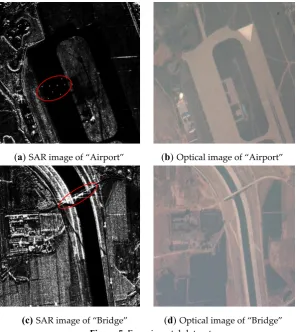
![Figure 1. Failure case of saliency detection of SAR image Amoon et al. [4] introduced it in SAR image ship detection, their method can remove disturbs come from the land](https://thumb-us.123doks.com/thumbv2/123dok_us/1002055.1600033/2.595.86.518.71.225/figure-failure-saliency-detection-introduced-detection-method-disturbs.webp)


Related documents
Three data mining methods including decision trees, NB and artificial neural network were used to classify and analyze the dataset of heart diseases.. The
The business performance of Punjab National Bank in relation to expansion of branches, recruitment of employees, deposits, advances and total business carried
The MEDLINE (via OVID, using keywords and MeSH in OVID), and PubMed (via NCBI using MeSH), and CINAHL databases were searched from January 2000 to April 2013 for results
The 1st Circuit addressed whether “a defendant who [is] sentenced pursuant to a binding C-type plea agreement. .” is “entitled to a sentence reduction by reason of
Therefore, in light of the theoretical principles concerning early maladaptive schemas and marital satisfaction and findings from previous studies, it can be concluded
We searched for patients with metastatic or recurrent pan- creatic adenocarcinoma diagnosed from March 1996 to July 2002 who were treated with gemcitabine in the out- patient clinic
A report by the Institute of Medicine#{176} of the National Academy of Sciences has estimated that current rates of low birth weight infants could be reduced by 15% among the
In vitro antifungal activity study of the prepared nano- liposomes (20 µ L) (F3, F6, and F9) was compared with that of both the flucytosine solution (20 µ L,
