Structural, Functional, and Metabolic Brain Differences as a Function of Gender
Identity or Sexual Orientation: A Systematic Review of the Human Neuroimaging
Literature
Alberto Frigerio1, Lucia Ballerini2, Maria del C. Valdés Hernández2
1 College of Medicine and Veterinary Medicine, University of Edinburgh, Edinburgh, UK 2 Department of Neuroimaging Sciences, Centre for Clinical Brain Sciences, University of
Edinburgh, Edinburgh EH16 4SB, UK; e-mail: [email protected]
ABSTRACT
Human sexuality is a complex reality, including gender identity and sexual orientation. A widespread approach to study human sexuality is to compare groups with opposite sexual
Keywords
INTRODUCTION
Sex, Gender Identity and Sexual Orientation
Human sexuality is a complex and multilevel structure made up of different components, and it is usually described by different perspectives and using different terminologies. Despite the terms sex and gender being used interchangeably (Shah, Jessell and Sanes, 2012), we refer “sex” to the biological condition (chromosomal, gonadal and phenotypic), “gender” to the inner
psychological perception of one’s own identity (gender identity) and to the outer cultural perception in behavior and habits attributed to and assumed by masculinity and femininity (gender role), and “sexual orientation” to sexual attraction (sexual preference).
The search for the origin of gender identity and sexual orientation is part of the debate on the impact of nature and culture on the human life (Lippa, 2002). This topic is highly controversial, due to its cultural, social and political implications, and it is widely debated within the scientific
community. Despite the efforts of scientists for conducting an objective research, researches on social problems are influenced by the cultural environment, and often reflect the dominant theories of their time (Jordan and Young, 2010). The vexata quaestio is: to what extent are gender and sexual orientation biologically determined and/or socially constructed by personal experiences and cultural expectations?
On the one hand, the so called “born the way theory” thinks that gender and sexual
orientation are innate and fixed properties (Swaab, 2007; Swaab, 2008; Savic et al., 2010). In this sense, transgenders’ and homosexuals’ brains would differ from those of cisgenders and
that such altered sexual differentiation in the brain causes an alteration in the development of the brain areas modulating body perception (in transgenderism) or sexual arousal (in homosexuality) (Burke et al. 2017). On the other hand, the so-called “gender theory” holds that human gender and sexual orientation are just cultural constructions, and it denies any kind of biological influences (Butler, 1990).
Actually, both gender and human sexual orientation seem to develop under two main types of influence: biological (genes, hormones and gene expression) and environmental (influences of parents, peers, partners and social models) factors (Balthazar, 2016; Jorge, 2010) as a result of the interaction between nature and culture (Hines, 2004). Evidence seems to suggest that biology contributes significantly to the development of both gender identity and sexual orientation (Roselli, 2018). Nevertheless, the idea that human sexuality is not biologically fixed is supported by
longitudinal studies, which reported a certain fluidity in both gender identity (Drummond et al., 2008) and sexual orientation (Savin, Williams and Ream, 2013). Eventually, the research on gender identity and sexual orientation is difficult because of the specificity of human sexuality, which makes difficult the use of animal models. Unlike animals, human beings receive and express their gender identity and role (Herbert, 2008), and their sexual behavior is, then, influenced by personal and social experiences and expectations (Maney, 2016).
Studies on Gender Identity and Sexual Orientation
involved in the development of gender identity and sexual orientation as a whole. But let us first clarify the relevant terminology.
While the term cisgender refers to people whose sense of gender identity corresponds to their birth sex, the term transgender refers to individuals who identify themselves with the gender opposite to that assigned at birth. If transgenders ask for a hormonal and/or surgical affirmation, they are called transsexuals (APA, 2013). According to recent evaluations, about 0.6 % of US adults would be transgender (Flores et al., 2016). While the term heterosexual refers to people who are emotionally, romantically or sexually attracted to people of the opposite sex, the term
homosexual refers to people who feel an emotional, romantic or sexual attraction toward subjects of the same sex (APA, 2005). According to recent estimates, about 1.9-2 % of US adults identify themselves as homosexuals and 2-4 % as bisexuals (Copen et al., 2016).
The neural bases of gender identity and sexual orientation have been studied through neural, hormonal and genetic investigations. Post mortem studies reported brain differences between cisgender and transgender people (Zhou et al. 1995; Kruijver et al., 2000; Garcia-Falgueras and Swaab, 2008) and between heterosexual and homosexual subjects (Swaab and Hofman, 1990; LeVay, 1991; Allen and Gorski, 1992). Hormonal researches suggest the involvement of prenatal hormones in the development of transgender identity (Dessens et al., 2005; Cohen-Kettenis, 2005) and homosexual orientation (Zucker et al., 1996; McFadden, 2002). Genetic investigations suggest a possible hereditary component for transgenderism (Green, 2000; Veale et al., 2010a; Segal, 2006; Heylens, 2012) and homosexuality (Wijchers and Festenstein, 2011; Drabant et al., 2012). Overall, biological factors seem to play a role in shaping both gender identity and sexual orientation. Nevertheless, no evidence allows experts to conclude that they are determined by any specific factor, and many scientists think that both biological and social factors are involved in the development of gender identity (APA, 2005) and sexual orientation (APA, 2014).
pointed out that, before hormonal treatment, in transgenders the most important brain parameters, namely intracranial, gray matter, white matter and cerebrospinal volumes, tend to be congruent with the gender assigned at birth - after hormone treatment they partly adjust to the characteristics of the desired gender -, although some structural, functional and metabolic brain features may exhibit signs of masculinization or feminization (Smith et al., 2015; Guillamon et al., 2016; Kreukels and Guillamon, 2016; Mueller et al., 2017). With regard to sexual orientation, the neuroimaging literature is scarce. Investigations have reported structural (Ponseti et al., 2007; Savic and Lindström, 2008; Witelson et al., 2008; Abé et al., 2014; Manzouri and Savic, 2018), functional (Hu et al., 2008; Paul et al., 2008; Ponseti et al., 2009; Zeki and Romaya, 2010; Hu et al., 2011; Kagerer et al., 2011; Zhang et al., 2011 Hu et al., 2013; Perry et al., 2013; Sylva, 2013; Hu et al., 2014; Manzouri and Savic, 2018; Safron et al., 2017; Safron et al., 2018) and metabolic (Kinnunen et al., 2004; Savic et al., 2005; Berglund et al., 2006; Savic and Lindström, 2008) differences between heterosexual and homosexual people, but an attempt to summarise and analyse these reports is, to the best of our knowledge, none-existent.
Overall, neuroimaging investigations on both gender identity and sexual orientation have reported conflicting results and some limitations such as the small size of samples and the
considerable overlap between transgender or homosexual people and control population, making it difficult to draw accurate conclusions. We conducted a systematic review and meta-analysis to investigate whether or not there are structural, functional, and metabolic neuroimaging features that differentiate groups of individuals with opposite sexual approaches: i.e. cisgender vs transgender people and heterosexual vs homosexual subjects in an attempt to provide the scientific community data gathered from the whole body of scientific literature that has been produced up to date, extracted and uniformly processed.
Aim. To document the scientific evidence from neuroimaging techniques on brain features that might be distinctive in groups of individuals with opposite sexual approaches, i.e. cisgenders vs transgenders (gender identity investigation) and heterosexuals vs homosexuals (sexual orientation investigation).
Hypotheses. Given the heterogeneity of the existent literature, and the small sample size of the studies on this theme of research, we hypothesise that it will not be possible to conclude on the specific brain phenotypes differential for each of the groups covered by this review.
MATERIALS AND METHODS
Systematic Literature Search
The literature search was conducted according to PRISMA (preferred reporting items for systematic reviews and meta-analyses) guidelines (Liberati et al., 2009). The search strategy, conducted in three different databases (Embase, Medline, PsycInfo), included articles published up to January 2018 comparing cisgenders vs transgenders and articles published up to April 2018 comparing heterosexuals vs homosexuals.
We analyzed only articles written in English and which published primary research output. Primary selection used title and abstract information. Authors where contacted if articles were not available online and/or if there was a question about the data presented in the article. After the initial selection, articles were checked for inclusion/exclusion criteria, and references were checked for possible further inclusions.
Selection Criteria
Inclusion criteria. The analysis of gender identity included articles which compared cisgender (non-transgender) population (male control = MC; female control = FC) with transgender people (male-to-female = MtF; female-to-male = FtM) before hormonal treatment, while the analysis of the sexual orientation included articles which compared heterosexual people (heterosexual man = HeM; heterosexual woman = HeW) with homosexual subjects (homosexual man = HoM; homosexual woman = HoW).
Exclusion criteria. Articles that investigated people affected by neurological diseases or by diseases associated with neurological outcome (e.g. HIV) were not included. As hormonal treatment may affect brain features (Rametti et al., 2012), studies and/or data on transsexuality after hormonal treatment were excluded.
Data Extraction
All quantitative outcomes including effect size and level of significance, regardless of whether or not they represented significant differences or not, were extracted from all papers included. In addition, we (i.e. all authors) independently, extracted sample size, subject
activated/relevant brain areas were extracted from studies that used fMRI (resting state or not) and voxel based morphometry (VBM).
Data Analysis
Microsoft Excel 2016 was used to represent the distribution of the demographic data and imaging modalities from all studies. The data extracted from each ROI were tabulated and
visualised to draw conclusions. GingerAle 2.3.6 software was used to meta-analyse the stereotaxic coordinates that showed relevance to our research question, for those studies that provided this information (i.e. those that used fMRI and VBM).
Due to the low number of studies conducted with metabolic neuroimaging techniques (i.e. PET and SPECT), it was not possible to carry out a meta-analysis of the brain regions that could metabolically differ between the groups of individuals involved in both of the analyses. The number of studies that used brain structural MRI to explore brain characteristics in relation to sexual
orientation was also reduced, not allowing to meta-analyse these data either. Instead, we summarised this information.
To calculate the risk of bias within and across studies we used the Quadas tool (Whiting et al., 2003). Quantitative results were converted to OR and CI using Practical Meta-Analysis Effect Size Calculator by David B. Wilson
(http://www.campbellcollaboration.org/escalc/html/EffectSizeCalculator-Home.php)64. After extracting all data available, it was not possible to do a meta-analysis per brain area due to the low number of studies with numerical data (see http://dx.doi.org/10.7488/ds/2412).
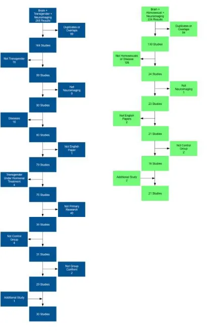
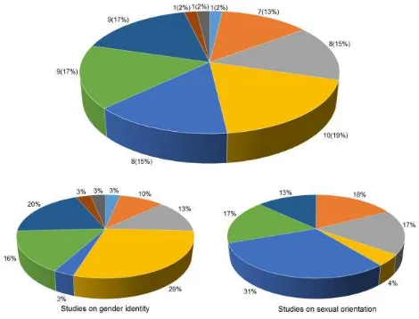
RESULTS
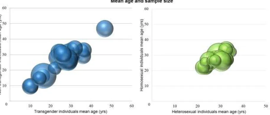
The search generated 492 publications from three different databases (Embase, Medline, and PsycInfo): 268 for the analyses of gender identity and 224 for the analyses on sexual orientation. Finally, 51 studies were included: thirty for the analyses of gender identity and twenty-one for the analyses on sexual orientation (Figure 1 and Appendices 1 and 2). All studies were conducted using different neuroimaging techniques: structural (MRI), functional (fMRI and rs-fMRI), and metabolic (PET and SPECT) (Figure 2). The majority of the studies included used functional MRI (i.e. 28/51 studies: 61% of the studies on sexual orientation and 45% of the studies on gender identity). Studies that used PET and SPECT modalities were few in both analyses (i.e. 13% of the studies on gender identity and 18% of the studies on sexual orientation). Figure 3 shows the mean age and sample size of the groups of individuals involved in the analyses. The analysis on gender identity involved individuals across a wider age range (mean ages 9.5 to 46.7 years old, i.e.
Figure 2. 3D pie charts summarising the number and percentage of studies included in the analyses, conducted with different neuroimaging techniques: overall information (top), analysis on gender identity (bottom left), and analysis on sexual orientation (bottom right).
Gender Identity Analyses. Study Selection
From 268 publications, 99 papers were duplicate or overlapped, 29 papers matched the inclusion criteria, and 140 were excluded. An additional study was included from the references (See Table 1 for studies information).
From 224 publications, 94 papers were duplicate or overlapped, 19 papers matched the inclusion criteria, and 111 were excluded. Two additional studies were included (See Table 2 for studies information).
Figure 3. 3D bubble charts of the mean age and sample size of: a) cisgenders and transgenders involved in the selected studies (left) (Note: the graph does not include the study by Yokota et al., 2005 because of the lack of data), and b) heterosexuals and homosexuals involved in the selected studies (right) (Note: the graph does not include the study by Hu et al., 2005 because of the lack of data. Four studies (Kagerer et al., 2011; Perry et al., 2013; Sylva et al., 2013; Zeki and Romaya, 2010) reported just the mean age of all the sample size, and we assumed that it was the same in heterosexual and homosexual subsamples).
Findings
ROI Analyses
Structural MRI was conducted in thirteen studies. However, only one of them conducted the analysis in specific stereotaxic coordinates (Simon et al., 2013). The twelve studies that conducted ROI analysis involved 229 FtM, 169 MtF, 478 FC, and 484 MC. Table 3 shows the ROI and the parameters investigated by each of these studies. Two studies involving 79 out of 229 FtM, 37 out of 169 MtF, 64 out of 478 FtM, and 57out of 484 MtF did not find differences in the mean
diffusivity of the hypothalamus (Kranz et al., 2018) nor in the volumes of cerebellum,
hypothalamus and medial frontal cortex (Hoekzema et al., 2015). Differences between cisgenders and transgenders were noticed by 10/12 studies in white matter microstructure (four studies), volumetric analysis (four studies), cortical thickness (two study), and corpus callosum shape (one study).
White matter microstructure of cisgender and transgender groups was analysed by four studies. Only one study (23 FtM, 21 MtF, 25 FC, and 25 MC) investigated the structural
Subcortical gray matter volume was investigated by four studies. All of them found that the volume of the putamen was consistently different between cisgender and transgender groups (Luders et al. 2009; Manzouri et al., 2017; Savic and Arver, 2011; Zubiaurre-Elorza, 2012).
Manzouri et al. (2017) found that the FtM’s left putamen was larger than that both female and male cisgenders (sample: 28 FtM, 34 FC, 34 MC), and Savic and Arver (2011) found that amongst all subcortical structures, MtF’s putamen and thalamus were smaller than those in both female and male cisgender groups (sample: 24 MtF, 24 MC, 24 FC). These two studies (Manzouri et al., 2017; Savic and Arver, 2011) also found that total gray matter volume did not differ between transgender and cisgender population. Zubiaurre-Elorza et al. (2012) also investigated subcortical gray matter in 24 FtM, 18 MtF, 23 FC and 29 MC; and reported that FtM had atypically increased right putamen volume. Luders et al. (2009) investigated gray matter volumes in 22 different regions, 12 in the right hemisphere and 10 in the left hemisphere (i.e. frontal, occipital and parietal lobes, superior frontal gyrus, midline, frontal pole, basal ganglia - caudate nucleus and putamen -, limbic system - subcallosum gyrus, mammillary body, amygdala, thalamus, hypothalamus, basal surface), in 24 MtF, 30 MC, 30 FC, and found an atypical reduction of the putaminal volume in MtF.
Cortical thickness was investigated by two studies, which reported differences between cisgender and transgender groups only in few non-overlapping regions. One (28 FtM - 34 FC - 34 MC) found differences between FtM and both FC and MC in the supramarginal, parietal, rostral middle frontal, inferior temporal gyrus, superior frontal gyrus and lingual-precalcarine cortex cuneus (Manzouri et al., 2017), and the other (24 FtM, 18 MtF, 23 FC, 29 MC) reported that MtFs have orbitofrontal, medial occipital and insular regions that resemble those typically seen in the female control group (Zubiaurre-Elorza et al., 2012).
Sexual Orientation
Five MRI studies were analysed (1/5 did not report the ROI analysis, but it provided stereotaxic coordinates using voxel based morphometry). The four studies that conducted the ROI analysis involved 81 HoM, 50 HoW, 96 HeM, and 86 HeW.
Due to the low number of studies conducted with MRI, it was not possible to do a meta-analysis on structural features in homosexual population compared to heterosexual subjects. However, findings contained in these studies offer data worth to be described. Table 4 shows the ROI and the parameters investigated by each of these studies.
Cortical thickness (CTh) was investigated by two studies (Abé et al., 2014; Manzouri and Savic, 2018). While Abé et al. (2014) found that HoM have a thinner CTh than HeM in visual area, Manzouri and Savic (2018) found that HoM have a thicker CTh than HeM in parietal lobe, while no differences were found between HoW and HeW.
Subcortical volumes were investigated by three studies (Abé et al., 2014; Manzouri and Savic, 2018; Witelson et al., 2008). Abé et al. (2014) found a smaller thalamus volume in HoM than HeM, while Witelson et al. (2008) found that HoM have a larger corpus callosum in the isthmus region. No other significant effects of sexual orientation were found.
A study measured cerebral and cerebellar hemispheres (Savic and Lindström, 2008). With regard to the cerebral hemisphere, they were symmetrical in HoM and in HeW, while they were asymmetrical in HoW and in HeM. With regards to the cerebellar hemisphere, no group reported asymmetry. Another study investigated white matter tracts of the whole brain (Manzouri and Savic, 2018), and no differences were found between heterosexual and homosexual population.
Stereotaxic Coordinates Analysis
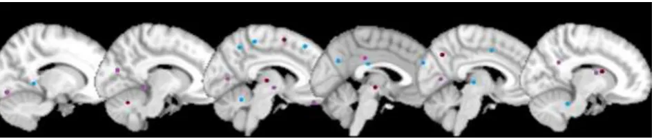
Six fMRI, eight rs-fMRI, and three VBM studies were analysed. fMRI studies were conducted under visual stimulation (2), smelling stimulation (1), vocal stimulation (1), mental rotation task (1), verbal fluency test (1). The seventeen studies that conducted stereotaxic coordinates analysis involved 195 FtM, 208 MtF, 347 FC, and 346 MC. Figure 4 displays six representative slices showing the foci resultant from the meta-analysis carried out using GingerAle 2.3.6 software using data from 12/17 studies (See Appendix 3 in Supplementary Materials for the labels of each foci and Table 5 for the number of foci related to different brain areas). The meta-analyses conducted (“Transgender_vs_Cisgender Natal Sex”, “Transgender_vs_Cisgender Opposite Sex”, and “Transgender_vs_Cisgender”) showed that transgender people’s brain activation differed more frequently in the Brodmann Areas (BA) 18 and 19, which include the occipital visual area along with BA 17, which is involved in visual processing.
gyrus (Spies et al., 2016). These four studies seem to suggest that transgenders tend to resemble their natal sex, even though some brain features present signs that resemble those of their experienced gender.
Figure 4. Six representative sagittal slices showing the foci resultant from the meta-analyses of stereotaxic coordinates where significant differences were found between groups with
opposite gender identity (result from GingerAle 2.3.6; figure generated with micron.exe) (Purple = Transgender_vs_Natal Sex; Red = Transgender_vs_Opposite Sex; Blue = Transgender_vs_Natal & Opposite Sex).
Sexual Orientation
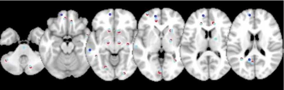
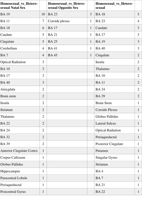
Eleven fMRI, 3 rs-fMRI and 1 VBM studies were analysed. fMRI studies were conducted under visual stimulation (10) and emotional judgement task (1). The 15 studies that conducted stereotaxic coordinates analysis involved 227 HoM, 94 HoW, 252 HeM, 117 HeW. Figure 5 shows six representative slices with the foci resultant from the meta-analysis carried out using GingerAle 2.3.6 software (See Appendix 4 in Supplementary Materials for the labels of each foci and Table 6 for the number of foci related to different brain areas).
The meta-analysis “Homosexual_vs_Heterosexual Natal Sex” showed different activations in visual area (BA 18 and 19). “Homosexual_vs_Heterosexual Opposite Sex” revealed differences in BA 23, which corresponds to the posterior cingular cortex, known to be involved in emotion, memory, meditation, and intrinsic control networks. Finally, the meta-analysis
Figure 5. Six representative axial slices showing the foci resultant from the meta-analyses of stereotaxic coordinates where significant differences were found between groups with opposite sexual orientation (result from GingerAle 2.3.6; figure generated with micron.exe) (Red = Homosexual_vs_Heterosexual Natal Sex; Cyan = Homosexual_vs_Heterosexual Opposite Sex; Blue = Transgender_vs_Natal & Opposite Sex; Indigo =
Homosexual_vs_Heterosexual).
Metabolic Analysis
Gender Identity
Three PET and 1 SPECT studies were analysed. They involved 25 FtM, 45 MtF, 41 FC, and 49 MC. A PET study investigated hypothalamic network in 12 gynephilic (sexual preference for women) MtF, 12 gynephilic MC, and 12 androphilic (sexual preference for men) FC under smelling stimulation with steroids. Transgender people reported an intermediate hypothalamic pattern of activation between males and females, with prevalent feminine features (Berglund et al., 2007). Another PET study investigated serotonin transporter distribution in 14 MtF of different sexual orientation, 13 MC and 9 FC with unspecified sexual orientation. While MC reported a rightward asymmetry in the midcingulate cortex, MtF and FC did not (Kranz et al., 2014a). Other PET study investigated serotonin transporter distribution in 19 MtF, 14 FtM, 24 MC and 11 FC. ROIs
Serotonin reuptake transporter non displaceable binding potential (BPnd) was lower in amygdala, caudate, insular cortex, hippocampus, and putamen in FtM respect to MC (Kranz et al., 2015). One SPECT study investigated regional cerebral blood flow (rCBF) in 11 gynephilic FtM and 9
androphilic FC. Transgender subjects reported an increase in rCBF in the right insula and a decrease in rCBF in the left anterior cingulate cortex (ACC) (Nawata et al., 2010). All together, these results seem to suggest that transgender people not under hormonal treatment have certain brain metabolic features which tend to be slightly different from their natal sex and which are either similar to the opposite sex or intermediate between the two sexes.
Sexual Orientation
Four PET studies were analysed. They involved 32 HoM, 24 HoW, 44 HeM, 37 HeW. Hypothalamic activation under smelling stimulation with AND (progesterone derivative) and EST (estrogen-like steroid) was investigated by two studies (Berglund et al., 2006; Savic et al., 2005). Berglund et al. (2006) found a different preoptic hypothalamus activation between HoW and HeM with AND, while Savic et al. (2005) found a different preoptic and ventromedial hypothalamus between HoW and HeM with AND. A study explored the brain activation 8 HoM and 7 HeM in response to fluoxetine (selective serotonin re-uptake inhibitor). With regards to the areas which are known to play a role in sexual behavior, HoM reported a lower decrease in hypothalamic glucose metabolism than HeM. With regard to the areas which are not known to play a role in sexual
results seem to suggest that homosexual individuals have certain brain metabolic features which tend to be slightly different from heterosexual individuals of their natal sex and in certain cases similar to the heterosexuals of the opposite sex.
Analysis of Bias
Appendix 5 and 6 show the risk of bias calculated using the Quadas tool (see
Supplementary Materials). Only 5/14 questions were applicable to our research. In all studies on
both gender identity and sexual orientation, the samples were not representative of the population. Selection criteria were described clearly in only 31/51 papers (i.e. 16/30 on gender identity and 15/21 on sexual orientation). Texts were explanatory enough so as it can be replicated in 44/51 papers (i.e. 24/30 on gender identity and 20/21 on sexual orientation). Intermediate results were reported in 46/51 papers (i.e. 26/30 on gender identity and 20/21 on sexual orientation).
Withdrawals from the studies included in this review were explained in all cases that referred it (i.e. 5/5 studies on gender identity and 3/3 studies on sexual orientation).
DISCUSSION
Main Findings
Consistent with our hypotheses, the results from systematic review and meta-analysis does not allow us to conclude on the specific brain phenotypes differential for each of the groups covered by this review. Although the analyses may suggest that before hormonal treatment transgenders’ brain features tend to be similar to those of their natal sex, some brain parameters might differ and in certain cases resemble those of their experienced gender. Also, altough homosexuals’s
individuals of their same sex, some brain features might be different and in certain cases may be similar to those of heterosexual individuals of the opposite sex. The compilation of the data from the studies included shows neural differences between the groups studied. However, brain functions are mediated by different brain areas and their interactions, rather than by single structures. The correlation or association between a certain brain function, volumetric change or activation, with a certain activity and/or behavior does not establish whether (or not) that structure/function is causally important for that activity/behavior (Koob, Everitt and Robins, 2013; Maney, 2016). It merely shows a possible involvement or apparent trend. Complex human behaviors (and few simple behaviors) cannot be entirely explained by phenomena occurring only on a single brain region. Therefore, the idea that brain sexual differences cause behavioral sexual differences, rather than being an assumption, still constitutes a hypothesis to verify.
ROI analysis
The lack of data did not allow us to meta-analyse the information obtained from the studies that conducted ROI analyses. From extracting and summarizing all the information available, differences were found between cisgender and transgender people in white matter microstructure, volumetric analyses, cortical thickness, and corpus callosum shape. Differences between
heterosexual and homosexual people were found in cortical thickness, subcortical volumes, and cerebral hemisphere, while no white matter tract reported differences. The studies included, in the rest of the ROIs analysed, either did not find differences between cisgender and transgender brains nor between heterosexual and homosexual; or found differences just between transgenders and opposite sex cisgenders, and between homosexuals and opposite sex heterosexuals (see Tables 3 and 4). Our findings on gender identity are consistent with previous studies that also attempted to summarise the literature findings on this topic, according to which gross morphology in
experienced gender (Smith et al., 2015; Guillamon et al., 2016; Kreukels and Guillamon, 2016; Mueller et al., 2017), even though white matter microstructure (Smith et al., 2015; Kreukels and Guillamon, 2016; Mueller et al., 2017), cortical thickness (Smith et al., 2015; Guillamon et al., 2016) and subcortical volumes (Mueller et al., 2017) may deviate from the biological sex towards values of experienced gender.
Stereotaxic coordinates analysis
Occipital brain regions, involved in visual processing, are the ones that more frequently reported a different activation in cisgenders respect to transgenders. This is not surprising given that, in general, most fMRI studies involved in our both analyses conducted visual stimulation. In addition to these regions, the BA 23 had different activations for heterosexuals with respect to homosexuals. In general, our meta-analysis found different brain activations between different groups scattered across the whole brain, but the low frequency with which these were reported did not allow us to conclude anything about them (see Table 5 and 6). Our results on gender identity are consistent with some of the previous studies mentioned above, according to which in certain brain areas transgenders’ activation is closer to those of their experienced gender (Smith et al., 2015; Guillamon et al., 2016), even though a clear picture has yet to emerge (Mueller et al., 2017).
Metabolic Analysis
Strengths and Limitations
Strengths
To the best of our knowledge, this is the first systematic review and meta-analysis of the neuroimaging literature on structural, functional, and metabolic differences between groups with opposite sexual attitudes. In addition, we carefully extracted and processed all data from all studies up-to-date and made them publicly available to facilitate further research in this important area.
Limitations
Several limitations regarding the small sample size of the meta-analysis and the
heterogeneity of the investigations must be acknowledged. Our analyses included a low number of studies (i.e. 30 on gender identity and 21 on sexual orientation), which were conducted with different neuroimaging techniques (1 SPECT, 3 PET, 6 fMRI, 8 rs-fMRI, and 13 MRI on gender identity; 4 PET, 5 MRI, 3 rs-fMRI, and 11 fMRI on sexual orientation). Different studies conducted with MRI investigated different brain structures (cortex, subcortical volumes, white matter, CSF, and ventricles in gender identity; analysis, cortex, subcortical volumes, and white matter in sexual orientation analysis). fMRI was conducted under different stimulations (1 smelling, 1 vocal stimulation, 1 mental rotation task, 1 verbal fluency test, and 2 visual in gender identity investigation; 10 visual stimulation, and 1 emotional judgment task in sexual orientation investigation). Metabolic analysis investigated different brain areas (hypothalamic network,
result, it was not possible to conduct a meta-analysis of the literature that fit our inclusion/exclusion criteria, and the main contribution of our work, therefore, is limited to the scientific compilation and synthesis of the data available.
Moreover, some studies had some limitations regarding the presentation of their data. First, some studies did not report statistical parameters and they just said whether or not there were significant differences between cisgenders and transgenders and between heterosexuals and homosexuals. Second, other studies reported statistical parameters only in case of significant differences between groups, and omitted reporting negative results (i.e. when no differences were found) (gender identity investigation: Burke et al., 2014; Kranz et al., 2014b; Kranza et al., 2015; Ku et al., 2013; Lin et al., 2014; Luders et al., 2009; Nota et al., 2017; Pol et al., 2006; Santarnecchi et al., 2012; Soleman et al., 2013; Spies et al., 2013; Yokota et al., 2005; Zubiaurre-Elorza et al., 2013; sexual orientation investigation: Hu et al., 2008; Ponsenti et al., 2007; Savic and Lindstrom, 2008; Sylva et al., 2013; Zecki et al., 2010; for more detailed information, please see analysis of bias in Appendix 5 and 6). A complete presentation of scientific data, including negative results, is an important element to precisely evaluate scientific investigations on a certain topic (Matosin et al., 2014).
Information on the biological sex of the studies’ participants is part of the scientific data we collect and make available. The data presented shows MtF and FtM transgender individuals do not have mirror images of brain differences. However, the heterogeneity of the design of the studies
involved, despite enriching the scope of this Review, due to the limited number of studies included and their sample sizes, made impossible to draw conclusions on specific biological sex differences for the attitudes covered in this Review. For example: some papers compared MtF with MC, others MtF with FC, others FtM with MC and others FtM with FC.
Finally, as Guillamon et al. (2016) noticed, some studies conducted on gender identity did not report the sexual orientation of the individuals that constituted their sample. Gender identity and sexual orientation are conceptually different, i.e. both cisgender and transgender people are either heterosexual or homosexual (Moser, 2010; Burke et al., 2017), and there are more gender identities other than cis-/transgender(ism) (such as genderqueer or non-binary) and other sexual orientations other than hetero-/homosexual(ism) (such as bi-, pan-, and asexual). Sexual orientation could be associated with brain structural specific features regardless and independently from gender identity. Thus, meaning that the structural, functional and metabolic variations found in homosexual
transgenders with respect to heterosexual cisgenders may be related to their sexual orientation rather than to their gender identity (Blanchard et al., 1987).
Conclusions and Future Direction
Due to conflicting results, it was not possible to identify specific brain features which consistently differ between cis- and transgender nor between hetero- and homosexual groups. Very small brain changes, to date undetectable using the current neuroimaging tools, may affect behavior. The small number of studies, the small sample size of each study, the heterogeneity of investigations, the lack of negative results reported by some studies, and the fact that some studies did not report the sexual orientation of the individuals that composed their sample did not allow drawing general
conclusions. Moreover, as the samples of the publications involved are not representative of the population analysed, caution should be taken in the interpretation of the results of this review.
To overcome the limitations mentioned above, future studies should: 1. keep investigating brain areas which are sexually dimorphic (e.g. hypothalamus, hippocampus, caudate, corpus
callosum, and serotonin transport) and brain areas involved in processing own-body perception (e.g. parietal, frontal, insular cortex, and its connections with thalamus and putamen) and sexual stimuli and arousal (e.g. hypothalamus and ventral striatum); 2. conduct Metabolic Analysis along with structural and functional to increase the number of data available; 3. report both positive and negative results to conduct an unbiased statistical analysis; 4. report sexual orientation of
individuals that conformed the sample size in studies on gender identity; 5. increase the sample size and expand the age range of the sample; 6. differentiate with respect between early or late onset gender dysphoria to reach a better understanding of the biological features underlying them. Future reviews in the topic should extend the inclusion criteria to distinguish between pre- vs.
post-pubertal and pre- vs. post-hormonal treatment, as well as include other advanced neuroimaging modalities such as magnetic resonance spectroscopy, and dynamic sequence acquisitions to increase the value and scope of the present report.
Declaration of interests
References
ABÉ, C., JOHANSSON, E., ALLZÉN, E., SAVIC, I. (2014). Sexual orientation related differences in cortical thickness in male individuals. PLoS ONE, 9:12, e114721.
ALLEN,L.S.,-GORSKI,R.A.(1992).Sexual orientation and the size of the anterior commissure in the human brain. PNAS, 89(15), 7199-7202.
AMERICAN PSYCHIATRIC ASSOCIATION. (2013). Diagnostic and Statistical Manual of Mental
Diseases V, American Psychiatric Publishing, Washington-London, 451-459.
AMERICAN PSYCHOLOGICAL ASSOCIATION.(2014). Answers to Your Questions About Transgender
People, Gender Identity & Gender Expression, http://www.apa.org/topics/lgbt/transgender.aspx, Accessed 13/09/2018.
AMERICAN PSYCHOLOGICAL ASSOCIATION. (2015). Answers to Your Questions for a better understanding of sexual orientation & homosexuality, http://www.apa.org/topics/lgbt/orientation.aspx, Accessed 13/09/2018.
BALTHAZAR,J. (2016)Sex differences in partner preferences in humans and animals. Philosophical Transactions of the Royal Society B 371.
BERGLUND,H.,LINDSTRÖM,P.,SAVIC,I. (2006). Brain response to putative pheromones in lesbian women, PNAS, 103(21), 8269-8274.
BERGLUND,H.,H.,LINDSTRÖM,P.,DHEJNE-HELMY,C.,SAVIC,I. (2008). Male-to-Female show sex-atypical hypothalamic activation when smelling odorous steroids, Cerebral Cortex 18(8), 1900-1908.
BLANCHARD,R.,CLEMMENSEN,L.H.,STEINER,B.W.(1987). Heterosexual and homosexual gender dysphoria, Archives of Sexual Behavior 16(2), 139-152.
BURKE, S. M., COHEN-KETTENIS, P. T., VELTMAN, D. J., KLINK, D. T., BAKKER, J. (2014). Hypothalamic response to the chemo-signal androstadienone in gender dysphoric children and adolescents, Frontier in Neuroendocrinology 5, 1-10.
BURKE,S.M.,KREUKELS,B.P.,COHEN-KETTENIS,P.T.,VELTMAN,D.J.,KLINK,D.T.,BAKKER,J. (2016). Male-typical visuospatial functioning in gynephilic girls with gender dysphoria. Organizational and activational effects of testosterone, Journal of Psychiatry and Neuroscience
41(6), 395-404.
BURKE,S.M.,MANZOURI,A.H.,SAVIC,I. (2017). Structural connections in the brain in relation to gender identity and sexual orientation, Scientific Reports 7(1), 17954-17965.
CLEMENS,B.,JUNGER,J.,PAULY,K.,NEUSCHAEFER-RUBE,C.,FRÖLICH,D.,MINGOIA,G.,DERNTI, B.,HABEL,U. (2017). Male-to-female gender dysphoria: Gender-specific differences in resting-state networks, Brain and Behavior 7(5), 1-10.
COHEN-KETTENIS,P.T. (2005). Gender change in 46,XY persons with 5alpha-reductase-2 deficiency and 17beta-hydroxysteroid dehydrogenase-3 deficiency, Archives of Sexual Behavior 34, 399-410.
COPEN, C.E., CHANDRA,A., FEBO-VAZQUEZ,I. (2016). Sexual Behavior, Sexual Attraction, and Sexual Orientation Among Adults Aged 18-44 in the United States: Data From 2011-2013 National Survey of Family Growth, National Health Statistics Reports, 1-14.
DESSENS, A. B., SLIJPER, F. M., DROP, S. L. (2005). Gender dysphoria and gender change in chromosomal females with congenital adrenal hyperplasia, Archives of Sexual Behavior 34, 389-397.
DRABANT,E.M.,KIEFER,A.K.,ERIKSSON,N.,MOUNTAIN,J.L.,FRANCKE,U.TUNG,J.Y.,HINDS,D. A.,DO,C.B. (2012). Genome-Wide Association Study of Sexual Orientation in a Large,
Web-based Cohort, 23andMe, Inc., Mountain View, Calif.
DRUMMOND,K.D.,BRADLEY,S.J.,PETERSON-BADALI,M.,ZUCKER,K.J. (2008). A follow-up study of girls with gender identity disorder, Developmental Psychology 44(1), 34-45.
FEUSNER,J.D.,LINDSTRÖM,A.,MOODY,T.D.,DHEJNE,C.,BOOKHEIMER,S.Y.,SAVIC,I. (2017). Intrinsic network connectivity and own body perception in gender dysphoria, Brain Imaging and Behavior 11(4), 964-976.
FLORES,A.R.,HERMAN,J.L.,GATES,G.J.,BROWN,T.N.T. (2016). How many adults identify as transgender in the United States?, William Institute, UCLA, Los Angeles, June 2016.
GARCIA-FALGUERAS, A., - SWAAB, D. F. (2008). A sex difference in the hypothalamic uncinate nucleus: Relationship to gender identity, Journal of Comparative Neurology, 3061-3084.
GIZEWSKI,E.R.,KRAUSE,E.,SCHLAMANN,M.,HAPPICH,F.,LADD,M.E.,FORSTING,M.,SENF,W. (2009). Specific cerebral activation due to visual erotic stimuli in male-to-female transsexual compared with male and female controls: An fMRI study, Journal of Sexual Medicine 6(2), 440-448.
GREEN,R. (2000). Family co-occurrence of ‘gender dysphoria’: Ten sibling or parent-child pairs,
Archives of Sexual Behavior 29, 499-507.
GUILLAMON,A., JUNQUE, C., GÓMEZ-GIL,E. (2016). A Review of the Status of Brain Structure Research in Transsexualism, Archives of Sexual Behavior 45, 1615-1648.
HERBERT,J. (2008). Who do we think we are? The brain and gender identity, Brain 131, 3115-3117. HEYLENS,G.,DE CUYPERE,G.,ZUCKER,K.J.,SCHELFAUT,C.,ELAUT,E.,VANDEN BOSSCHE,H.,DE BAERE,E.,T’SJOEN,G. (2012). Gender identity disorder in twins: a review of the case report literature, The Journal of Sexual Medicine 9, 751-757.
HINES, M. (2004). Brain Gender, Oxford University Press, Oxford.
HOEKZEMA, E., SCHAGEN, S. E., KREUKELS, B. P., VELTMAN, D. J., COHEN-KETTENIS, P. T., DELEMARRE-VAN DE WAAL,H., BAKKER,J. (2015). Regional volumes and spatial volumetric distribution of gray matter in the gender dysphoric brain, Psychoneuroendocrinology 55, 55-59. HU,S.,XU,D.,PETERSON,B.S.,WANG,Q.,HE,X.,HU,J.,XU,X.,WEI,N.,LONG,D.,HUANG,M., ZHOU,W.,XU,W.,ZHANG,M.,XU,Y. (2013). Association of Cerebral Networks in Resting State with Sexual Preference of Homosexual Men: A Study of Regional Homogeneity and Functional Connectivity, PLOS ONE 8(3), e59426.
HU,S., XU,D., PETERSON,B.S., WANG,Q., LAI, J.,HU,J., WEI,N., ZHANG,M., XU,Y. (2014). Differing default mode network activities in men with homosexual or heterosexual preferences,
Journal of Sexual Medicine 11(10), 2474-2484.
HU,S.,WANG,Q.,XU,Y.,LIAO,Z.L.,XU,L.J.,XU,X.J.,WEI,E.Q.,YAN,L.Q.,HU,J.B.,WEI,N., ZHOU,W. H., HUANG,M.L.,ZHANG,M.M. (2011). Haemodynamic brain response to visual stimuli is different between homosexual and heterosexual men, Journal of International Medicine Research 39(1), 199-211.
HU,S.,WEI,E.Q.,WANG,Q.,YAN,L.Q.,WEI,E.Q.,ZHANG,M.M.,HU,J.B.,HUANG,M.L.,ZHOU, W.H.,XU,Y. (2008).Patterns of brain activation during visually evoked sexual arousal differ between homosexual and heterosexual men, American Journal of Neuroradiology 29(10), 1890-1896.
JORDAN-YOUNG,R. M.(2010). Brainstorm: The flaws in the science of sex differences, Harvard University Press, Cambridge.
JORGE,J.C.(2010). The Embriology of Gender, Journal of LGBT Youth 7, 310-319.
JUNGER,J.,HABEL,U.,BRÖHR,S.,NEULEN,J.,NEUSCHAEFER-RUBE,C.,BIRKHOLZ,P.,KOHLER,C., SCHNEIDER,F.,DERNTL,B.,PAULY,K.(2014). More than just two sexes: The neural correlates of voice gender perception in gender dysphoria, PLoS ONE 9(11), e111672.
KAGERER,S.,KLUCKEN,T.,WEHRUM,S.,ZIMMERMANN,M.,SCHIENLE,A.,WALTER,B.,VAITL,D., STARK,R. (2011). Neural activation toward erotic stimuli in homosexual and heterosexual males,
Journal of Sexual Medicine 8(11), 3132-3143.
KOOB, G. F., EVERITT, B.J., ROBBINS, T.W. (2013). Reward, Motivation and Addition, 874, in SQUIRRE, L. R., BERG, D., BLOOM, F.E., DU LAC, S., GHOSH, A., SPITZER, N.C. (EDS),
Fundamental Neuroscience (fourth edition), Elsevier, Waltham-Oxford, 871-898.
KRANZ,G.S.,HAHN,A.,BALDINGER,P.,HAEUSLER,D.,PHILIPPE,C.,KAUFMANN,U.,WADSAK,W., SAVLI, M., HOEFLICH, A., KRAUS, C., VANICEK, T., MITTERHAUSER, M., KASPER, S., LANZENBERGER,R. (2014a). Cerebral serotonin transporter asymmetry in females, males and male-to-female transsexuals measured by PET in vivo, Brain Structure and Function 219(1), 171-183.
KRANZ,G.S.,WADSAK,W.,KAUFMANN,U.,SAVLI,M.,BALDINGER,P.,GRYGLEWSKI,G.,HAEUSLER,
D., SPIES, M., MITTERHAUSER, M., KASPER, S., LANZENBERGER, R. (2015). High-dose
testosterone treatment increases serotonin transport biding in transgender people, Brain Structure and Function 223(1), 321-328.
KRANZ, G. S., HAHN, A., KAUFMANN, U., TIK, M., GANGER, S., SEIGER, R., HUMMER, A., WINDISCHBERGER,C.,KASPER,S.,LANZENBERGER,R. (2018). Effects of testosterone treatment on hypotalamic neuroplasticity in female-to-male transgender individuals, Brain Structure and Function 223(1), 321-328.
KRANZ,G. S., HAHN,A., KAUFMANN,U., KÜBLBÖCK, M.,HUMMER, A.,GANGER, S.,SEIGER, R., WINKLER, D. SWAAB, D. F., WINDISCHBERGER, C., KASPER, S., LANZENBERGER, R.(2014b). White matter microstructure in transsexuals and controls investigated by diffusion tensor imaging, Journal of Neuroscience 34(46), 15466-15475.
KREUKELS, B. P. C., - GUILLAMON, A. (2016). Neuroimaging studies in people with gender incongruence, International Review of Psychiatry 28(1), 120-128.
KRUIJVER,F.P.M.,ZHOU,J.N.,POOL,C.W.,HOFMAN,M.A.,GOOREN,L.J.,SWAAB,D.F. (2000). Male-to-female transsexuals have female neuron numbers in a limbic system, Journal of Clinical Endocrinology and Metabolism 85, 2034-2041.
KU,H.L.,LIN,C.S.,CHAO,H.T.,TU,P.C.,LI,C.T.,CHENG,C.M.,SU,T.P.,LEE,Y.C.HSIEH,J.C. (2015). Brain signature Characterizing the Body-Brain-Mind Axis of Transsexuals, PLoS ONE
8(7), e70808.
LEVAY,S.(1991). A difference in hypotalamic structure between heterosexual and homosexual men,
Science 253, 1034-1037.
LIN,C.S.,KU,H.L.,CHAO,H.T.,TU,P.C.,LI,C.T.,CHENG,C.M.,SU,T.P.,LEE,Y.C.,HSIEH,J. C. (2014). Neural network of body representation differs between transsexuals and cissexuals,
PLoS ONE 9(1), e85914.
LIPPA,RICHARD A.(2002). Gender, Nature, and Nurture, LEA, New Jersey.
LUDERS,E.,SÁNCHEZ,F.J.,GASER,C.TOGA,A.W.,NARR,K.L.HAMILTON,L.S.,VILAIN,E.(2009). Regional gray matter variation in male-to-female transsexualism, NeuroImage 46(4), 904-907. MANEY,DONNA L. (2016), Perils and pitfalls of reporting sex differences, Philosophical Transactions
of the Royal Society B 371.
MANZOURI,A.,KOSIDOU,K.,SAVIC,I. (2017). Anatomical and functional findings in female-to-male transsexuals: Testing a new hypothesis, Cerebral Cortex 27(2), 998-1010.
MANZOURI,A.,-SAVIC,I. (2018). Cerebral sex dimorphism and sexual orientation, Human Brain
Mapping, 39(3), 1175-1186.
MATOSIN,N.,FRANK,E.,ENGEL,M.,LUM,J.S.,NEWELL,K.A.(2014). Negativity towards negative results: a discussion of the disconnect between scientific worth and scientific culture, Dis Model Mech 7(2), 171-173.
MCFADDEN,D. (2002). Masculinization effects in the auditory system, Archives of Sexual Behavior 31, 99-111.
MOSER,C.(2010). Blanchard’s autogynephilia theory: a critique, Journal of Homosexuality 57(6), 790-809.
MUELLER,S.C.,DE CUYPERE,G.,T’SJOEN,G. (2017). Transgender Research in the 21st Century: A Selective Critical Review From a Neurocognitive Perspective, The American Journal of Psychiatry 174(12), 1155-1162.
NAWATA,H.,OGOMORI,K.,TANAKA,M., NISHIMURA,R.,URASHIMA,H.,YANO,R.,TAKANO,K., KUWABARA,Y. (2010). Regional cerebral blood flow changes in female to male gender identity,
Psychiatry & Clinical Neuroscience 64(2), 157-161.
NOTA,N.M.,KREUKELS,B.P.C., DEN HEIJER,M.,VELTMAN,D.J.,COHEN-KETTENIS,P.T.,BURKE, S.M.,BAKKER,J. (2017). Brain functional connectivity patterns in children and adolescent with gender dysphoria: Sex-atypical or not?, Psychoneuroendocrinology 86, 187-195.
PAUL,T.,SCHIFFER,B.,ZWARG,T.,KRÜGER,T.H.,KARAMA,S.,SCHEDLOWSKI,M.,FORSTING,M., GIZEWSKI,E.R. (2008). Brain response to visual sexual stimuli in heterosexual and homosexual males, Human Brain Mapping 29, 726-735.
PERRY,D.,WALDER,K.,HENDLER,T.,SHAMAY-TSOORY,S.G. (2013). The gender you are and the gender you like: Sexual preference and empathic neural response, Brain Research 1534, 66-75. POL,H.E.H.,COHEN-KETTENIS,P.T., VAN HAREN,N.E.M.,PEPER,J.S.,BRANS,R.G.H.,CAHN,
brain; Influences of testosterone and estrogen on adult human brain structure, European Journal of Endocrinology 155(1), S107-S114.
PONSETI,J.,GRANERT,O.,JANSEN,O.,WOLFF,S.,MEHDORN,H.,BOSINSKI,H.,SIEBNER,H.(2009). Assessment of sexual orientation using the hemodynamic brain response to visual stimuli,
Journal of Sexual Medicine 6(6), 1628-1634.
PONSETI, J., SIEBNER, H., KLÖPPEL, S., WOLFF, S., GRANERT, O., JANSEN, O., MEHDORN, H. M., BOSINSKI,H.A.(2007).Homosexual Women Have Less Grey Matter in Perirhinal Cortex than Heterosexual Women, PLoS ONE 2(8), e762.
RAMETTI,G.,CARILLO,B., GÓMEZ-GIL,E.,JUNQUE,C.,SEGOVIA,S.,GOMEZ,Á., GUILLAMON, A. (2011a). White matter microstructure in female to male transsexuals before cross-sex hormonal treatment. A diffusion tensor imaging, Journal of Psychiatric Research 45(2), 199-204.
RAMETTI, G., CARILLO, B., GÓMEZ-GIL, E., JUNQUE, C., ZUBIAURRE-ELORZA, L., SEGOVIA, S., GOMEZ, Á., GUILLAMON, A. (2011b). The microstructure of white matter in male to female transsexuals before cross-sex hormonal treatment. A DTI study, Journal of Psychiatric Research
45(2), 949-954.
G. RAMETTI, B. CARRILLO, E. GÓMEZ-GIL, C. JUNQUE, L. ZUBIAURRE-ELORZA, S. SEGOVIA, Á. GOMEZ, K. KARADI, A. GUILLAMON, (2012). Effects of androgenization on the white matter microstructure of female-to-male transsexuals. A diffusion tensor imaging study.
Psychoneuroendocrinology 37, 1261-1269.
ROSELLI, C. E. (2018). Neurobiology of gender identity and sexual orientation, Journal of Neuroendocrinology 730, e12562.
SAFRON,A.,SYLVA,D.,KLIMAJ,V.,ROSENTHAL,A.M.,LI,M.,WALTER,M.,BAILEY,J.M.(2017). Neural Correlates of Sexual Orientation in Heterosexual, Bisexual, and Homosexual Men,
Scientific Reports 7,41314.
SAFRON,A.,KLIMAJ,V.,SYLVA,D.,ROSENTHAL,A.M.,LI,M.,WALTER,M.,BAILEY,J.M. (2018). Neural Correlates of Sexual Orientation in Heterosexual, Bisexual, and Homosexual Women,
Scientific Reports 8,673.
SANTARNECCHI,E.,VATTI,G.,DÉTTORE,D.,ROSSI,A. (2012). Intrinsic cerebral connectivity analysis in an untreated female-to-male transsexual subject: A first attempt using resting-state fMRI,
Neuroendocrinology 96(3), 188-193.
SAVIC, I., BERGLUND, H., LINDSTRÖM, P. (2005). Brain response to putative pheromones in homosexual men, PNAS 102(20), 7356-7361.
SAVIC,I.,-ARVER,S.(2011). Sex dimorphism of the brain in male-to-female transsexuals, Cerebral
SAVIC,I.,-GARCIA-FALGUERAS,A.,SWAAB,D.F.(2010). Sexual differentiation of the human brain in relation to gender identity and sexual orientation, Progress in Brain Research 186, 41-62. SAVIC, I., - LINDSTRÖM, P. (2008). PET and MRI show differences in cerebral asymmetry and
functional connectivity between homo- and heterosexual subjects, PNAS 105(27), 9403-9408. SAVIN-WILLIAMS, R. C., - REAM, G. L. (2003). Prevalence and Stability of Sexual Orientation
components During Adolescence and Young Adulthood, Journal of Adolescent Health 52(4), 465-472.
SCHÖNING,S.,ENGELIEN,A.,BAUER,C.,KUGEL,H.,KERSTING,A.,ROESTEL,C.,ZWITSERLOOD,P., PYKA, M., DANNLOWSKI, U., LEHMANN, W. HEINDEL, W., AROLT, V., KONRAD, C. (2010). Neuroimaging Differences in Spatial Cognition between Men and Male-to-Female Transsexuals Before and During Hormone Therapy, Journal Sex of Medicine 7, 1858-1867.
SEGAL,N. L. (2006). Two monozygotic twin pairs discordant for female-to-male transsexualism,
Archives of Sexual Behavior 35, 347-358.
SHAH,NIRAO M.,-JESSELL,THOMAS M.,-SANES,JOSHUA R., “Sexual Difference of the Nervous System”, in ERIC R. KANDEL - JAMES H. SCHWARTZ - THOMAS M. JESSELL - STEVEN A. SIEGELBAUM -A.J.HUDSPETH (ed.), Principles of Neural Science [Fifth Edition], McGraw-Hill Medical, New York 2012, 1306-1327.
SIMON,L.,KOZÁK,L.R.,SIMON,V.,CZOBOR,P.,UNOKA,Z.,SZABÓ,Á.,CSUKLY,G. (2013). Regional grey matter structure differences between transsexuals and healthy controls. A voxel based morphometry study, PLoS ONE 8,12.
SMITH,E.S.,JUNGER,J.,DERNTL,B.,HABEL,U.(2015). The transsexual brain - A review of findings on the neural basis of transsexualism, Neuroscience and Biobehavioral Reviews 59, 251-266. SOLEMAN, R. S., SCHAGEN, S. E., VELTMAN, D. J., KREUKELS, B. P., COHEN-KETTENIS, P. T.,
LAMBALK,C. B., WOUTERS,F., DELEMARRE-VAN DE WAAL,H. A. (2013). Sex differences in verbal fluency during adolescence: A functional magnetic resonance imaging study in gender dysphoric and control boys and girls, Journal of Sexual Medicine 10(8), 1969-1977.
SPIES,M.,HAHN,A.,KRANZ,G.S.,SLADKY,R.,KAUFMANN,U.,HUMMER,A.,GANGER,S.,KRAUS, C.,WINKLER,D.,SEIGER,R.,COMASCO,E.,WINDISCHBERGER,C.,KASPER,S.LANZENBERGER, R. (2016). Gender transition affects neural correlates of empathy: A resting state functional connectivity study with ultra high-field 7T MR imaging, NeuroImage 138, 257-265.
SWAAB,D.F. (2007). Sexual differentiation of the brain and behavior, Best Practice & Research
Clinical Endocrinology & Metabolism 21, 1431-444.
SWAAB,D.F.,-HOFMAN,M.A.(1990).An enlarged suprachiasmatic nucleus in homosexual men,
Brain Research 537, 141-148.
SYLVA,D.(2013). Neural correlates of sexual arousal in heterosexual and homosexual women and men, Hormones and Behavior 64(4), 673-684.
VEALE,J.F.,CLARKE,D.E.,LOMAX,T.C.(2010a) Biological and psychological correlates of adult gender-variant identities: New findings, Personality and Individual Differences 49, 252-257. WHITING,P.,RUTJES,A.W.,REITSMA,J.B.,BOSSUYT,P.M.,KLEIJNEN,J.(2003). The development
of QUADAS: a tool for the quality assessment of studies of diagnostic accuracy included in systematic reviews, BMC Medical Research Methodology 25(3), 141-148.
WIJCHERS,P.J.,-FESTENSTEIN,R.J.(2011). Epigenetic regulation of autosomal gene expression by sex chromosomes, Trends in Genetics 27, 132-140.
WITELSON,S.F.,KIGAR,D.L.SCAMVOUGERAS,A.,KIDECKEL,D.M.,BUCK,B.,STANCHEV,P.L., BRONSKILL,M.,BLACK,S.(2008).Corpus callosum anatomy in right-handed homosexual and heterosexual men, Archives of Sexual Behavior 37(6), 857-863.
YOKOTA,Y.,KAWAMURA,Y.,KAMEYA,Y.(2005). Callosal shapes at the midsagittal plane: MRI
differences of normal males, normal females, and GID, Proceedings of the 27th Annual Conference of IEEE Engineering in Medicine and Biology, September 1-4, Shanghai, China 2005.
ZEKI,S.,- ROMAYA,J.P.(2010). The brain reaction to viewing faces of opposite- and same-sex romantic partners, PLoS ONE 5(12), e15802.
ZHANG,T.Y.,-MEANEY,M.J.(2010). Epigenetics and the environmental regulation of the genome and its function, Annual Review of Psychology 61, 439-466.
ZHOU,J.N.,HOFMAN,M.A.,GOOREN,L. J.,SWAAB,D.F.(1995). A sex difference in the human brain and its relation to transsexuality, Nature 378, 68-60.
ZUBIAURRE-ELORZA, L., JUNQUE, C., GÓMEZ-GIL, E., SEGOVIA, S., CARRILLO, B., RAMETTI, G., GUILLAMON,A. (2013). Cortical thickness in untreated transsexuals, Cerebral Cortex 23(12), 2855-2862.
Table 1. Studies included in the analysis of gender identity (abbreviations: FtM = Female-to Male; MtF = Male-to-Female; FC = Female Control; MC = Male Control)
Study Year Sample Mean Age Technique
BERGLUND 2008 12 MtF - 12 MC - 12 FC 32 - 26 - 33 PET
BURKE 2014 17 FtM - 19 MtF - 19 FC - 20 MC 9.6 - 10.4 - 9.7 - 9.5 fMRI
BURKE 2016 21 FtM - 21 FC - 20 MC 16.1 - 16.3 - 15.9 fMRI
CLEMENS 2017 15 MtF - 21 MC - 20 FC 35.5 - 32.32 - 32.5 rs-fMRI
FEUSNER 2017 27 FtM - 27 FC - 27 MC 24.2 - 32.1 - 31 rs-fMRI
GIZEWSKI 2009 12 MtF - 12 MC - 12 FC 36 - 29 - 29 fMRI
HAHN 2015 23 FtM - 21 MtF - 25 FC - 25 MC 26.9 - 30.9 - 25.3 - 25.6 MRI
HOEKZEMA 2015 54 FtM - 37 MtF - 52 FC - 44 MC 16.92 - 16.05 - 16.29 - 16.42 MRI
JUNGER 2014 16 MtF - 21 MC - 20 FC 36.38 - 32.35 - 33.16 fMRI
KRANZ 2014a 14 MtF - 13 MC - 9 FC 31.4 - 29.8 - 29 PET
KRANZ 2014b 23 FtM - 21 MtF - 23 FC - 22 MC 25.91 - 30.86 - 25.96 - 25.45 MRI
KRANZ 2015 14 FtM - 19 MtF - 11 FC - 24 MC 28.21 - 31.79 - 30.43 - 34.14 PET
KRANZ 2018 25 FtM - 12 FC - 13 MC 27.24 - 24.42 - 28.77 MRI
KU 2013 12 FtM - 11 MtF - 12 FC - 11 MC All Trans 25.4 - All Cis 24.4 rs-fMRI
LIN 2014 12 FtM - 11 MtF - 12 FC - 11 MC All Trans 25.4 - All Cis 24.4 rs-fMRI
LUDERS 2009 24 MtF - 30 MC - 30 FC 46.73 - 46.57 - 46.77 MRI
MANZOURI 2017 28 FtM - 34 FC - 34 MC 23.5 - 27.6 - 28.8 MRI &
rs-fMRI
NAWATA 2010 11 FtM - 9 FC 23.4 - 23.6 SPECT
NOTA 2017 13 FtM - 18 MtF - 18 FC - 21 MC 9.7 - 10.5 - 9.6 - 9.4 rs-fMRI
POL 2006 6 FtM - 8 MtF - 6 FC - 9 MC 28 - 25 - 23 - 25 MRI
RAMETTI 2011a 18 FtM - 19 FC - 24 MC 28.24 - 31.22 - 33 MRI
RAMETTI 2011b 18 MtF - 19 MC - 19 FC 24.74 - 31.94 - 33 MRI
SANTARNECCHI 2012 1 FtM - 25 FC - 25 MC 22 - 21 - 21 rs-fMRI
SAVIC AND ARVER 2011 24 MtF - 24 MC - 24 FC 32 - 33 - 35 MRI
SCHÖNING 2010 11 MtF - 11 MC 37.55 - 33.09 fMRI
SIMON 2013 7 FtM - 10 MtF - 7 FC - 11 MC 24.8 - 28.5 - 23.9 - 27.1 MRI
SOLEMAN 2013 11 FtM - 6 MtF - 26 FC - 24 MC 14.64 - 14.25 - 14.44 - 14.68 fMRI
SPIES 2016 33 FtM - 24 MtF - 44 FC - 33 MC 26.79 - 30.25 - 26.16 - 27.48 rs-fMRI
Study Year Sample Mean Age Technique
Table 2. Studies included in the analysis of sexual orientation (abbreviations: HoM = Homosexual Men; HoW = Homosexual Women; HeM = Heterosexual Men; HeW = Heterosexual Women)
Study Year Sample Mean Age Technique
ABÉ ET AL. 2014 19 HoM - 21 HeM - 21 HeW 33.5 - 31.9 - 33.2 MRI
BERGLUND ET AL. 2006 12 HoW - 12 HeW - 12 HeM 33 - 26 - 28 PET
HU ET AL. 2013 26 HoM - 26 HeM 22.27 -23.46 rs-fMRI
HU ET AL. 2014 26 HoM - 26 HeM 22.27 -23.46 rs-fMRI
HU ET AL. 2011 14 HoM - 14 HeM Not reported fMRI
HU ET AL. 2008 10 HoM - 10 HeM 26.5 - 27.9 fMRI
KAGERER ET AL. 2011 11 HoM - 10 HeM All sample 28 fMRI
KINNUNEN ET AL. 2004 8 HoM - 7 HeM 29 - 28 PET
MANZOURI -SAVIC 2018 30 HoM - 30 HoW - 40 HeM - 40 HeW 31.4 - 27.9 - 29.5 - 29.3 MRI & rs-fMRI
PAUL ET AL. 2008 12 HoM - 12 HeM 32 - 34.8 fMRI
PERRY ET AL. 2013 12 HoM - 12 HoW - 13 HeM - 15 HeW All sample 28.46 fMRI
PONSETI ET AL. 2009 14 HoM - 12 HeM 27.4 - 26.8 fMRI
PONSETI ET AL. 2007 16 HoM - 15 HoW - 24 HeM - 25 HeW 27.3 - 24.9 - 25.3 - 24.9 MRI [VBM]
SAFRON ET AL. 2017 22 HoM - 23 HeM 22 HoM - 19 HeM
33.2 - 32.3 Not reported
fMRI [picture] fMRI [video]
SAFRON ET AL. 2018 20 HoW - 20 HeW 20 HoW - 18 HeW
29 - 29.7 Not reported
fMRI [picture] fMRI [video]
SAVIC ET AL. 2005 12 HoM - 12 HeM - 12 HeW 33 - 28 - 26 PET
SAVIC -LINDSTRÖM 2008 20 HoM - 20 HoW - 25 HeM - 25 HeW 12 HoM - 12 HoW - 13 HeM - 13 HeW
32 -20 - 30 -31 Not reported
MRI PET
SYLVA ET AL. 2013 12 HoM - 11 HoW - 12 HeM - 11 HeW All sample 22.1 fMRI
WITELSON ET AL. 2008 12 HoM - 10 HeM 25.4 - 23.3 MRI
ZEKI -ROMAYA 2010 6 HoM - 6 HoW - 6 HeM - 6 HeW All sample 26.3 fMRI
Table 3. ROI analysis on gender identity (Legend: Cis = Natal & Opposite Sex; GM = Gray Matter; WM = White Matter; CSF = Cerebrospinal Fluid; CST = Corticospinal Tract; SFG = Superior Frontal Gyrus; SLF = Superior Longitudinal Fasciculus; R = Right; L = Left; HCR = Hemispheric Connectivity Ratio; LCW = Lobar Connectivity Weight; MD = Mean Diffusivity; FA = Fractional Anisotropy; CTh = Cortical Thickness; SA = Surface Area; GMV = Gray Matter Volume; TIV = Total Intracranial Volume; v = differences; x = non differences; Nd = Not definable; Nr = Not reported)
Reference ROI Parameters Trans_vs_Natal Sex Trans_vs_Opposite Sex Trans_vs_Cis M_vs_F
Hahn et al., 2015 Subcortical L Subcortical R
Subcortical R - Frontal R Subcortical L - Parietal L
HCR HCR LCW LCW Nr Nr Nr Nr Nr Nr Nr Nr Nd v Nd Nd Nd v Nd Nd
Hoekzema et al., 2015
Cerebellum L Cerebellum R Hypothalamus Medial Frontal Cortex
Volume Volume Volume Volume Nd Nd x Nd Nr Nr Nr Nr Nr Nr Nr Nr v v v v
Kranz et al., 2018 Hypothalamus MD x x x v
Kranz et al., 2014b GM WM CSF TIV CST R CST L Forceps Major Forceps Minor CST R CST L Forceps Major Forceps Minor Volume Volume Volume Volume MD MD MD MD FA FA FA FA
x (MtF) - x (FtM) x (MtF) - x (FtM) x (MtF) - x (FtM) x (MtF) - x (FtM) v (MtF) - v (FtM) v (MtF) - v (FtM) x (MtF) - x (FtM) v (MtF) - v (FtM) x (MtF) - x (FtM) x (MtF) - x (FtM) x (MtF) - x (FtM) v (MtF) - v (FtM)
v (MtF) - v (FtM) v (MtF) - v (FtM) v (MtF) - v (FtM) v (MtF) - v (FtM) v (MtF) - v (FtM) v (MtF) - v (FtM) x (MtF) - x (FtM) v (MtF) - v (FtM) x (MtF) - x (FtM) x (MtF) - x (FtM) x (MtF) - x (FtM) v (MtF) - v (FtM)
x x x x v v x v x x x x v v v v v v x v x x x x
Luders et al., 2009 Frontal Lobe Occipital Lobe Parietal Lobe SFG Midline Frontal Pole Caudate Nucleus Putamen Subcallosum Gyrus Mammillary Body Amygdala Thalamus Hypothalamus Basal Surface Volume Volume Volume Volume Volume Volume Volume Volume Volume Volume Volume Volume Volume Volume x x x x x x x v x x x x x x v v v v v v v x v v v v v v Nd Nd Nd Nd Nd Nd Nd Nd Nd Nd Nd Nd Nd Nd v v v v v v v v v v v v v v
Reference ROI Parameters Trans_vs_Natal Sex Trans_vs_Opposite Sex Trans_vs_Cis M_vs_F
Pol et al., 2006 Intracranial Total Brain Hypothalamus 3rd Ventricle Lateral Ventricle GM WM Volume Volume Volume Volume Volume Volume Volume x x x x x x x v v x x x x x x x x x x x x v v x x x x x
Rametti et al., 2011a SLF R Forceps Minor CST FA FA FA v v v x x v Nd Nd v v v v
Rametti et al., 2011b GM WM CSF Total Intracranial SLF R Forceps Minor CST
Anterior Cingulum R
Volume Volume Volume TIV FA FA FA FA x x x x v v v v v v v v v v v v Nd Nd Nd Nd v v v v v v v v v v v v
Savic and Arver, 2011 Hippocampus Thalamus Caudate Putamen Total Tissue Total Brain GM WM Volume Volume Volume Volume Volume Volume Volume Volume x v x v x x x x Nd v x v Nd x x x Nd v x v Nd x x x v x x x v x x x
Yokota et al., 2005
Corpus Callosum Shape v v v v
Zubiaurre-Elorza et al., 2013
Cortex Putamen R Putamen L Thalamus Caudate Pallidum Hippocampus Amygdala CTh Volume Volume Volume Volume Volume Volume Volume
v (MtF) - x (FtM) x (MtF) - v (FtM) x (MtF) - x (FtM) x (MtF) - x (FtM) x (MtF) - x (FtM) x (MtF) - x (FtM) x (MtF) - x (FtM) x (MtF) - x (FtM)
x (MtF) - v (FtM) x (MtF) - x (FtM) x (MtF) - x (FtM) x (MtF) - x (FtM) x (MtF) - x (FtM) x (MtF) - x (FtM) x (MtF) - x (FtM) x (MtF) - x (FtM)
Table 4. ROI analysis on sexual orientation (Legend: WM = White Matter; CC = Corpus Callosum; CTh = Cortical Thickness; TIV = Total Intracranial Volume; FA = Fractional Anisotropy; v = differences; x = non differences; Nr = Not reported)
Reference ROI Prameters Homo_vs_Hetero Natal Sex Homo_vs_Hetero Opposite Sex
M_vs_F
Abé et al., 2014 Middle Temporal Cortex L Superior Temporal Cortex L Inferior Temporal Cortex R Lateral Orbitofrontal Cortex R Pars Triangularis R
Lingual Cortex R Cuneus Cortex R Pericalcarine Cortex R Amygdala Cerebellum Hippocampus Putamen Thalamus CTh CTh CTh CTh CTh CTh CTh CTh Volume Volume Volume Volume Volume x x v v v v v v x x x x v v x v x v x x x v v v v v v v v v v v v v v v v v v
Manzouri and Savic, 2018
Parietal Lobe Cortex
Superior Temporal Gyrus Cortex Amygdala Caudate Hippocampus Putamen Total Intracranial WM CTh CTh Volume Volume Volume Volume TIV FA
v (HoM) - x (HoW) x (HoM) - x (HoW) x (HoM) - x (HoW) x (HoM) - x (HoW) x (HoM) - x (HoW) x (HoM) - x (HoW) x (HoM) - x (HoW) x (HoM) - x (HoW)
x (HoM) - v (HoW) v (HoM) - x (HoW) v (HoM) - v (HoW) x (HoM) - x (HoW) v (HoM) - v (HoW) x (HoM) - x (HoW) x (HoM) - x (HoW) v (HoM) - v (HoW)
v v v x v x x v Savic and Lindström, 2008
Cerebral Hemisphere R Cerebral Hemisphere L Cerebellar Hemisphere R Cerebellar Hemisphere L
Volume Volume Volume Volume
v (HoM) - v (HoW) x (HoM) - x (HoW) x (HoM) - x (HoW) x (HoM) - x (HoW)
x (HoM) - x (HoW) x (HoM) - x (HoW) x (HoM) - x (HoW) x (HoM) - x (HoW)
v x x x
Witelson et al., 2008 Anterior Half CC Posterior Mid-Body CC Isthmus CC
Table 5. Stereotaxic coordinates analysis on gender identity (number of foci related to different brain areas) (Legend: BA = Brodmann Area; Transgender_vs_Cisgender Natal Sex = MtF_vs_MC + FtM_vs_FC; Transgender_vs_Cisgender Opposite Sex = MtF_vs_FC + FtM_vs_MC)
Transgender_vs_Cis-gender Natal Sex
Transgender_vs_Cis-gender Opposite Sex
Transgender_vs_Cisgender
BA 18 7 BA 19 4 BA 18 9
Thalamus 2 Crebellum 2 BA 10 6
BA 10 2 BA 9 2 BA 19 6
BA 22 2 Insula 2 Insula 4
BA 23 2 Anterior Cingulate 1 BA 32 4
Insula 1 Frontal Gyrus 1 Thalamus 3
Caudate 1 Posterior Cingulate 1 BA 22 3
Gyrus Precuneus 1 Putamen 1 Brainstem 2
Hypothalamus 1 Thalamus 1 Cerebellum 2
Midbrain 1 BA 10 1 Frontal Gyrus 2
Parahippocampal Gyrus 1 BA 22 1 Hypothalamus 2
Perisylvian 1 BA 24 1 Posterior Cingulate Cortex 2
Substantia 1 BA 39 1 BA 9 2
BA 6 1 BA 31 2
BA 9 1 BA 23 1
BA 11 4 Anterior cingulate Cortex 1
BA 17 3 Caudate 1
BA 19 2 Fusiform 1
BA 31 5 Hippocampus 1
BA 32 1 Perisylvian 1
Precentral Gyrus 1
Putamen 1
Temporal Gyrus 1
BA 4 1
BA 5 1
BA 7 1
BA 21 1
BA 24 1
BA 37 1
Table 6. Stereotaxic coordinates analysis on sexual orientation (number of foci related to different brain areas) (Legend: BA = Brodmann Area; Homosexual_vs_Heterosexual Natal Sex = HoM_vs_HeM + HoW_vs_HeW; Homosexual_vs_Heterosexual Opposite Sex = HoM_vs_HeW + HoW_vs_HeM)
Homosexual_vs_Hetero-sexual Natal Sex
Homosexual_vs_Hetero-sexual Opposite Sex
Homosexual_vs_Hetero-sexual
BA 19 10 BA 23 3 BA 18 5
BA 11 7 Coroide plexus 1 BA 23 4
BA 18 6 BA 17 1 Caudate 3
Caudate 5 BA 21 1 BA 17 3
Cingulate 5 BA 25 1 BA 19 3
Cerebellum 4 BA 41 1 BA 40 3
BA 7 4 BA 45 1 Cingulate 2
Optical Radiation 3 Insula 2
BA 10 3 Thalamus 2
BA 17 3 BA 10 2
BA 40 3 BA 11 2
Amygdala 2 BA 24 2
Brain stem 2 BA 39 2
Insula 2 Brain Stem 1
Striatum 2 Coroide Plexus 1
Thalamus 2 Globus Pallidus 1
BA 22 2 Lateral Sulcus 1
BA 24 2 Optical Radiation 1
BA 32 2 Periaqueductal 1
BA 39 2 Posterior Cingulate 1
Anterior Cingulate Cortex 1 Putamen 1
Corpus Callosum 1 Singular Gyrus 1
Globus Pallidus 1 Striatum 1
Hippocampus 1 BA 4 1
Paracentral Lobule 1 BA 7 1
Periaqueductal 1 BA 21 1
Posterior Cingulate 1 BA 25 1
Precentral Gyrus 1 BA 31 1
Putamen 1 BA 32 1
Temporal Pole 1 BA 41 1
Temporo Parietal Junction 1 BA 45 1
BA 6 1 BA 47 1
BA 23 1
BA 25 1
BA 30 1