Maslivec, Amy (2018) Age related changes in the mechanisms contributing to head stabilisation, and whole body stability during steady state gait and gait initiation. Doctoral thesis, University of Cumbria (awarded by Lancaster University).
Downloaded from: http://insight.cumbria.ac.uk/id/eprint/3752/
Usage of any items from the University of Cumbria’s institutional repository ‘Insight’ must conform to the following fair usage guidelines.
Any item and its associated metadata held in the University of Cumbria’s institutional repository Insight (unless stated otherwise on the metadata record) may be copied, displayed or performed, and stored in line with the JISC fair dealing guidelines (available here) for educational and notforprofit activities
provided that
• the authors, title and full bibliographic details of the item are cited clearly when any part of the work is referred to verbally or in the written form
• a hyperlink/URL to the original Insight record of that item is included in any citations of the work
• the content is not changed in any way
• all files required for usage of the item are kept together with the main item file.
You may not
• sell any part of an item
• refer to any part of an item without citation
• amend any item or contextualise it in a way that will impugn the creator’s reputation
• remove or alter the copyright statement on an item.
The full policy can be found here.
AGE RELATED CHANGES IN THE
MECHANISMS CONTRIBUTING TO HEAD
STABILISATION, AND WHOLE BODY
STABILITY DURING STEADY STATE GAIT
AND GAIT INITIATION
Amy Maslivec
A Thesis submitted in partial fulfilment of the
requirements of
Lancaster University for the degree of
Doctor of Philosophy
November 2017
ABSTRACT
ACKNOWLEDGEMENTS
Anyone who has researched and written a PhD Thesis understands how challenging the process can be, and I certainly have had my ups and downs over the past four years. It would be remiss of me not to acknowledge the people who have helped me get to this point.
Firstly, I would like to express my heartfelt gratitude to my supervisor Dr Theodoros Bampouras for the continuous support, patience, superb work ethic, and tireless expertise he has shown me, and for keeping his faith in me, even when I lost it in myself (numerous times). His guidance has helped me on my journey from Undergraduate to PhD and I am extremely grateful for the time and energy he has devoted to me over the past seven years. I would also like to offer my sincere thanks to my supervisor Dr Susan Dewhurst, for her understanding, support, advice and encouragement.
TABLE OF CONTENTS
Page
Chapter 1
Introduction 1
Chapter 2
Literature review 5
Chapter 3
Ethical procedures and considerations 17
Chapter 4
Thesis structure and presentation 19
Chapter 5
Experimental study 1 22
Chapter 6
Experimental study 2 42
Chapter 7
Experimental study 3 69
Chapter 8
Experimental study 4
Chapter 9
General discussion
Chapter 10
Conclusion and original contribution to knowledge
92
117
134
References Appendices 1-6
FIGURES AND TABLES
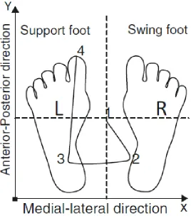
Figure 1 A schematic diagram of a typical COP path during gait initiation 8
Figure 2 Sagittal plane angles of the head and trunk 29
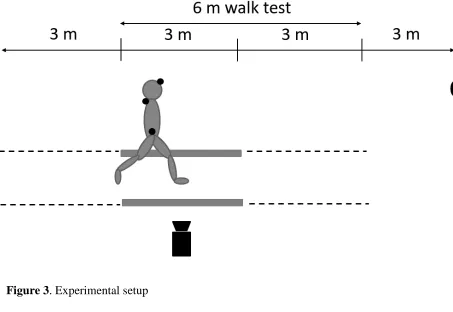
Figure 3 Experimental Setup 30
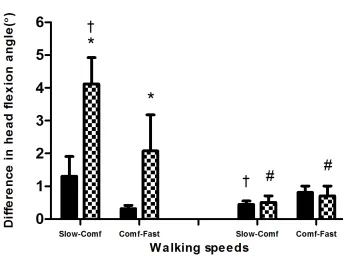
Figure 4 Delta values in head flexion angle between walking speeds and visual condition
34
Figure 5 Experimental Setup 50
Figure 6 Marker and electrode placement 51
Figure 7 Variability of upper body segment angular displacement between young and older
57
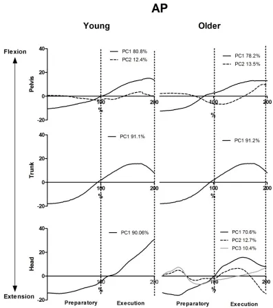
Figure 8 Principal component analysis on the data set of AP angular displacement of the upper body segments
59
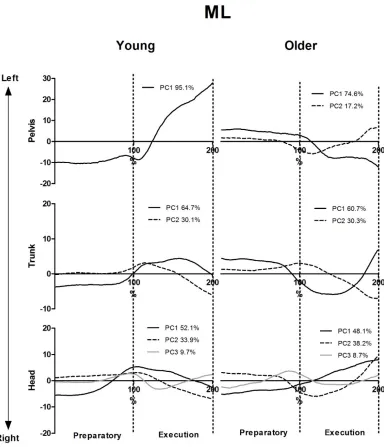
Figure 9 Principal component analysis on the data set of ML angular displacement of the upper body segments
60
Figure 10 AP Acceleration attenuation between young and older 62
Figure 11 ML Acceleration attenuation between young and older 63
Figure 12 Margin of stability between young and older 66
Figure 13 Variability of upper body segment angular displacement between target conditions
Figure 14 Acceleration attenuation between target conditions 86
Figure 15 Variability of head angular displacement between speed conditions 105
Figure 16 Acceleration attenuation between speed conditions 107
Figure 17 Margin of stability between speed conditions 112
Table 1 Gait parameters between walking speeds and visual condition
37
Table 2 Onset and relative amplitude of muscle activation 65
Table 3 Gait spatiotemporal parameters, angular displacement and margin of stability between target conditions
81
Table 4 Onset and amplitude of muscle activation between target conditions for young
88
Table 5 Onset and amplitude of muscle activation between target conditions for older
89
Table 6 Onset and amplitude of muscle activation between speed conditions for young
109
Table 7 Onset and amplitude of muscle activation between speed conditions for older
110
Table 8 Variables contributing to whole body stability for young 113
ABBREVIATION LIST ANOVA AP APA AvgSd BOS COM COP EMG ES GSR ML MOS NE NVT PCA RMS SCM VOR VT
Analysis of Variance Antereoposterior
Anticipatory Postural Adjustment Average Standard Deviation Base of Support
Centre of Mass Centre of Pressure Electromyography Erector Spinae Gait Stability Ratio Margin of Stability Mediolateral Neck Extensors No Visual Target
Principal Component Analysis Root Mean Sqaure
PUBLICATIONS AND CONFERENCE PROCEEDINGS
Publications
Maslivec A, Bampouras T.M, Dewhurst S. Head flexion and different walking speeds do not affect gait stability in older females, Human Movement Science. 55 (2017) 87– 93.
Maslivec A, Bampouras TM, Dewhurst S, Macaluso A, Vanozzi G, Laudani L. Mechanisms of head stability during gait initiation in young and older women: a neuro-mechanical analysis. Journal of Electromyography and Kinesiology. 38 (2018) 103-110.
Conferences
Maslivec A. Proposed PhD Outline: Age-related changes in gait and posture. Presented at Connections: Data, Publication and Defence, 25th March 2014, University of Western Scotland, Scotland.
Maslivec A. Mechanisms of head stability during gait initiation in young and older women: a neuro-mechanical analysis. BASES Biomechanics Interest Group meeting, 3rd March 2016, Liverpool John Moore’s University, UK.
Maslivec A. Effects of a visual target on head stabilisation and upper body balance strategies during gait initiation in young and older females. Presented at Research and Impact in Active Ageing Symposium 24th June 2016 University of Cumbria, UK.
1
CHAPTER 1
Introduction
With age, there is a decline in the functional capacity of the neuromuscular system (Prince, Corriveau, Hébert, & Winter, 1997), which has serious implications for the performance of simple tasks, especially the control of posture and balance during gait and gait related tasks. Gait is a habitual activity, requiring transition from a stable to an unstable position, i.e. from double to single leg support. Such movement results in a continuous perturbation in the balance equilibrium, as the centre of mass (COM) alters in relation to the concurrently changing base of support (BOS) (Woollacott & Tang, 1997). This can prove challenging for older individuals (Ihlen et al., 2012; Prince et al., 1997), reflected by the fact that a substantial number of falls occur during walking in older individuals (Rubenstein, 2006).
2 Early gait studies consider the upper body as one rigid segment (Winter, MacKinnon, Ruder, & Wieman, 1993), however as the upper body has many degrees of freedom, the upper body should be considered as separate components. The goal of the human postural system is to control the motion of the upper body in order to stabilise the head during gait by attenuating gait related oscillations of the lower body. Analysis of the head and trunk interaction have identified controlled movement strategies that enhance head stability during walking in young adults, (Cromwell et al., 2004; Cromwell & Wellmon, 2001) therefore it is important to understand if this is reflected in older adults to maintain head stability.
It has been reported that older individuals typically implement head flexion as a behavioural adaptation to help to identify lower limb trajectory to enable footfall vision during gait tasks (Marigold & Patla, 2008). However, it has also been suggested that head flexion may not be a behavioural adaptation, but a physiological adaptation, associated with the age related loss of the number of motor units per muscle (Enoka et al., 2003) and weakness of the neck flexors and neck extensors (Griegel-Morris, Larson, Mueller-Klaus, & Oatis, 1992). The understanding of whether head flexion is a safe behavioural adaptation implemented by older adults or whether it is a physiological adaptation attributed with the ageing process, which could be undermining head stabilisation and subsequently, whole body stability is warranted.
3 requires movement of the head on trunk to compensate for external perturbations or from the trunk (Cromwell, Newton, & Carlton, 2001). Perhaps surprisingly, only one study has examined the control of head stability during gait initiation which reported older females to have increased head movement variability with a more anterior angular displacement and therefore decreased head stability, compared to young (Laudani, Casabona, Perciavalle, & Macaluso, 2006). However, no studies have investigated the mechanisms underpinning such decreased head stability and the effect on whole body stability.
In addition to the lack of research on the mechanisms underpinning head stability during gait related tasks in older individuals, there are some methodological issues that need to be examined, to ensure correct interpretation of results and advice provided. Firstly, to account for head movement, studies investigating gait typically use a fixed visual target at eye level e.g. (Caderby, Yiou, Peyrot, Begon, & Dalleau, 2014; Hirasaki, Moore, Raphan, & Cohen, 1999; Laudani et al., 2006). If indeed older individuals do adopt an increased flexed head position, then walking whilst focussing on a fixed visual target at eye level does not illustrate a context-specific activity, as experimental controls restrict natural head movement (Hirasaki, Kubo, Nozawa, Matano, & Matsunaga, 1993). To date, no studies have challenged the use of fixed visual implementation during gait tasks. The evaluation of this will help to provide more realistic and ecologically valid results to provide useful information for clinical and research assessments.
4 as an outcome measure of gait ability (Montero-Odasso et al., 2004) but it is rarely used as the subject of investigation, particularly within the older population. However, regardless of age, environmental demands sometimes require different walking speeds to an individuals perceived ‘comfortable’, for example, walking fast when wanting to catch a bus. Given that the majority of falls occur during walking (Winter, 1995), a better understanding of different walking speeds and the effect on head stability and subsequently whole body stability is warranted. One study has examined head stability during different steady state walking speeds and found that young were able to maintain control of head stabilisation regardless of walking speeds, but this was compromised in older females at fast walking (Mazzà, Iosa, Pecoraro, & Cappozzo, 2008). However there was no measure of whole body stability. Further examination of the effect of different speeds on head stabilisation and the effect it has on whole body stability in older adults is needed.
5
CHAPTER 2
Literature Review
This literature review will draw on the key studies involving gait and gait initiation in older adults. The section will commence with the discussion of gait and gait initiation, before going into more detail on anticipatory postural adjustments (APA’s) and their contribution to overall balance. A discussion on head position and head stability along with the mechanisms underpinning head stabilisation will be presented and methodological considerations for gait assessment will be highlighted.
Gait
6 adaptation in gait parameters, older adults’ cadence is reported to remain similar to that of young adults (Rogers, Cromwell, & Grady, 2008). This means that as older adults shorten their step length, they cover less distance with the same number of steps. By adapting gait in this manner, older adults spend more time in the double limb support phase and less in the single limb support phase, thus creating a more stable walking pattern (Cromwell et al., 2001). This increased stability however, proves less effective for moving the body forward.
To account for the change in velocity and step length altering interdependently, the Gait Stability Ratio (GRS) was developed as a measure that reflects changes in both of those parameters (Cromwell & Newton, 2004). Through 2D analysis, GSR i.e. the ratio of steps to velocity is calculated to provide an indication of walking stability. An increase in GSR indicates that more steps per unit of distance and therefore a greater portion of the gait cycle is spent in the double support phase.
Gait initiation
8
9
Anticipatory control
APA’s are considered to be under prospective control (von Hofsten, 2004) and, as such, they are used to assess the ability to feedforward control motor actions, in other words preparing for a movement. It is likely that dynamic instabilities during step transitions may be more critical factors to reach a steady state walking speed than standing postural control. The transition from standing to steady state gait, therefore, is an ideal paradigm to examine the impact of advancing age on balance (Muir, Rietdyk, & Haddad, 2014) since those changes may not be apparent during quiet support tests. It has been shown that older adults demonstrate a reduced ability to generate APAs in gait initiation compared to their younger counterparts, through a delayed muscle onset time of the leading limb as well as a slower velocity and lesser backward displacement of COP, which consequently negatively affects the performance of the task (Khanmohammadi et al., 2015). APAs of the lower limbs have been extensively researched during gait initiation, (Hyodo et al., 2012; Kubicki, Mourey, & Bonnetblanc, 2015; O’Kane, McGibbon, & Krebs, 2003; Woollacott & Manchester, 1993) however less is known about APAs of the upper body during gait initiation in older adults. There is a need to examine this phenomenon at head level to gain an understanding if any anticipatory adjustments of the head are either beneficial or detrimental to overall balance in older during gait initiation.
10 postural adjustments use afferent feedback to control the position of the body when the initial setting is disturbed (Woollacott & Manchester, 1993). In contrast, anticipatory postural control is based on predictive control to prepare the body for movements as disturbances are anticipated using higher-order processing rather than feedback (Malouin & Richards, 2000). Thus, this mechanism involves muscle activation prior to the disturbance. APA’s operate when individuals deal with disturbances generated by their own movements, such as the transition of standing to walking during gait initiation (Prince et al., 1994).
In older adults, APA’s have been seen not to be used effectively due to the inability of their to use feedforward control, possibly relating to the deleterious motor and sensory modifications associated with ageing (Ene et al., 2003). A reduced ability to produce APAs relates to an increased likelihood of falls in older populations, whereas older adults demonstrating APAs show no difference in stability as compared to young adults during stepping with lateral perturbations (Hyodo et al., 2012). If these anticipatory adjustments are not effective, the disturbance will be greater, and compensatory postural adjustments may not be enough to recover balance during gait initiation.
Head position
11 al., 1992). Lee, Han, Cheon, Park, & Yong, (2015) found that head flexion reduces the muscle activity of the neck flexors which is problematic, as this may compromise overall balance in older adults. It is important to understand whether head flexion is a safe behavioural adaptation implemented by older adults or whether it is a physiological adaptation attributed with the ageing process which could be undermining head stabilisation and therefore overall balance. Activation of the neck flexors has been reported to be critical in the role of maintaining head stability (Dos Santos, Degani, & Latash, 2007) and therefore overall balance.
Whichever the reason for head flexion in the older, it remains that a considerable segment ~7% of overall body mass (de Leva, 1996) is moved forwards, and is, thus, likely to affect the COM. For example, during quiet standing in older individuals, Buckley, Anand, Scally, & Elliott (2005) reported a destabilising effect of head flexion with an increased AP sway as a result of an increased shift of the AP COM. If stability is reduced in static conditions with head flexion, it can be reasonably hypothesised that a flexed head can exacerbate the already forward shifting of the centre of mass during gait or during the fall forward when initiating gait, which will threaten balance further.
Head stabilisation
The movement of the large mass of the head, (de Leva, 1996), can lead to a larger tendency to head instability and can therefore negatively influence whole body stability.
12 movement alters visual input and the vestibular by the endolymph fluid in the semicircular canals exerting pressure against the canal’s sensory receptor. This can result in a perturbation to the postural system, which may be avoided through stabilisation of the head in space. The Vestibular Occular Reflex (VOR) is important for stabilising gaze on regions of interest during head movement, and is critical for gaining estimates of self-motion based on visual information. Studies have reported age related declines in VOR which has been correlated to fall risk (Baloh et al., 2003). It is possible that since the VOR promotes stabilisation of gaze relative to movements of the head, head flexion, along with the age related deterioration of the VOR, may disrupt this function and may affect components of balance, however, the consequences of this are yet to be discerned.
Given that head stabilisation presumably relies heavily on visual information, the interaction between head stability and postural control on the other hand is an interesting area of research. Although this area has been widely explored in young adults, it is not well researched in older adults.During dynamic situations head stabilisation is said to be achieved through coordinated movements of the head on trunk, to compensate for oscillations from the lower parts of the body (Cromwell et al., 2001). To date, only one study to date has investigated control of head stability in young and older individuals during gait initiation. Laudani et al., (2006) found decreased head stability of older compared to young, however the mechanisms causing such instability is not well examined.
13 There is an age related loss of the number of motor units per muscle, and an increase in the variability of motor unit firing that together reduce the precision with which older adults can generate muscle force (Enoka et al., 2003). From the literature available concerning upper body muscle activation during locomotion, studies have highlighted the importance of the different levels of the erector spinae in the organisation of locomotor tasks (Anders et al., 2007; de Sèze et al., 2008). A ‘top down approach’, with the aim of attenuating postural perturbations from the lower body has been identified during dynamic tasks. This entails control of the paraspinal muscles, which stabilises the head first, and subsequently inferior parts of the upper body, in young individuals during locomotion (Winter et al., 1993).
Ceccato et al., (2009) reported anticipatory muscular activity during the anticipatory phase of gait initiation in young individuals, propagated from the superior to inferior sections of the trunk (C7 – L3), confirming a feedforward mechanism. It may be suggested that maintenance of head stability primarily relies on a feedforward command, with anticipatory activation of neck muscles at C7 level during the anticipatory phase of gait initiation in healthy young individuals (Ceccato et al., 2009). However, this mechanism remains relatively unexplored in older individuals during gait initiation.
Acceleration Attenuation
14 2001). Consequently the ability to attenuate acceleration is considered a strong balance control indicator for children, adults, and elderly individuals during steady gait (Mazzà et al., 2008; Mazzà, Zok, & Cappozzo, 2010). It has been reported that whilst young healthy adults are able to attenuate the accelerations from pelvis to head even when increasing their walking speed (Latt, Menz, Fung, & Lord, 2007), this ability is challenged in older adults (Kavanagh et al., 2003). Older adults, in particular, typically develop axial rigidity, which can impair their ability to attenuate the accelerations that are from the lower limbs during gait to the upper body, impacting on head stability. Further , difficulties in controlling the upper body accelerations have also been reported to be associated with the risk of fall (Marigold & Patla, 2008).
There is inconsistency within the literature regarding the amount of attenuation that each acceleration component undergoes. Menz et al., (2003) found higher ML accelerations at head level in older male/female adults as compared with young adults, despite smaller accelerations at pelvis level. Kavanagh et al., (2006) found significant differences between the older and young males/females only in the AP direction. Walking at faster gait speeds to comfortable reduces the ability to attenuate accelerations at head level (Mazzà, Iosa, Pecoraro, & Cappozzo, 2008), resulting in a reduced ability to maintain head stability. While it is known that head stability is threatened at fast steady state walking, studies fail to address what happens during transitory locomotive tasks such as gait initiation.
15 acceleration attenuation of the upper body during the task of gait initiation, however this was in a pathological population (Buckley, Galna, Rochester, & Mazzà, 2015). The results demonstrated impaired attenuation of accelerations from the pelvis and neck to the head in adults living with Parkinson’s disease. How well healthy older adults are able to attenuate such accelerations during gait initiation is yet to be examined.
Visual target implementation
Stationary visual targets are commonly used in gait studies, in both young and older populations. Although the reason for using a visual target during walking is not fully understood, one likely reason of directing gaze at a target during gait is to provide a focal point that will consequently reduce head movement, thus providing better head stability. Given that gait induces linear and angular head perturbations, causing head movements to occur across the gait cycle (Hirasaki et al 1993; Pozzo et al., 1990) and that older adults tend to implement head flexion during gait (Marigold & Patla, 2008), implementing a visual target may restrict what is naturally occurring.
16 a fixed visual target. In response to fixing their gaze on a stationary target, head-on-trunk with respect to head-on-trunk movements were similar to that of young adults, however there was no measure of whole body stability. While the effects of focussing on a stationary target has on stability measures is known in static and dynamic conditions in older adults, little is known about fixating on a target has on head movements and whole body stability during the transitory task of gait initiation.
17
CHAPTER 3
Ethical procedures and considerations
Integral to any process of investigation is the requirement to conduct the research process in an ethical manner. During the experimental procedures, it was ensured that research participants came to no harm when engaging with the research. This included getting consent from participants, respecting anonymity and confidentiality of participant’s data and to fully inform the participant on how and where the information that they contribute is likely to be used.
To ensure that the older participants were comfortable with the demands of the study and could execute the tasks required, in addition to the standard ethical procedures, a different approach to the conventional one was followed. Initially, the Active Ageing Research Group approached the Lancaster and Morecambe University of Third Age branch to deliver a talk on topics of U3A’s membership interest while promoting the Group’s research. During that presentation, the present study was also presented to the attendees (>80) and an expression of interest contact was proposed. Once the participants made contact, the study demands were explained in detail and opportunities for questions were provided. Following that conversation, the Participant Information Sheet and Informed Consent Form (see Appendix 1 and 2) were e-mailed to them, to allow them to read them at their own pace and ask any more questions.
18 explained thoroughly, to ensure any concerns were removed. Prior to starting the experiment, participants were asked to fill out a health screening form to ensure they were medically stable to participate in the study (Appendix 3). Several trials were allowed to gain familiarisation and confidence. Participants were encouraged to visit with a friend (regardless of whether the friend would take part or not) to ensure comfort during the process.
19
CHAPTER 4
Thesis structure and presentation
To examine the effect of head position and the strategies underpinning head stability, and whole body stability during steady state gait and gait initiation, four studies were carried out. A summary for each is presented below and the studies are presented in this Thesis as experimental standalone papers in the next four chapters.
Study 1
This study aimed to examine A) if head position and gait spatiotemporal parameters were altered when walking with free head movement and with a visual target, and B) how the effect of using a visual target may change head position at different walking speeds. Walking trials were performed on an unobstructed 9m flat walkway under two visual conditions: walking with no visual target, and walking focusing on a fixed visual target set at eye level. All trials were completed at three walking speeds (slow, comfortable and fast).
20
Study 2
This study aimed to examine neuromechanical strategies underpinning head stabilisation and whole body stability during gait initiation between young and older females. Participants initiated gait in a straight line for at least three steps at their comfortable walking speed. In addition, they were asked to focus on a fixed visual target set at eye level.
Head stabilisation and whole body stability was assessed using 3D analysis and EMG. Variability of angular displacement (AvgSD and principal component analysis (PCA)), acceleration attenuation coefficient and the onset and activation amplitude of the neck (SCM and NE) and trunk (ES) muscles were used as a measure of upper body stability. Whole body stability was quantified using an adapted version of the margin of stability. Independent sample t–tests were used to examine for differences between groups.
Study 3
This study aimed to examine the effect of free head movement on head stability and the neuromechanical strategies underpinning head stability, and whole body stability in young and females during gait initiation. Participants initiated gait in a straight line for at least three steps at their comfortable walking speed under two target conditions: with no visual target, and focussing on a fixed visual target set at eye level.
21 of upper body stability. Whole body stability was quantified using an adapted version of the margin of stability. Paired comparisons were made to examine for differences between the two target conditions.
Study 4
This study aimed to examine the effect of initiating gait at different speeds on neuromechanical mechanisms underpinning head stability, and whole body stability in young and older individuals. Participants initiated gait in a straight line for at least three steps under two speed conditions: comfortable and fast.
22
CHAPTER 5
Head flexion and different walking speeds do not affect gait stability in
older females
A version of the work from this chapter has been published as:
23
Abstract
24
Introduction
Walking is a habitual activity, requiring transition from a stable to an unstable position, i.e. from double to single leg support. Such movement results in a continuous perturbation in the balance equilibrium, as the centre of mass (COM) alters in relation to the also changing base of support (BOS) (Woollacott & Tang, 1997). This can prove challenging for older individuals (Ihlen et al., 2012; Prince et al., 1997), reflected by the fact that the majority of falls occur during walking in older individuals (Rubenstein, 2006).
25 the effect head flexion could have to either consider it in future studies and interventions or reject it as a contributor to gait instability.
Head flexion has also been shown to be influenced by gait speed in young individuals. Hirasaki, Moore, Raphan, & Cohen (1999) reported that at speeds >1.2m.s-1, there was an increased magnitude of head pitch displacement, such that a greater amount of head flexion was observed. Although gait speed is commonly assessed as an outcome measure of functional capacity and gait ability in the older population (Bongers et al., 2015; Montero-Odasso et al., 2004; Toots et al., 2013; Verghese, Holtzer, Lipton, & Wang, 2009), it has rarely been considered the subject of investigation. In other words the effect of different walking speeds on head flexion has rarely been examined in older adults. During day to day life, however, walking at different speeds is required, for example, when walking faster due to being late for an appointment, or in contrast, walking slower to negotiate a busy shopping centre. If the findings by Hirasaki et al., (1999) in young also hold true for older adults, it is feasible that as walking speed increases, concurrently increasing head flexion, postural control may also be increasingly challenged.
26 potentially reaching to erroneous results and less specific intervention advice. Therefore, understanding differences between a natural head position and a typical standardised head position, at different walking speeds, is warranted.
The aim of the study was twofold; to examine A) if head flexion and gait parameters were altered when walking without and with a visual target, and B) how the effect of using a visual target may change at different walking speeds. It was hypothesised that the implementation of a visual target would restrict head flexion, which in turn, would alter gait pattern. Females were the focus of the study as it has been reported that females whole body stability declines to a greater extent than males (Wolfson, Whipple, Derby, Amerman, & Nashner, 1994) and tend to fall more often (Schultz, Ashton-Miller, & Alexander, 1997).
Methods
Participants
Sixteen healthy older females (age 75.5 ± 6.2 years, height 1.62 ± 0.04 m, body mass 74 ± 6.8 kg) and 15 healthy young females (age 23±3.5years, height 1.67±0.04m, body mass 63.3 ± 6.0 kg) participated in the study. Older females were recruited from local community groups while young were students at the Institution. All participants had no known neuromuscular disorders, impaired postural alignment such as kyphosis, osteoarthritis or neck related pain, while older participants were community residing, functionally independent, considered medically stable (Greig et al., 1994). All participants were able to perform all conditions without the use of bifocal or multifocal
27 Ethical approval was obtained from the Institutional Ethics Committee and written informed consent was obtained prior to testing.
Protocol
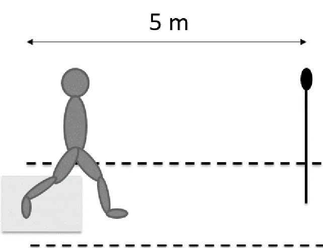
Walking trials were performed on an unobstructed 9m flat walkway under two visual conditions; walking with no visual target and walking with a visual target. In the no visual target condition, no instructions were given to participants as to where to orient their gaze whereas in the visual target condition, participants were instructed to focus on a stationary target located at eye level, 3 m directly ahead of the end of the walkway. The visual target consisted of a black circle (15 cm diameter) on a white background. The position, size and distance of the visual target were decided following pilot testing, which allowed a target which could be comfortably seen by the participants without excessive eye focusing effort. All participants underwent familiarisation with each visual condition and speed, and confirmed they were able to clearly see the target from the beginning of the walkway without the use of glasses.
28
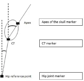
Head flexion
To measure head flexion, a marker was placed on the apex of the skull (attached to a headband secured around the participant’s head, horizontal to the ground, during standing in the reference body position) and a marker placed on the seventh cervical vertebrae (C7). The angle formed by the vertical axis (passing through the C7 marker) and the straight line between the C7 and the apex of the skull markers, was measured as head flexion angle.
Trunk flexion
29
30
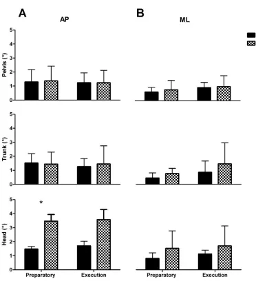
31 Head and trunk flexion angles were measured in the sagittal plane using 2D video analysis (Kinovea for Windows, Version 0.8.15, www.kinovea.org) with a sampling frequency of 100 Hz, at the first heel strike of the left foot (first frame the heel made contact with the ground) as soon as the participant crossed the 6 m marker. Το obtain a realistic understanding of changes in head and trunk flexion angles by avoiding ‘postural adjustments’ during standing measurements (Thomas, Bampouras, Donovan, & Dewhurst, 2016) the difference in angle from comfortable to slow walking speed and comfortable to fast walking speed (Δ values) were calculated for both age groups and both visual target conditions. Positive angle values indicated greater head and trunk flexion of the given walking speed in comparison to comfortable walking speed.
Walking velocity
The 6m walk test was used to measure walking velocity at each walking speed as it has been shown to have high reliability for comfortable and fast walking (ICC=.97 and .96, respectively) (Steffen, Hacker, & Mollinger, 2002). Walking velocity was calculated from the time taken to walk between 3m and 9m (6m) of the walkway using wireless timing gates (Brower timing gates, Draper, UT, USA) set at hip height.
Gait parameters
32 of two infrared photocell bars that can derive contact time of each foot from the breaking of the transmitted beam. Gait stability ratio (GSR, calculated as cadence / velocity) has been developed from 2D gait analysis of flat walking and was used as a measure of walking stability. A higher GSR indicates a greater proportion of the gait cycle is spent in contact with the floor, thus avoiding the dynamic components of walking (Ronita L Cromwell & Newton, 2004), as one would do when a greater need for stability is required.
Statistical analysis
To assess intrarater reliability of angle and gait measurements, sensitivity (typical error
(TE), calculated as standard deviation of the change scores between measurement / √2)
and intraclass correlation coefficient (ICC, calculated as 1 – TE2 / mean between-subject standard deviation between measurements) between the three trials were obtained from a customised spreadsheet (Hopkins, 2000).
33 effect respectively (Fritz, Morris, & Richler, 2012). An alpha level was set at p < 0.05. Data are presented as mean ± standard deviation (SD).
Results
Data for gait parameters are presented in Table 1. For clarity, effect sizes for significant differences are reported only if they were below moderate (0.05).
Reliability
Head and trunk flexion ICCs for both age groups in all measurements ranged from 0.89 – 0.90, indicating high reliability, whilst only a small TE (<1.12°) was present. Similarly, ICC for step length ranged from 0.83-0.95, with only fast walking with visual target for the young exhibiting a lower ICC (0.77).
Head flexion
34
35
Trunk flexion
There was a significant main effect of age (p = 0.013) and visual condition (p = 0.02) on trunk angle. There were significant interactions for age × visual condition (p = 0.026). There was no difference in trunk flexion at any walking speed or visual condition between young and older Delta values showed older displayed a greater increase in trunk flexion angle from the fast walking to comfortable walking (p = 0.001, ES = 0.08) in the no visual target compared to the visual target condition, while there were no differences between visual conditions in young.
Walking velocity
There was no difference in gait velocity between visual conditions for either group. Predictably, walking velocity significantly increased with walking speed in both age groups (p = 0.019, ES = 0.24-0.49 and p = 0.038, ES = 0.28–0.39 for young and older respectively), indicating participants successfully followed walking speed instructions. Young were significantly faster at comfortable and fast walking compared to older (p = 0.008), however there was no difference in gait velocity at slow walking speed between groups.
Gait parameters
37
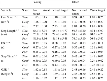
Table 1. Gait parameters during both visual conditions for each walking speed for young and older. Data is presented as mean ± SD.
Young Older
Variable Speed No visual target
Visual target No visual target
Visual target Gait Speed a, b Slow 1.05 ± 0.15 1.10 ± 0.20 0.94 ± 0.21 1.01 ± 0.26 (m.s-1) Comf 1.58 ± 0.20 1.51 ± 0.10 1.32 ± 0.28 1.42 ± 0.29
Fast 2.37 ± 0.30 2.23 ± 0.20 1.81 ± 0.20 1.81 ± 0.32 Step Length a, b Slow 64.1 ± 3.94 65.16 ± 4.77 59.3 ± 5.20 65.4 ± 5.90 (cm) Comf 73.8 ± 5.03 74.40 ± 4.38 68.9 ± 4.90 70.6 ± 4.20
Fast 86.9 ± 4.96 86.18 ± 5.48 76.0 ± 5.10 76.6 ± 5.40 DST a, c Slow 0.39 ± 0.05 0.38 ± 0.07 0.45 ± 0.13 0.43 ± 0.11
(s) Comf 0.27 ± 0.04 0.27 ± 0.03 0.35 ± 0.21 0.31 ± 0.06
Fast 0.15 ± 0.04 0.16 ± 0.03 0.20 ± 0.03 0.22 ± 0.04 Step Time a, b Slow 0.61 ± 0.04 0.57 ± 0.04 0.42 ± 0.07 0.41 ± 0.07
(s) Comf 0.49 ± 0.03 0.49 ± 0.03 0.29 ± 0.04 0.29 ± 0.02
Fast 0.38 ± 0.05 0.42 ± 0.05 0.21 ± 0.03 0.22 ±0.030
GSR a Slow 1.62 ± 0.18 1.61 ± 0.16 2.48 ± 0.16 2.41 ± 0.22
(Step.m-1) Comf 1.41 ± 0.12 1.39 ± 0.14 2.45 ± 0.70 2.53 ± 0.12
Fast 1.16 ± 0.07 1.17 ± 0.12 2.92 ± 0.23 2.62 ± 0.26
38
Discussion
The purpose of the present study was to examine if head flexion and gait parameters were altered when walking without and with a visual target, and how the effect of using a visual target may change at different walking speeds. Findings showed that older individuals adopted greater head flexion at all walking speeds in the no visual target condition compared to young. Head flexion was constrained to that similar of young when walking with a visual target with no changes in gait parameters.
39 found. Hirasaki et al., (1993), however, reported increased head extension whilst the present study found increased head flexion. The reasons for this can possibly be attributed to a difference in population characteristics between studies. Interestingly, DST was not significantly different between young and older in Hirasaki et al., while
the present results showed that older did have significantly longer DST. Thus, results
show that differences were not only seen for head position but actually for gait variables,
lending to the speculation that differences may be due to differences in population
characteristics. Hirasaki et al., proposed that older may have reduced flexibility of the
vertebral column preventing flexion of the head, however very little information is
given about the older participants in the study. Older participants in the present study
were healthy and physically active, therefore flexibility of the vertebral common may
not have been a problem, allowing a more unrestricted head movement.
The hypothesis of focusing on a visual target (to reduce head flexion) altering gait parameters, can be rejected as gait parameters remained similar in both visual conditions. Sway has been seen to be affected by head flexion during static conditions (Buckley et al., 2005). Despite differences in head flexion between visual conditions for the older, there was no difference in GSR values, suggesting head flexion whilst walking did not pose any additional fall risk. In the present study, head flexion was measured independent of the trunk. Previous studies have shown that the trunk flexion can influence gait results (Saha, Gard, & Fatone, 2008), however the present study found no trunk flexion, demonstrating that trunk was not responsible for head flexion.
40 head position during walking (e.g. Cromwell et al., 2002; Hirasaki et al., 1999). Such instructions, which constrain head movement, may have masked a true effect, as they would reduce the naturally occurring head flexion, supported by the findings in the present study. It was hypothesised that this in turn, this would impact on gait stability and postural control, most likely underestimating the true balance challenge walking poses on older individuals and potentially reaching to erroneous results and specific intervention advice. From the findings of the present study, however, this does not appear to be so for the population studied.
Hirasaki et al., (1999) reported that at speeds >1.2 m.s-1, there was an increased
magnitude of head pitch displacement and a further increase when walking at speeds of 2 m·s-1 in young individuals. These results are reflected in the present study as young had greater head flexion between comfortable – fast (2.53 m·s-1 compared to slow-comfortable (1.51 m·s-1). The present results support Hirasaki et al., previous reports that head displacement changes with walking speed for young adults, however the opposite effect was found in older as older produced greater head flexion at slow – comfortable walking speed compared to comfortable – fast walking speed (with low effect sizes, however). Despite trivial differences in head flexion at different speeds, overall gait stability was unaffected in both age groups.
41 older population. The present data supports the notion that other measures are contributing to gait instability and that ageing effects on speed are not straight forward, thus a more holistic assessment is warranted.
Older individuals can have a kyphotic posture, an exaggerated anterior curvature which tends to increase with age (Ailon, Shaffrey, Lenke, Harrop, & Smith, 2015; Katzman, Wanek, Shepherd, & Sellmeyer, 2010). This impaired postural alignment affects physical functioning and can have implications on fall risk for the elderly (Ailon et al., 2015; Katzman et al., 2010). The participants in the present study were free from such condition and it would be expected that kyphotic individuals would present different findings to the current participants. Further, to ensure that changes in head flexion angle could be attributed to head movement rather than trunk flexion, the two segments were examined separately. The results showed that trunk flexion did not change in any substantial way (as indicated by the very small effects sizes), suggesting that trunk flexion remained stable when changing between visual target conditions and walking speeds.
A limitation to the study was that although a visual target approach was used, it was not quantified using an eye tracking device to examine whether participants was fixating on the target. However, the use of the target was not aimed to fixate gaze, but rather to adjust the head position by fixing the gaze. This was achieved, even if eyes were not always on the target, as the instruction of keeping the head up was followed.
Conclusion
43
CHAPTER 6
Mechanisms of head stability during gait initiation in young and older
women: a neuromechanical analysis
Maslivec A, Bampouras TM, Dewhurst S, Macaluso A, Vanozzi
G, Laudani L. Mechanisms of head stability during gait initiation
in young and older women: a neuro-mechanical analysis. Journal
of Electromyography and Kinesiology. 38 (2018) 103-110
.
A version of the work from this chapter was presented at the BASES
Biomechanics Interest Group meeting, 3
rdMarch 2016, Liverpool John
Moore’s University, UK.
44
Abstract
45
Introduction
Stabilisation of the head in space is fundamental to optimise inputs from the visual, vestibular, and somatosensory systems and, therefore, to maintain whole body balance during locomotion (Kavanagh et al., 2005; Pozzo et al., 1990). Decreased head stability has been reported in older individuals during different types of locomotion, including steady-state walking (Cromwell et al., 2001) and locomotor transitions such as gait initiation (Laudani et al., 2006). Transitory locomotor tasks, in particular, involve complex interactions between neural and mechanical factors which may challenge whole body balance to a greater extent than unconstrained walking (Nagano et al., 2013). This challange may help to explain why the number of falls in older individuals are frequent during locomotor transitions such as gait initiation and termination (Winter, 1995).
46 upper body accelerations during the transitory task of gait initiation in healthy young and older individuals.
From a neuromuscular point of view, electromyography (EMG) studies have highlighted the importance of trunk paraspinal muscle activation in actively attenuating postural perturbations from the lower body during locomotor tasks (Anders et al., 2007; de Sèze et al., 2008). A ‘top down’ anticipatory control of erector spinae muscles, which stabilises the upper trunk first and subsequently the lower trunk, has been reported in young individuals during gait (Winter et al., 1993; Prince et al., 1994). In line with that, Ceccato et al., (2009) have reported a metachronal activation of erector spinae muscle occurring during the preparation of the first step for gait initiation. To date, most of the studies on older individuals have revealed characteristic age related changes of muscle recruitment in the lower limb during gait initiation. For instance, older individuals have been shown to initiate walking with greater co-contraction of the lower leg muscles (Khanmohammadi et al., 2015a) and a delayed activation of the tibialis anterior muscle compared to young individuals (Khanmohammadi et al., 2015b). It is not known, however, whether older individuals would effectively recruit the trunk muscles and/or adopt an anticipatory control in order to actively aid stabilisation of the head during the transitory phase of gait initiation.
47 dynamic balance in young and older participants was investigated by evaluating whether the conditions for dynamical stability were met within each age group. It was hypothesised that older women would a) demonstrate reduced ability to attenuate acceleration from lower to upper parts of the upper body, b) have impaired muscle activation pattern of the trunk and neck and c) have reduced whole body stability, compared to the younger women.
Methods
Participants
Elevenhealthy young (age: 23.1 ± 1.1 years, height: 1.64 ± 0.71 m, body mass: 57.5 ± 6.7 kg) and 12 healthy older (age: 73.9 ± 2.4 years, height: 1.63 ± 0.45 m, body mass: 66.2 ± 10.2 kg) females volunteered to participate in the study. Women were the focus of the study as it has been reported that their whole body stability declines to a greater extent than males (Wolfson et al., 1994) and tend to fall more often (Schultz et al., 1997). Older participants were considered ‘medically stable’ to participate in the study, according to exclusion criteria for older people in exercise studies (Greig et al., 1994). No participants had any history of neurological disorders that would affect their balance or gait ability, and were able to complete the task without the use of bifocal or multifocal spectacles. Written informed consent was provided by all participants and ethical approval was given by the institution’s ethics committee.
Experimental protocol and equipment
48 accord from a single force platform (Bertec Corp, Worthington, OH) and to continue to walk forwards in a straight line for at least three steps at their comfortable walking speed. In addition, they were instructed to focus on a fixed visual target, which was set at eye level for each participant and located five metres ahead of the starting position. The position, size and distance of the visual target were decided following pilot testing, which allowed a target which could be comfortably seen by the participants. The right leg was used as the starting (swing) leg for all trials. Starting feet position at shoulder width apart was marked on the force platform and participants repositioned themselves in that position for each trial. In total five trials were completed and analysed.
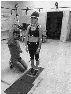
A seven camera motion analysis system (VICON, Oxford Metrics, London, England) was used to record and reconstruct the 3D position of 35 reflective markers placed on body landmarks, following the Davis protocol (Davis et al., 1991) with a sampling rate of 100 Hz. The VICON whole body plug-in-gait model was used to define a local anatomical reference frame for the pelvis (markers on the left and right anterior and posterior superior iliac spines), trunk (markers located at the clavicle and sternum level as well as at C7 and at T10), and head (four markers, placed on the left and right side of the front and back of the head) and then calculating the relevant kinematic data. The force platform was used to track COP motion with a sampling frequency of 1000 Hz and then resampled.
49 performed as a whole movement and divided into two phases: 1) anticipatory phase, which lasted from the onset of COP motion to the instant of toe off of the swing limb 2): execution phase, which lasted from toe off of the swing limb to the instant of toe off of the stance leg. Temporal events of gait initiation were obtained from both position and velocity curves derived from markers placed on the calcaneus and fifth metatarsal bones (Mickelborough et al., 2000). These events corresponded to the instants of heel off, toe off and heel contact of the swing limb. Angular displacement and the motion of the upper body segments (pelvis, trunk, and head) were measured in the AP and ML direction. Additionally, whole body COM was recorded as a weighted sum of all body segments using the whole plug-in-gait model in the AP and ML direction.
50
51
52
Data analysis
Variability of angular displacement
Angular displacement of the pelvis, trunk, and head was filtered using a second-order low-pass Butterworth filter with a cut-off frequency of 5 Hz and re-scaled to the first value of the anticipatory phase. To quantify variability of the pelvis, trunk, and head motion during gait initiation, the average standard deviation (AvgSD) was calculated using the following equation:
𝐴𝐴𝐴𝐴𝐴𝐴𝐴𝐴𝐴𝐴= �∑ θ1002
θ= Angular displacement of the segment.
53 corresponding to 1% intervals (anticipatory phase: 1-100%, execution phase: 101-200%).
Attenuation of upper body accelerations
Acceleration of the pelvis, trunk and head segments was calculated by double derivative of the 3D position of the origin of each upper body segment reference frame in the AP, ML and cranio-caudal (CC) direction. It was computed by a customised Matlab 7.5 script (Mathworks, Inc, USA) and filtered using a second-order low-pass Butterworth filter with a cut- off frequency of 5Hz. The magnitude of acceleration of each segment was calculated using the root mean square (RMS) in the AP, ML and CC direction. RMS acceleration values are known to be influenced by gait velocity (Kavanagh & Menz, 2008), thus AP and ML RMS acceleration were normalized by CC acceleration RMS as proposed by Iosa et al., (2012). The ability to attenuate accelerations through the upper body segments was quantified using the attenuation coefficient expressed as a percentage. The attenuation coefficient describes the ability to reduce accelerations from inferior to superior segments, with reduced linear acceleration from inferior to superior parts of the upper body used as an indicator of upper body stability (Summa et al., 2016). The attenuation coefficients were calculated using RMS values of each segment as follows (for both AP and ML direction):
Cxy =�1−RMSRMS𝑦𝑦𝑥𝑥� ∗100
𝑥𝑥 = inferior segment y = superior segment
Each coefficient represents the attenuation froma lower to an upper body level. CPH
54 from the pelvis to the trunk, and CTH representing the attenuation from the trunk to the
head. A positive coefficient value indicated a reduced acceleration whilst a negative coefficient value indicated a greater acceleration between the two specified segments.
Activation patterns of the trunk and neck muscles
Raw EMG signals were first high-pass filtered at 20 Hz to remove movement artifacts, then full-wave rectified and filtered using a second-order high-pass Butterworth filter with a cut- off frequency of 50 Hz using a custom Matlab script. Figure 6 shows a typical example of the filtered data split into the baseline (defined as the stance, 500ms prior to COP onset), anticipatory phase and execution phase, as described previously. The onset of muscular activity was visually estimated by the same experimenter for all calculations, which has been shown to be reliable to achieve muscle onset (Micera et al., 2001), and was expressed as a percentage from COP onset to the end of the anticipatory phase. The relative amplitude of muscular activity was calculated from the area under the EMG curve of each muscle using a customised Matlab script and further expressed as a percentage normalised to the EMG activity of the execution phase of gait initiation.
Whole body stability during gait initiation
55 the calculation of the margin of stability, the positions of the COM and BOS need to be known. COM was recorded as a weighted sum of all body segments using the whole plug-in-gait model while BOS was calculated from the distance between the position of the swing heel marker at heel-contact and the position of the stance heel marker at toe off represented the step length and width, and was representative AP and ML BOS respectively. MOS was taken at heel contact of the swing limb, as it has previously been shown that foot strike was systematically made with the heel (Caderby et al., 2014).
The position of the 𝑒𝑒𝑥𝑥𝑒𝑒𝑒𝑒𝑒𝑒 was then calculated as follows
𝑒𝑒𝑥𝑥𝑒𝑒𝑒𝑒𝑒𝑒=𝑥𝑥𝑒𝑒𝑒𝑒𝑒𝑒+ 𝑥𝑥′𝑒𝑒𝑒𝑒𝑒𝑒
�𝐴𝐴𝑙𝑙
With 𝑥𝑥𝑒𝑒𝑒𝑒𝑒𝑒 and 𝑥𝑥′𝑒𝑒𝑒𝑒𝑒𝑒representing the COM position and velocity respectively, 𝐴𝐴 = 9.81m.s-1, the gravitational acceleration, and 𝑙𝑙 corresponding to the limb length, taken
from anthropometric measurements prior to data collection (inverted pendulum eigenfrequency). The MOS corresponded to the difference between the AP and ML boundary of support (BOS) and the AP and ML position of the ‘extrapolated COM’ (𝑒𝑒𝑥𝑥𝑒𝑒𝑒𝑒𝑒𝑒) at heel contact and defined as BOS - 𝑒𝑒𝑥𝑥𝑒𝑒𝑒𝑒𝑒𝑒. The lower the MOS value, the closer the 𝑒𝑒𝑥𝑥𝑒𝑒𝑒𝑒𝑒𝑒 is to the BOS, indicating reduced whole body stability.
Statistical analysis
56 for difference between young and older groups for the AvgSD of angular displacement of each upper body segment, RMS of acceleration at each upper body segment and attenuation of such acceleration and MOS values, with Bonferroni correction for multiple comparisons applied. Finally, for the onset of muscular activity and relative amplitude of muscle activity of the anticipatory phase. Statistical significance was assessed with an alpha level of 0.05. All data are presented as mean ± SD unless otherwise stated.
Results
Variability of angular displacement
57
58 PCA of angular displacement is presented in Figure 5 and 6 in the AP and ML direction respectively. In the AP direction, both groups demonstrated a similar amount of variability of pelvis angular displacement as two PCs explained over 90% of the movement pattern variance in both groups. Both groups demonstrated low variability of trunk angular displacement, as only one PC was needed to explain over 90% of the movement pattern variance. Young showed low variability of angular head displacement as only one PC was needed to explain over 90% of variance. Older however, demonstrated high variability in head angular displacement indicated by the requirement of three PCs to explain over 90% of variance (Figure 5).
59
60
61
Attenuation of upper body accelerations
During the anticipatory and execution phase, young displayed significantly greater AP RMS acceleration for the pelvis, trunk and head compared to older (p < 0.05) (Figure 7A and B). During the anticipatory phase, AP CTH was significantly lower in older
compared to young (-1.9 ± 20.2% versus 10.1± 21.6%, p = 0.02, respectively) (Figure 7C). During the execution phase, there were no significant differences in acceleration attenuation between groups (Figure 7D).
During the anticipatory and execution phases, there was no difference in ML RMS acceleration for the pelvis, trunk or head between age groups (Figure 8A and B). During the anticipatory phase, ML accelerations were attenuated for both groups, with the exception of older not able to attenuate CPT, however there were no significant
62
63
64
Muscle activity
EMG data for young were analysed for all muscles and six out of 12 older was analysed for the neck muscles. It was not possible to gain muscle onset data for the neck extensors for older due to difficulty in identifying an onset point because of noisy signal. Older displayed a significantly delayed muscle activity onset of the SCM compared to young (p < 0.05) (Table 2). There were no differences in muscle activity onset time for the ES (T9) or ES (L3) between groups. Both groups activated all muscles to a greater extent in the execution phase compared to the anticipatory phase as shown by less than 100% EMG values. Older had significantly greater relative muscle activity in the anticipatory phase compared to young for the SCM, ES (T9) and ES (L3) (p < 0.05) (Table 2).
Whole body stability
65
Table 2. The time of the onset of muscle activity given as a percentage of total duration of the anticipatory phase of gait initiation. Amplitude is given as a percentage, normalised to the execution phase of gait initiation. P value (p < 0.05) indicates significance between groups.
Young (n =11) Older (n = 6) P-value SCM
Onset (%) Amplitude (%)
20.5 ± 13.2 49.3 ± 20.7
50.5 ± 15.4 88.2 ± 19.8
0.028 0.002
Extensor Onset (%) Amplitude (%)
54.8 ± 22.1 32.3 ± 16.2
N/A
59.6 ± 16.1
N/A 0.32
Upper spine (T9) Onset (%) Amplitude (%)
42.2 ± 20.5 28.3 ± 22.6
63.3 ± 24.7 52.9 ± 22.1
0.182
0.005
Lower spine (L3) Onset (%) Amplitude (%)
53.1 ± 25.6 23.8 ± 11.5
60.7 ± 22.5 41.5 ± 15.4
0.192
66
67
Discussion
The purpose of the study was to examine any age related change in the neuromechanical strategies underpinning head stabilisation and whole body stability during gait initiation. Older displayed lower AP acceleration of the upper body segments compared to younger and were less able to attenuate AP accelerations between trunk and head compared to young. Older revealed a greater relative magnitude and delayed anticipatory activation of the neck muscles compared to young. Finally, older demonstrated reduced ML whole body stability, while there was no difference between age groups for AP whole body stability. Older participants showed greater variability of head angular displacement in AP direction compared to young participants during both the anticipatory and execution phase of gait initiation, which is in agreement with a previous study by Laudani et al., (2006).
In the present study, young displayed greater AP RMS acceleration at each upper body segment compared to older, indicating older may adopt a more cautious strategy in order to move from a standing posture to forward walking (Menz, Lord, & Fitzpatrick, 2003). No difference between groups existed for ML acceleration attenuation, and similar to previous studies (Kavanagh et al., 2005; Mazzà et al., 2008), both groups found it difficult to attenuate ML accelerations during the execution phase.
68 were able to attenuate accelerations from trunk to head, aiding protection of the head, older could not, suggesting acceleration did not decrease from the trunk to the head. The inefficiency in attenuating these accelerations may be attributed to deleterious age related changes to passive structures of the spinal column or to sequential activation of the axial musculature (Doherty, 2003) .
69 Instability during walking in older populations is commonly considered in the ML plane, while loss of ML stability can have a profound effect on walking function (Maki, 1997b). Interestingly, differences in upper body stabilisation between young and older were only observed in the AP direction during the present thesis. Even though differences in upper body stabilisation were apparent between age groups, there were no differences in AP MOS between groups. A possible explanation is that upper body differences were not considerable enough to alter AP whole body stability. AP MOS has previously been described as similar between young and older females during steady state walking (McCrum et al., 2016). Despite no differences between groups in the ML direction of upper body variability or attenuation of acceleration, older demonstrated significantly reduced MOS, indicating reduced ML whole body stability. This may have implications for fall risk as whole body stability is an indicator of fall risk (Lockhart & Liu, 2008; Toebes, Hoozemans, Furrer, Dekker, & van Dieën, 2012). Caderby et al., (2014) observed that young were able to maintain ML whole body stability during gait initiation, while ML whole body stability in older during gait initiation warrants further research to generate an understanding of why ML whole body stability declines during gait initiation in older females.
Conclusion
70
CHAPTER 7
Effects of free head movement on head stability and whole body
stability during gait initiation in young and older women
71
Abstract
72
Introduction
The ageing process can make walking a challenging process, due to reduced neuromuscular performance in several aspects e.g. strength, balance, functional ability (Boyer et al., 2017). Indeed, gait and gait disorders are an important contributor to falls in older adults (Rubenstein, 2006). As a result, several studies have investigated gait mechanics in older adults and the changes that occur due to ageing (Boyer et al., 2017; Aboutorabi et al., 2016, Prince et al., 1997). The vast majority of these studies, however, focus on lower limbs and the effects of ageing on e.g. step width, step length, or walking speed, with little attention given to upper body movements.
Head flexion, for example, is a movement frequently implemented by older adults during gait to identify lower limb trajectory to enable better footfall vision or to gather more information when walking towards an obstacle (Muir, Haddad, Heijnen, & Rietdyk, 2015). Walking results in a continuous perturbation in the balance equilibrium, as the centre of mass (COM) alters in relation to the also changing base of support (BOS) (Woollacott & Tang, 1997), this head flexion may exacerbate the forward shifting of the COM closer to the BOS, further threatening stability.
73 that head movement in a dynamic situation did not have an effect (Thomas et al., 2017). Head stability is thought to be critical in the control of whole body stability (Pozzo et al., 1990), however, how head movement relates to the mechanisms underpinning head stability warrants further understanding. While the effects of head movement and focusing on a stationary target on stability measures have received some attention in static and dynamic conditions in older adults, little is known about the effect of head movement and head stability has on whole body stability during the transition from a standing position to walking (gait initiation).
Therefore, the aim of the present study was to examine the effect of free head movement during gait initiation on upper body, lower body and whole body stability parameters of older females. To achieve this, there was two conditions; gait initiation with free head movement and an instructed gait initiation (head focusing on a visual target). It was hypothesised that participants would have better head stabilisation with a visual target (VT) compared the free head movement condition with no visual target (NVT). Females were the focus of the study as it has been reported that their whole body stability declines to a greater extent than males (Wolfson et al., 1994) and they tend to fall more often (Schultz et al., 1997).
Methods
Participants
74 for older people in exercise studies of Greig et al., (1994). In addition to these criteria, no participants had any history of neurological disorders that would affect their balance or gait ability, and were able to complete the task without the use of bifocal or multifocal spectacles. Written informed consent was provided by all participants and ethical approval was given by the institution’s ethics committee.
Experimental Protocol
Participants wore their everyday flat shoes. Instructions were to stand as still as possible with their feet in a comfortable position at shoulder width apart, and with the arms alongside the trunk. Participants were verbally instructed to start walking on their own accord from a single force platform (Bertec Corp, Worthington, OH