S e v e r e S y s t e m i c C a l c i p h y l a x i s i n a Y o u n g C a t
K.P. Anfinsen, R.J. Piercy, C. Massey, K.C. Smith, P.J. Kenny, and O.A. Garden
Key words: Idiopathic hypercalcemia; Kitten; Mineralization.
A
7-month-old female intact domestic shorthair/ British Blue crossbreed cat was referred to the Queen Mother Hospital for Animals (QMHA) of the Royal Veterinary College for further investigation of progressive lethargy preceded by 48 hours of mixed bowel diarrhea, moderate pyrexia (39.9°C; 103.8°F), and unproductive retching. Laboratory tests performed at the primary practice identified marked hypercalce-mia (serum total calcium concentration). Despite treat-ment with IV fluids and potentiated amoxicillin, the cat deteriorated clinically, prompting referral. On pre-sentation to the QMHA, the cat was dull but respon-sive with poor body condition (3.5/9) and pyrexia (39.2°C; 102.6°F). Firm white lingual plaques were observed (Fig 1). Both kidneys were subjectively mildly enlarged. The remainder of the physical examination was unremarkable. With the exception of the cat’s decreased mentation, neurologic examination was con-sidered normal. No neurologic deficits suggesting a structural intracranial lesion were found at any point throughout the time the cat was hospitalized, but the level of obtundation would wax and wane. No associa-tion between the cat’s mentaassocia-tion and the treatments administered was apparent.Initial investigations confirmed marked total hyper-calcemia (19.28 mg/dL; 4.82 mmol/L; reference inter-val [RI], 8.28–10.72 mg/dL) and ionized hypercalcemia (9.0 mg/dL; 2.25 mmol/L; RI, 4.52–5.32 mg/dL), with mild hyperphosphatemia (7.68 mg/dL; 2.48 mmol/L; RI, 2.85–6.69 mg/dL). Markedly increased serum CK activity was identified (21,322 U/L; RI, 52–506 U/L) increasing to 43,616 U/L upon repeated measurement after 3 days. Serum ALT activity was within normal limits. At the time of presentation, serum urea nitro-gen concentration was moderately increased (49.0 mg/ dL; 17.5 mmol/L; RI, 17.1–33.6 mg/dL), whereas serum creatinine concentration was within normal limits (1.36 mg/dL; 118 lmol/L; RI, 0.84–2.1 mg/dL).
Interpretation of complete blood cell count was consis-tent with a stress leukogram. Mild leukocytosis (26.29 109/L; RI, 5.5–19.59109/L) with mild neu-trophilia (23.89109/L; RI, 2.5–12.59 109/L) and mild lymphopenia (0.589 109/L; RI, 1.5–7.0 9109/L) were present. Urinalysis disclosed the presence of gran-ular casts (10 per 81 high-power fields, 4009), but was otherwise unremarkable. Urine specific gravity was 1.015 (after IV fluid therapy).
Venous blood gas, electrolyte, and metabolite analy-ses were performed throughout hospitalization to mon-itor plasma creatinine, urea, and ionized calcium concentrations. Plasma ionized calcium concentration gradually decreased from 9.0 mg/dL to 7.44 mg/dL 24 hours after admission, whereas blood urea nitrogen and creatinine concentration increased to 142.6 mg/dL (50.9 mmol/L) and 3.0 mg/dL (268lmol/L), respec-tively. The azotemia was believed to be at least partly prerenal, because it gradually resolved over the next 48 hours, with concurrent decreases in packed cell vol-ume and total protein concentration (from 30% and 6.4 g/dL to 20% and 5.4 g/dL, respectively). During the first 3 days of hospitalization, treatment included IV fluid therapy (up to 8 mL/kg/h by the second day), furosemidea (0.5–1.0 mg/kg IV q6h), and salmon calci-toninb (4 IU/kg SC q8h), all of which were initiated on the first day of hospitalization. Furosemide proba-bly contributed to dehydration and development of azotemia, supported by a 100 g (4.8%) decrease in body weight over the first 24 hours. Owing to persis-tent hypercalcemia despite treatment, IV pamidronatec infusion (1.75 mg/kg diluted in 16 mL 0.9% NaCl, infused at a rate of 4 mL/h) was administered on the third day of hospitalization, at which time salmon calcitonin was discontinued. These treatments failed to substantially alter the plasma ionized calcium concen-tration, but approximately 48 hours after initiating prednisoloned treatment (0.5 mg/kg PO q12h) on day 6 of hospitalization, plasma ionized calcium
con-From the Department of Clinical Sciences and Services, Queen Mother Hospital for Animals (Anfinsen, Kenny, Garden); the Comparative Neuromuscular Diseases Laboratory, Department of
Clinical Sciences and Services, Royal Veterinary College,
University of London, London, UK (Piercy, Massey); and the Department of Pathology & Pathogen Biology (Smith), Royal Veterinary College, University of London, Hatfield, UK.
Corresponding author: K.P. Anfinsen, Department of Compan-ion Animal Clinical Sciences, NMBU School of Veterinary Science, N-0033 Oslo, Norway; e-mail: kristin.anfinsen@nmbu.no.
Submitted November 5, 2013; Revised March 18, 2014; Accepted April 22, 2014.
Copyright©2014 by the American College of Veterinary Internal
Medicine
DOI: 10.1111/jvim.12378
Abbreviations:
ALT alanine aminotransferase Ca9P calcium9phosphate CK creatine kinase DSH domestic shorthair H&E hematoxylin and eosin
IRIS international renal interest society PTH parathyroid hormone
PTHrP PTH-related protein
QMHA Queen Mother Hospital for Animals RI reference interval
centration was within normal limits (4.76 mg/dL; 1.19 mmol/L). Plasma ionized calcium concentration subsequently increased to 6.04 mg/dL (1.51 mmol/L) and remained at that concentration for the remainder of hospitalization.
Thorough investigations did not identify an underly-ing cause for the hypercalcemia. Plasma parathyroid hormone (PTH)e concentration was 10 pg/ml (RI, <40 pg/mL; sample obtained at the time of ionized hypercalcemia), whereas plasma PTH-related protein (PTHrP)f concentration was equivocally increased (14.25 pg/mL; 1.5 pmol/L; RI, <9.5 pg/mL). Plasma PTHrP concentrations >20.9 pg/mL are considered suggestive of malignancy. Thoracic and abdominal imaging identified only nonspecific changes consistent with tissue mineralization, and neoplastic disease hence was considered unlikely. Thoracic radiography dis-closed a diffuse bronchial pattern with mild bronchial mineralization. Abdominal radiography identified loss of serosal detail, consistent with lack of intra-abdomi-nal fat; both kidneys appeared slightly enlarged. Abdominal ultrasound examination confirmed slight bilateral renomegaly (both kidneys measuring 4 cm in length), with hyperechoic speckled cortices and bilat-eral medullary rim sign. Bilatbilat-eral pyelectasia (1–2 mm) was present, and attributed to IV fluid therapy. Fine-needle aspirates of the kidneys obtained under ultra-sound guidance to investigate the possibility of renal lymphoma did not identify any lymphocytes or micro-organisms. Because vitamin D toxicosis was a differen-tial diagnosis for hypercalcemia in this patient (usually associated with mildly increased serum inorganic phosphorus concentration), plasma 25-hydroxyvitamin D concentration was measured, yielding a value of 22.8 ng/mL; 57 nmol/L (RI, 26.0–68.1 ng/mL). Granulomatous diseases are thought to cause
hypercal-cemia through synthesis of vitamin D analogs,1 and could have explained the increased serum CK activity in this patient. However, a serum Toxoplasma gondii
microagglutination test, a serum cryptococcal latex agglutination test, a feline coronavirus antibody titer, and a Baermann flotation for feline lungworm ( Aeluro-strongylus spp.) all were negative. Serum cortisol con-centration was 6.8lg/dL (188.0 nmol/L), probably ruling out glucocorticoid-deficient hypoadrenocorti-cism as a cause of the hypercalcemia. Looking for other explanations for the decreased and somewhat waxing and waning mentation, the possibility of a por-tosystemic vascular anomaly (PSVA) was explored. Plasma ammonia concentration was moderately increased (211lg/dL; 124lmol/L; RI, 0–102 lg/dL), whereas serum concentrations of pre- and postprandial bile acids were within normal limits, as were results of biochemical liver function tests in combination with an ultrasonographically normal appearance of the liver. These results suggested that a PSVA was highly unli-kely. The hyperammonemia was considered unlikely to be of clinical relevance, because a direct association with the more clinically relevant findings in this patient (ie, marked hypercalcemia and widespread mineraliza-tion) was not apparent.
The cat’s mentation did not change substantially despite transient normalization of the plasma ionized calcium concentration. In light of the poor quality of life and poor prognosis, the owner elected euthanasia. Postmortem examination confirmed calcification of the lungs, kidneys, major blood vessels, and skeletal mus-cles, the latter in combination with marked dystrophic lesions.
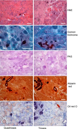
The most prominent features of the histopathologic examination were multifocal metastatic calcifications affecting multiple tissues, and multifocal degenerative myopathy (Fig 2). Several sections of skeletal muscle had multifocal myofiber atrophy, degeneration, and calcification with interstitial fibroplasia. Foci of ische-mic necrosis affecting whole muscle fascicles occasion-ally were noted (eg, triceps brachii), demonstrating swelling and hypereosinophilia of affected myofibers. Multinucleated regenerative myofibers were inter-spersed with atrophic, degenerate, and calcified fibers. Lakes of calcified debris forming nodules at the lingual margins were present, as was multifocal gastric mucosal calcification. Radial cortical calcification, centered on tubular basement membranes, was demonstrated in the kidneys, providing an explanation for the granular casts. Multifocal basement membrane calcification of small airways and blood vessels also was observed, and the adrenal glands had focal calcification. Representa-tive sections of the brain (cerebrum and medulla oblon-gata), spinal cord (with spinal nerve and dorsal root ganglion), and pituitary gland were microscopically normal, whereas parathyroid tissue was atrophic.
In light of the multiple muscle fiber abnormalities and markedly increased serum CK activity, a form of muscular dystrophy was considered as a differential diagnosis in this cat once a number of more common granulomatous diseases had been eliminated, but the Fig 1. Firm white lingual plaques observed at the time of
hypercalcemia was considered unusual for a primary muscular dystrophy. Samples of quadriceps and triceps muscles were obtained immediately postmortem and snap-frozen in isopentane, cooled in liquid nitrogen. An extended staining panel conducted on 8 lm cryosections included hematoxylin and eosin, periodic
and large areas of infiltrating mononuclear cells (probably macrophages). The Alizarin red stain con-firmed the presence of prominent accumulations of calcium in sarcoplasmic and extracellular regions, and there also was a marked accumulation of calcium deposits in the smooth muscle of blood vessels. Oil red O stain identified fine punctate lipid accumulation in many fibers.
Extensive diagnostic evaluation failed to disclose a definitive diagnosis for this cat. Neither of the described muscular dystrophies was considered to fit the clinical or histopathologic picture. The cat was female, hence X-linked dystrophin deficiency would be highly unlikely,2and the cat did not display the typical extensor contracture of the pelvic limbs or lipid accu-mulation in the muscle fibers seen with laminin a2 (merosin) deficiency,2 nor the ventroflexed neck and dorsally protruding scapulae described in a -dystrogly-can deficiency; moreover, the latter cases had normal CK activity.3 Of the muscular dystrophies described in companion animals, b-sarcoglycan deficiency initially was considered a possibility in this cat. This condition has been associated with hypercalcemia in a domestic shorthair cat and a Boston Terrier, associated in both cases with markedly increased serum CK activity, but these patients had mild total hypercalcemia attributed to young age.4,5In light of the absence of muscle fiber hypertrophy, the marked hypercalcemia, and the wide-spread calcium deposition in other organs and blood vessels in our patient, we considered the dystrophic features of the muscle fibers likely to be an effect rather than a primary cause of the disease.
No underlying cause for this patient’s hypercalcemia was identified, rendering idiopathic hypercalcemia our presumptive diagnosis. Although serum PTHrP concen-tration was mildly increased, no evidence of neoplasia was found on postmortem examination. Despite the detectable plasma PTH concentration, primary hyper-parathyroidism was ruled out on the basis of the atro-phic parathyroid glands. Furthermore, this is a rare condition in cats, usually presenting in older animals and associated with a low-normal to decreased serum phosphate concentration.6 Although increased concen-trations of plasma ionized calcium, PTH, and phos-phate would be expected in so-called ‘tertiary hyperparathyroidism’, this condition has only been described infrequently in cats with advanced renal dis-ease (IRIS stages 3–4), after a period of renal secondary hyperparathyroidism.7This was not consistent with the mild, transient azotemia in this patient, and would have resulted in hyperplasia of the parathyroid glands.
Although idiopathic hypercalcemia is relatively com-mon in cats, reportedly accounting for an increasing proportion of hypercalcemia in this species,8 severe metastatic calcification has hitherto not been described in these patients. Serum phosphate concentration was above the RI in this patient, resulting in a markedly increased Ca9P product. Young animals normally have higher calcium or phosphate concentrations or both than adults and are thought to be more resistant to tissue mineralization.9 However, the magnitude of
the electrolyte derangements in this patient accounted for the severe, widespread mineralization.
Ca 9P product. The potentially deleterious conse-quences of an increased Ca9P product emphasize the importance of monitoring the Ca 9P product in hypercalcemic patients, and suggest that every effort should be made to decrease it (and thereby probably the risk of mineralization) as soon as possible.
This report describes, to our knowledge, the first documentation of severe systemic calciphylaxis with hypercalcemia in a cat. On the basis of the clinical pre-sentation and thorough ante- and postmortem investi-gations, we conclude that the markedly increased Ca 9P product in this patient accounted for the min-eralization, with the extensive muscle fiber calcification explaining the markedly increased serum CK activity. We consider the patient’s decreased mentation to be a result of the systemic effects of the severe tissue miner-alization, which did not resolve within a matter of days15,22 despite transient normocalcemia. Pain is one of the initial signs of calciphylaxis in humans,23 and the cat may have responded to painful sensation with decreased mentation, although the cat did not appear painful when handled. The hypercalcemia itself also may have contributed to this patient’s decreased men-tation. Human patients with hypercalcemia related to primary hyperparathyroidism can present with head-aches, fatigue, nausea, and mental disturbances, including coma.24These signs have been found to cor-relate with the degree of hypercalcemia rather than plasma PTH concentration, with clinical improvement corresponding to decreasing calcium concentration despite persistently high PTH concentration.24 The lack of clinical improvement in our patient at the time of normocalcemia, however, does not support hyper-calcemia as the sole cause of obtundation. Hyperam-monemia contributing to the patient’s decreased mentation was considered unlikely, but could not be eliminated. Because mammalian muscle produces ammonia,25 muscle necrosis may have caused the hyperammonemia in this patient.
Footnotes
aFurosemide monoethanolamine (Dimazon, injectable, 5% w/v
Furosemide monoethanolamine; per 1 mL: 50 mg Furosemide monoethanolamine, 15 mg benzyl alcohol, 1 mg Disodium Ede-tate Dihydrate, and 1.8 mg sodium Sulphite Anhydrous), Inter-vet UK Ltd, Walton Manor, Milton Keynes, Bucks, MK7 7AJ, UK
b
Calcitonin Salmon (Miacalcic, injectable, 100 IU/mL), Sandoz Pharmaceuticals, Frimley Business Park, Camberley, Surrey, GU16 7SR, UK
c
Pamidronate disodium pentahydrate (Aredia, 15 mg/5 mL), Ciba Laboratories, Frimley Business Park, Camberley, Surrey, GU16 7SR, UK
d
Prednisolone (Prednicare tablets, 1 mg), Animal Care ltd, York, YO19 5RU, UK
eEnzyme-linked immunosorbent assay, IDS limited, Cambridge
Specialist Laboratories, UK. Canine PTH assay validated for felines by parallelism (unpublished results, personal communica-tion)
f
Radioimmunoassay, Beckman Coulter, Cambridge Specialist Laboratories, UK. Human PTHrP assay validated for felines by the laboratory (unpublished results, personal communication)
Acknowledgment
Conflict of Interest Declaration: The authors disclose no conflict of interest.
References
1. Stern JA, Chew DJ, Schissler JR, Green EM. Cutaneous and systemic blastomycosis, hypercalcemia, and excess synthesis of calcitriol in a domestic shorthair cat. J Am Anim Hosp Assoc 2011;47:e116–e120.
2. Shelton GD, Engvall E. Muscular dystrophies and other inherited myopathies. Vet Clin North Am Small Anim Pract 2002;32:103–124.
3. Martin PT, Shelton GD, Dickinson PJ, et al. Muscular dystrophy associated with alpha-dystroglycan deficiency in Sph-ynx and Devon Rex cats. Neuromuscul Disord 2008;18:942– 952.
4. Deitz K, Morrison JA, Kline K, et al. Sarcoglycan-deficient muscular dystrophy in a Boston Terrier. J Vet Intern Med 2008;22:476–480.
5. Salvadori C, Vattemi G, Lombardo R, et al. Muscular dys-trophy with reduced beta-sarcoglycan in a cat. J Comp Pathol 2009;140:278–282.
6. Feldman EC. Disorders of the parathyroid glands. In: Ett-inger SJ, Feldman EC, eds. Textbook of Veterinary Internal Medicine, vol 2. 7th ed. St. Louis, MO: Saunders Elsevier; 2010:1743–1744.
7. Kruger JM, Osborne CA. Calcium disorders. In: Bartges J, Polzin DJ, eds. Nephrology and Urology of Small Animals, 1st ed. West Sussex: Wiley-Blackwell; 2011:642–656.
8. Midkiff AM, Chew DJ, Randolph JF, et al. Idiopathic hypercalcemia in cats. J Vet Intern Med 2000;14:619–626.
9. Stockham SL, Scott MA. Fundamentals of Veterinary Clin-ical Pathology, 2nd ed. Oxford: Blackwell Publishing; 2008.
10. Selye H, Gentile G, Jean P. An experimental model of “dermatomyositis” induced by calciphylaxis. Can Med Assoc J 1961;85:770–776.
11. Nigwekar SU, Wolf M, Sterns RH, Hix JK. Calciphylaxis from nonuremic causes: A systematic review. Clin J Am Soc Nephrol 2008;3:1139–1143.
12. Budisavljevic MN, Cheek D, Ploth DW. Calciphylaxis in chronic renal failure. J Am Soc Nephrol 1996;7:978–982.
13. Morita T, Awakura T, Shimada A, et al. Vitamin D toxi-cosis in cats: Natural outbreak and experimental study. J Vet Med Sci 1995;57:831–837.
14. Hilbe M, Sydler T, Fischer L, Naegeli H. Metastatic calci-fication in a dog attributable to ingestion of a tacalcitol oint-ment. Vet Pathol 2000;37:490–492.
15. Nakamura Y, Gotoh M, Fukuo Y, et al. Severe calcifica-tion of mucocutaneous and gastrointestinal tissues induced by high dose administration of vitamin D in a puppy. J Vet Med Sci 2004;66:1133–1135.
16. Declercq J, Bhatti S. Calcinosis involving multiple paws in a cat with chronic renal failure and in a cat with hyperthyroid-ism. Vet Dermatol 2005;16:74–78.
18. Yabuzoe A, Yokoi S, Sekiguchi M, et al. Fibrodysplasia ossificans progressiva in a Maine Coon cat with prominent ossifi-cation in dorsal muscle. J Vet Med Sci 2009;71:1649–1652.
19. Shelton GD, Engvall E. Canine and feline models of human inherited muscle diseases. Neuromuscul Disord 2005;15:127–138.
20. Tan JY, Valberg SJ, Sebastian MM, et al. Suspected sys-temic calcinosis and calciphylaxis in 5 horses. Can Vet J 2010;51:993–999.
21. Thom N, Er E, Reinacher M. Nonuraemic nonfatal idio-pathic calciphylaxis in a kitten. Vet Dermatol 2013;24:547–e131.
22. Demetriou ET, Pietras SM, Holick MF. Hypercalcemia and soft tissue calcification owing to sarcoidosis: The sunlight-cola connection. J Bone Miner Res 2010;25:1695–1699.
23. Brandenburg VM, Cozzolino M, Ketteler M. Calciphylax-is: A still unmet challenge. J Nephrol 2011;24:142–148.
24. Petersen P. The psychiatry of primary hyperparathyroid-ism. A contribution to the psychopathology of disorders of cal-cium metabolism. Monogr Gesamtgeb Neurol Psychiatr 1967;120:1–86.