저작자표시-비영리-변경금지 2.0 대한민국 이용자는 아래의 조건을 따르는 경우에 한하여 자유롭게
l 이 저작물을 복제, 배포, 전송, 전시, 공연 및 방송할 수 있습니다. 다음과 같은 조건을 따라야 합니다:
l 귀하는, 이 저작물의 재이용이나 배포의 경우, 이 저작물에 적용된 이용허락조건 을 명확하게 나타내어야 합니다.
l 저작권자로부터 별도의 허가를 받으면 이러한 조건들은 적용되지 않습니다.
저작권법에 따른 이용자의 권리는 위의 내용에 의하여 영향을 받지 않습니다. 이것은 이용허락규약(Legal Code)을 이해하기 쉽게 요약한 것입니다.
Disclaimer
저작자표시. 귀하는 원저작자를 표시하여야 합니다.
비영리. 귀하는 이 저작물을 영리 목적으로 이용할 수 없습니다.
理學博士學位論文
분열성
효모에서
철
흡수
억제
전사인자
Fep1
의
특성
분석
Characterization of iron uptake repressor Fep1
in Schizosaccharomyces pombe
2017
年
2
月
서울大學校
大學院
生命科學部
Characterization of iron uptake repressor
Fep1
in Schizosaccharomyces pombe
By
Hyo-Jin Kim
under the supervision of
Professor Jung-Hye Roe, Ph.D.
A Thesis Submitted in Partial Fulfillment
of the Requirements for the Degree of
Doctor of Philosophy
February, 2017
School of Biological Sciences
Graduate School
i
ABSTRACT
Iron is an important cofactor for a wide variety of proteins involved in the major life processes, such as respiration and tricarboxylic acid cycle, DNA replication and repair, and nitrogen fixation. The essential redox-active property of iron that enables facile switch between ferric (Fe3+) and ferrous iron (Fe2+) also renders
toxicity via generating reactive oxygen species. Therefore, most organisms are equipped with regulated mechanisms to maintain optimal intracellular levels of iron.
In the fission yeast Schizosaccharomyces pombe, two repressors Php4 and Fep1 regulate iron-dependent expression of genes for iron usage/storage and acquisition, respectively. The iron-sensing depends on the CGFS-type monothiol glutaredoxin Grx4 that binds Fe-S cluster in a homodimer or in a heterodimer with a BolA-type protein Fra2. Under iron-rich condition, Grx4 binding to Php4 causes its cytosolic sequestration, resulting in the induction of iron usage/storage genes. Grx4 also binds Fep1 regardless of iron availability. Under iron-starved condition, Grx4 and Fra2 inhibit Fep1 repressor activity, resulting in derepression of iron acquisition genes.
Fep1, a GATA-family transcription factor, binds to the promoter regions of genes for iron acquisition, such as reductive iron import (fio1+, frp1+), siderophore transport (str1+, str2+, str3+), and vacuolar transport (abc3+), to avoid iron overload under iron-rich conditions. The N-terminal DNA-binding domain of Fep1 contains four conserved cysteines located between the two zinc finger motifs. It has been demonstrated that the N-terminal 241 aa residues of Fep1 can bind to the target promoters in vivo under iron-replete condition. Through this N-terminal
ii
domain, Fep1 is known to interact with the monothiol glutaredoxin domain of Grx4 under iron-starved condition. Under iron-starved condition, Fep1 is released from binding to its target sites, inducing iron uptake genes. Fep1 orthologs are well conserved, especially in the DNA binding domain, across filamentous fungi, such as Ustilago maydis (Urbs1), Neurospora crassa (SRE), Histoplasma capsulatum (Sre1), Aspergillus spp. (SREA), Candida albicans (Sfu1), and Cryptococcus neoformans (Cir1).
Even though the function and interaction partners of Fep1 have been elucidated extensively in S. pombe, the molecular basis by which Fep1 is inactivated under iron starvation remains unknown. In this study, to elucidate the mechanism behind iron-sensing by Fep1, I pursued biochemical and spectroscopic analyses of Fep1, in full length and truncated forms, as isolated or reconstituted proteins, with the wild type or substituted cysteine mutations. Evidences are presented that Fep1 binds iron, in the form of Fe-S cluster. Spectroscopic and biochemical analyses of as isolated and reconstituted Fep1 suggest that the dimeric Fep1 binds Fe-S clusters. Iron to acid-labile sulfide stoichiometry of purified Fep1-N238 was roughly 1 : 1 in both before and after chemical reconstitution. Furthermore, the reconstitution of the His-tagged Fep1-N238 requires not only iron donor but also sulfide donor, indicating that Fep1 binds [Fe-S] clusters. The mutation study revealed that the cluster-binding depended on the conserved cysteines located between the two zinc fingers in the DNA binding domain. EPR analyses revealed [Fe-S]-specific peaks indicative of mixed presence of [2Fe-2S], [3Fe-4S], or [4Fe-4S]. Overall, the finding that Fep1 is an Fe-S protein fits nicely with the model that the Fe-S-trafficking Grx4 senses intracellular iron environment and modulates the activity of Fep1.
Key Words
iii
CONTENTS
ABSTRACT ... IIII
CONTENTS ... III
LIST OF FIGURES ... VI
LIST OF TABLES ... IX
LIST OF ABBREVIATIONS ... X
CHAPTER I. INTRODUCTION ... 1
I.1. Iron homeostasis and disease ... 2
I.2. Regulation of iron homeostasis in Schizosaccaromyces pombe 3
I.2.1. Fep1, the iron-regulatory GATA-type repressor ... 3
I.2.2. Php4, a key regulator for iron economy ... 6
I.2.3. Roles for Grx4 in iron homeostasis in S. pombe ... 8
I.3. CGFS monothiol glutaredoxins
... 10
I.3.1. Mitochondrial monodomain Grxs in the maturation of Fe-S
protein ... 13
I.3.2. Nucleo-cytosolic multidomain Grxs in iron metabolism ... 14
I.4. CGFS monothiol glutaredoxins and BolA proteins
... 18
iv
I.4.2. Roles of Grx3/4 and Fra2 in iron homeostasis in S. cerevisiae ... 20
I.4.3. Role of Grx4 and Fra2 in iron homeostasis in S. pombe ... 23
I.5. Biology of Schizosaccharomyces pombe
... 24
I.5.1. The early research and phylogeny of S. pombe ... 24
I.5.2. Life cycle of S. pombe ... 25
I.5.3. Genomic information of S. pombe ... 27
I.6. Aims of this study
... 30
CHAPTER II. MATERIALS AND METHODS ... 32
II.1. Strains and plasmids construction ... 33
II.2. Protein expression and purification ... 33
II.3. Transformation of Escherichia coli and Yeast ... 33
II.4. Western blot analysis ... 34
II.5. Preparation of apoprotein ... 34
II.6. Fe-S cluster reconstitution in vitro ... 35
II.7. Assays for quantification of iron, sulfide and protein ... 35
II.7.1. Determination of iron concentration ... 35
II.7.2. Determination of sulfide concentration ... 36
II.7.3. Determination of protein concentration ... 36
v
CHAPTER III. RESULTS & DISCUSSION ... 38
III. 1. Purification and UV-visible absorption spectroscopy of
full-length Fep1
... 39
III. 1. 1. Purification of full-length Fep1... 39
III. 1.2. UV-visible absorption spectroscopy of full-length Fep1 ... 43
III.2. Fep1-N238 binds an Fe-S cluster via four conserved cysteine
residues
... 44
III.2.1. Purification and UV-visible absorption spectroscopy of
Fep1-N238 ... 44
III. 2.2. Purification and UV-visible absorption spectroscopy of Fep1
cysteine mutants... 53
III. 2.3. Both iron and sulfide are required to reconstitute iron-bound
Fep1 ... 56
III. 2.4. Iron and acid labile sulfide content of purified Fep1-N238 ... 56
III. 3. EPR analyses of Fep1-N238
... 62
CHAPTER IV. CONCLUSION ... 67
REFERENCES ... 72
vi
LIST OF FIGURES
FIG. I-1. MODELS FOR THE REGULATORY ACTION OF
IRON-RESPONSIVE TRANSCRIPTION FACTORS IN RESPONSE TO LEVELS OF IRON. ... 5 FIG. I-2. DOMAIN COMPARISONS OF GATA-TYPE IRON REGULATORY
TRANSCRIPTION FACTORS. ... 7 FIG. I-3. A SCHEMATIC MODEL OF IRON-DEPENDENT REGULATION OF FEP1 REPRESSOR ACTIVITY. ... 9 FIG. I-4. DOMAIN STRUCTURE OF CGFS MONOTHIOL GRXS FROM E.
COLI, S. CEREVISIAE, S. POMBE, AND H. SAPIENS. ... 11
FIG I-5. INTRACELLULAR IRON TRAFFICKING AND THE REGULATION OF CELLULAR IRON UPTAKE IN EUKARYOTES... 17 FIG I-6. MODELS FOR [2FE-2S]2+ GRX HOMODIMERS AND [2FE-2S]2+
GRX-BOLA HETEROCOMPLEXES CHARACTERIZED FROM E. COLI, S.
CEREVISIAE, AND H. SAPIENS. ... 19
FIG I-7. PROPOSED MODEL FOR IRON REGULATION VIA AFT1 AND AFT2 UNDER IRON REPLETE CONDITIONS ... 21 FIG. I-8. LIFE CYCLE OF FISSION YEAST S. POMBE ... 28 FIG II-1. DOMAIN STRUCTURE OF FEP1 IN S. POMBE ... 40
vii
FIG II-2. PURIFICATION OF FULL-LENGTH HIS-TAGGED FEP1. ... 42 FIG II-3. EFFECT OF OXYGEN ON STABILITY OF FE BINDING ON
FULL-LENGTH FEP1 ... 45 FIG II-4. UV-VISIBLE ABSORPTION SPECTROSCOPY OF REDUCED
LENGTH FEP1 ... 46 FIG II-5. UV-VISIBLE ABSORPTION SPECTROSCOPY OF
RECONSTITUTED FULL-LENGTH FEP1 ... 46 FIG II-6. SCHEMATIC PRESENTATIONS OF THE WILD-TYPE AND THE
CYSTEINE MUTANT FEP1 USED IN THIS STUDY ... 48 FIG II-7. REDDISH BROWN COLOR OF FEP1-OVEREXPRODUCED CELL 49 FIG II-8. SDS-PAGE ANALYSIS OF TRUNCATED VARIANT FEP1
PROTEINS ... 50 FIG II-9. UV-VISIBLE ABSORPTION SPECTRUM OF FEP1-N238 & OXYGEN
STABILITY ... 51 FIG II-10. UV-VISIBLE ABSORPTION SPECTRUM OF REDUCED &
RECONSTITUTED FEP1-N238 ... 52 FIG II-11. UV-VISIBLE ABSORPTION SPECTRUM OF AEROBICALLY
PURIFIED FEP1-N238 & FEP1-N238 4CS (C70S, C76S, C85S, C88S) 54 FIG II-12. UV-VISIBLE ABSORPTION SPECTRUM OF TRUNCATED FEP1 &
viii
TRUNCATED FEP1 2CS (C85S, C88S) ... 56 FIG II-13. IRON AND SULFIDE REQUIREMENT TO RECONSTITUTE
IRON-BOUND FEP1-N238. ... 59 FIG II-14. IRON AND SULFIDE REQUIREMENT TO RECONSTITUTE
IRON-BOUND FULL-LENGTH FEP1. ... 60 FIG II-15. MOST IMPORTANT TYPES OF FE-S CLUSTERSENCOUNTERED
IN NATURE AND THEIR CORRESPONDING ELECTRONIC PROPERTIES. ... 65 FIG II-16. EPR SPECTRA OF AS-ISOLATED FEP1-N238 ANALYZED AT 123
K... 66 FIG II-17. EPR SPECTRA OF AS-ISOLATED FEP1-N238 ANALYZED AT 5 K
... 67 FIG II-18. BIFC ANALYSIS OF GRX4 WITH BOLA PROTEINS ... 68 FIG II-19. A PROPOSED MODEL OF IRON-DEPENDENT REGULATION OF
FEP! REPRESSOR ACTIVITY………...71 FIG II-20. A PROPOSED MODEL OF IRON-DEPENDENT REGULATION OF FEP! REPRESSOR ACTIVITY………...72
ix
LIST OF TABLES
TABLE II-1. IRON AND ACID LABILE SULFIDE CONTENT OF
PURIFIED FEP1-N238 ... 61
TABLE II-2. MOLECULAR MASS ANALYSIS OF FEP1 ... 62
x
LIST OF ABBREVIATIONS
EPR Electron Paramagnetic Resonance
BiFC
Bimolecular flouorescence complementation
DTT
dithiothreitol
DNA
deoxyribocunleic acid
SDS
sodium dodecyl sulfate
EDTA
ethylenediamine tetraacetate
EtBr
ethidium bromide
GFP
green fluorescence protein
IPTG
isopropyl-
-D-thiogalactopyranoside
Kb
kilo base pair
kDa
kilo Dalton
OD
optical density
ORF
open reading frame
PAGE
polyacrylamide gel electrophoresis
PCR
polymerase chain reaction
WT
wild-type
Grx glutaredoxin
Trx thioredoxin
1
CHAPTER I.
INTRODUCTION
2
I.1. Iron homeostasis and disease
Iron is an important cofactor for a wide variety of proteins involved in the major life processes, such as respiration, tricarboxylic acid cycle, DNA replication and repair, and nitrogen fixation (Kaplan & Ward, 2013). In high concentrations, however, the essential redox-active property of iron that enables facile switch between ferric (Fe3+) and ferrous iron (Fe2+) also renders toxicity via generating
reactive oxygen species. Therefore, most organisms are equipped with regulated mechanisms to maintain optimal intracellular levels of iron. Because of the low bioavailability of iron, iron deficiency is the most common and widespread nutritional disorder in the world (Zimmermann & Hurrell, 2007). At the other extreme, iron overload diseases are common systemic iron disorders primarily caused by mutations in proteins that sense, regulate, or mediate iron absorption from the gastrointestinal tract (Fleming & Ponka, 2012, Nairz & Weiss, 2006). Increased iron absorption and storage leads to excessive iron accumulation in various organs (mainly liver, heart, and pancreas) causing progressive organ damage and increased mortality as a consequence of elevated oxidative stress. In addition to systemic iron disorders, numerous human diseases have also been linked with iron dysregulation at the cellular level. For example, specific defects in Fe-S cluster biogenesis factors lead to Friedreich’s ataxia, X-linked sideroblastic anemia, sideroblastic-like microcytic anemia, and myopathy (Sheftel, Stehling et al., 2010, Ye & Rouault, 2010).
The fungi Saccharomyces cerevisiae and Schizosaccharomyces pombe have proven to be effective models for studying eukaryotic iron homeostasis at the cellular level. Despite their relative simplicity, biochemical and genetic studies in yeast have been critical for identifying proteins required for iron uptake, intracellular iron transport and mobilization, and heme and Fe-S cluster biogenesis in higher eukaryotes (Askwith & Kaplan, 1998, De Freitas, Wintz et al., 2003, Lill &
3
Muhlenhoff, 2008, Wingert, Galloway et al., 2005). Furthermore, genome-wide studies in yeast have revealed how eukaryotic cells adapt to both iron deficiency (Philpott, Leidgens et al., 2012) and iron overload (Jo, Loguinov et al., 2008, Lin, Li et al., 2011). In addition, yeast studies have been pivotal in defining the pathophysiology of human diseases of iron metabolism, such as Friedreich’s ataxia and aceruloplasminemia (Bleackley & Macgillivray, 2011, Lodi, Tonon et al., 2006, Pandolfo & Pastore, 2009, Rouault & Tong, 2008).
I.2.
Regulation
of
iron
homeostasis
in
Schizosaccaromyces pombe
I.2.1. Fep1, the iron-regulatory GATA-type repressor
In the fission yeast S. pombe, two repressors Php4 and Fep1 regulate iron-dependent expression of genes for iron usage/storage and acquisition, respectively (Brault, Mourer et al., 2015, Labbe, Khan et al., 2013, Mercier, Pelletier et al., 2006, Pelletier, Beaudoin et al., 2003). Fep1, a GATA-family transcription factor, binds to the promoter regions of genes for iron acquisition, such as reductive iron import (fio1+, frp1+), siderophore transport (str1+, str2+, str3+), and vacuolar transport (abc3+), to avoid iron overload under iron-rich conditions (Fig I-1). The N-terminal DNA-binding domain of Fep1 contains four conserved cysteines located between the two zinc finger motifs (Brault et al., 2015, Labbe et al., 2013). It has been demonstrated that the N-terminal 241 aa residues of Fep1 can bind to the target promoters in vivo under iron-replete condition (Jbel, Mercier et al., 2009). Through this N-terminal domain, Fep1 is known to interact with the monothiol glutaredoxin domain of Grx4 under iron-starved condition (Jbel, Mercier et al., 2011). Repression by DNA-bound Fep1 requires recruitment and binding of the co-repressors Tup11 and Tup12 in an iron-independent manner, and deletion mapping studies have delineated the Tup11 interaction domain in the
4
Fig I-1. Models for the regulatory action of iron-responsive transcription
factors in response to levels of iron (Brault, A et al., 2015)
A. In S. pombe, Fep1 interacts with GATA elements in promoters of php4+ and iron
transport genes. Fep1 represses target gene transcription with the help of the corepressor Tup11 or Tup12. In this context, Fra2 may participate in the dissociation of the GRX domain (of Grx4) from the N-terminal portion of Fep1 by forming a [2Fe-2S] (red circle) Fra2-GRX domain heterodimer, allowing the N-terminus of Fep1 to be free and available for binding to chromatin and repressing transcription. Under conditions of iron excess (Fe+), Grx4 acquires an [2Fe-2S] cluster that would in turn trigger the interaction between the GRX domain (of Grx4) and the C-terminal region of Php4. This interaction between the GRX domain and Php4 would disrupt the Php4/Php2/Php3/Php5 heteromeric complex, leading to Php4 release and inactivation. The Grx4-mediated inactivation of Php4 would lead to its recruitment by the exportin Crm1 and then to its subsequent export from the nucleus to the cytoplasm. The absence of Php4 enables fep1+ and iron utilization
genes to be expressed through the CCAAT-binding core complex that is composed of Php2, Php3, and Php5.
B. Grx4 and Fra2 are inhibitory partners of Fep1, blocking its association with
chromatin and preventing its repressive effect on target gene expression. In the absence of iron (Fe-), Php4 forms a complex with Php2, Php3, Php5, and Grx4. Because only the TRX domain of Grx4 interacts with Php4, this leaves the repression domain of Php4 available to repress several genes, including fep1+ and
several others encoding iron-using proteins. Php4 nuclear import is mediated by distinct karyopherins, namely Imp1, Cut15, and Sal3.
6
C-terminus of Fep1 (Fig I-2) (Znaidi, Pelletier et al., 2004). The C-terminus of Fep1 also contains a dimerization domain that is required for efficient repression of Fep1 target genes (Pelletier, Trott et al., 2005). Under iron-starved condition, Fep1 is released from binding to its target sites, inducing iron uptake genes (Jbel et al., 2009). Fep1 orthologs are well conserved(Fig I-2), especially in the DNA binding domain, across filamentous fungi, such as Ustilago maydis (Urbs1) (An, Mei et al., 1997), Neurospora crassa (SRE) (Zhou, Haas et al., 1998), Histoplasma capsulatum (Sre1) (Chao, Marletta et al., 2008), Aspergillus spp. (SREA) (Haas, Zadra et al., 1999, Schrettl, Kim et al., 2008), Candida albicans (Sfu1)(Lan, Rodarte et al., 2004), and Cryptococcus neoformans (Cir1) (Jung, Sham et al., 2006).
I.2.2. Php4, a key regulator for iron economy
The transcriptional repressor Php4 also controls iron homeostasis in S. pombe by regulating expression of genes involved in iron-dependent metabolic pathways. Php4 binds to a heterotrimeric protein complex composed of Php2, Php3, and Php5. Under iron replete conditions, Php4 is not expressed and the Php2/Php3/Php5 complex activates expression of its target genes by binding to CCAAT sequences in their promoters (Fig I-1A). Php2/Php3/Php5-regulated genes encode proteins involved in iron-dependent metabolic pathways such as iron-sulfur cluster biogenesis, heme biosynthesis, the mitochondrial electron transport chain, and the tricarboxylic acid cycle (Mercier et al., 2006). Under low iron conditions, Php4 is expressed and binds to the Php2/Php3/Php5 complex, causing it to switch from an activator to a repressor (Fig I-1B). Thus, iron-utilizing pathways are downregulated as an iron sparing response to lower bioavailable iron.
Php4 itself is regulated at the transcriptional level by Fep1 since the php4+ gene contains GATA elements within its promoter (Mercier et al., 2006). When iron
7
Fig I-2. Domain comparisons of GATA-type iron regulatory transcription
factors (Brault, A et al., 2015)
.ZF1 and ZF2 (red) are the N-terminal and C-terminal GATA-type zinc finger motifs, respectively. Four highly conserved Cys residues, denoted Cys-rich region (blue). A conserved coiled-coil region (black). Putative NLSs are indicated in green. Regions of Fep1 that interact with the GRX and TRX domains of Grx4 as well as with the corepressor Tup11 are indicated within brackets.
8
levels are high, Fep1 binds to the promoter of php4+ and represses the transcription of php4+ (Fig I-1A). When iron levels are low, Fep1 is unable to inhibit php4+ transcription and Php4 is subsequently expressed, allowing it to repress iron-utilizing pathways via the Php2/Php3/Php5 complex (Fig I-1B). A genome-wide DNA microarray study also revealed that the fep1+ gene contains CCAAT cis-acting elements in its promoter region and is downregulated in a Php4-dependent manner in response to iron deprivation (Fig I-1B) (Mercier, Watt et al., 2008). This reciprocal regulatory loop between two iron-responsive repressors through mutual control of each other’s expression allows direct crosstalk between iron acquisition and iron utilization pathways to fine tune iron homeostasis in S. pombe.
I.2.3. Roles for Grx4 in iron homeostasis in S. pombe
The activities of both Fep1 and Php4 are controlled at the post-translational level by S. pombe Grx4, a member of the multidomain CGFS Grx subfamily. Grx4 is required to regulate the activity of both Fep1 and Php4 through specific protein-protein interactions.
Fep1 is regulated through Grx4 at the post-translational level (Jbel et al., 2011, Kim, Kim et al., 2011). Grx4 was shown to constitutively interact with Fep1; however, the Fep1-Grx4 complex resides in the nucleus regardless of iron status inside the cell. Under iron-starved condition, Grx4 and Fra2 inhibit Fep1 repressor activity, resulting in derepression of iron acquisition genes. When iron is abundant, Grx4 is unable to inhibit Fep1 repressor function, although it still physically interacts with Fep1 (Fig I-1A) (Jbel et al., 2011, Kim et al., 2011).
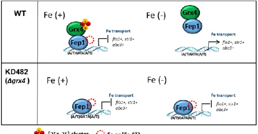
Deletion of grx4 leads to constitutive repression of both Php4- and Fep1-regulated genes and constitutive nuclear localization of Php4 (Fig I-3) (Jbel et al., 2011, Kim et al., 2011, Mercier & Labbe, 2009). Yeast two-hybrid and bimolecular
9
Fig I-3. A schematic model of iron-dependent regulation of Fep1
repressor activity.
[Fe-S]-binding Grx4-Fra2 has been reported to bind Fep1. In the presence of sufficient iron, Fep1 represses genes for iron acquisition. Under iron starvation, Fep1 repressor activity is inhibited by Grx4 and Fra2, de-repressing iron acquisition genes. Whether iron binds to Fep1 as a mononuclear form or as an Fe-S cluster is under question.
10
fluorescence complementation experiments established that Grx4 physically interacts with Php4 regardless of cellular iron levels. However, under iron-replete conditions Grx4 promotes Php4 export to the cytosol by facilitating direct interaction with the nuclear exportin Crm1 (Fig I-1A).
I.3. CGFS monothiol glutaredoxins
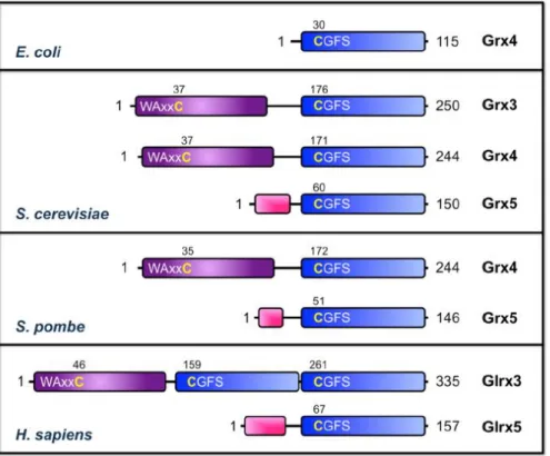
Glutaredoxins (Grxs) were initially identified as members of the thioredoxin (Trx)-fold family that catalyze thiol-disulfide exchange reactions in a glutathione (GSH)-dependent manner via a conserved CPY/FC active site (Lillig, Berndt et al., 2008). Classical dithiol Grxs that utilize a dithiol mechanism to reduce intramolecular disulfide bonds require both cysteines for catalytic activity, while Grxs that catalyze glutathionylation/deglutathionylation reactions via a monothiol mechanism require only the N-terminal active site Cys (Fernandes & Holmgren, 2004). With the increasing number of sequenced genomes, several Grxs with divergent active site sequences have been identified, thus requiring an updated phylogenetic classification for the Grx family (Couturier, Jacquot et al., 2009). The most widespread Grxs present in both prokaryotes and eukaryotes are grouped in Class I, which includes the classical dithiol Grxs, and in Class II, which are defined as monothiol Grxs with a conserved CGFS active site. CGFS-type monothiol Grxs can be further classified into two groups: single domain CGFS Grxs and multidomain CGFS Grxs with an N-terminal Trx-like domain and one or more Grx-like domains (Fig I-4).
Unlike Class I Grxs, CGFS Grxs have little or no thiol-disulfide oxidoreductase activity when tested with standard Grx model substrates (Fernandes, Fladvad et al., 2005, Ken, Chen et al., 2011, Mesecke, Mittler et al., 2008, Tamarit, Belli et al., 2003, Zaffagnini, Michelet et al., 2008). However, the CGFS active site is required
11
Fig I-4. Domain structure of CGFS monothiol Grxs from E. coli, S.
cerevisiae, S. pombe, and H. sapiens (Li, H et al., 2012)
The Trx-like domains and Grx-like domains are shown as purple and blue boxes, respectively. The conserved cysteines in the active sites of the Trx and Grx domains are numbered and shown in yellow. Predicted or known mitochondrial targeting signals are shown as pink boxes.
12
for a different purpose: coordination of a [2Fe-2S] cluster. Both single and multidomain CGFS Grxs form [2Fe-2S]2+-bridged homodimers with all-cysteinyl
ligation provided by the two CGFS active sites and two GSH molecules (Bandyopadhyay, Gama et al., 2008, Haunhorst, Berndt et al., 2010, Iwema, Picciocchi et al., 2009, Li, Mapolelo et al., 2009, Picciocchi, Saguez et al., 2007, Ye, Jeong et al., 2010, Yeung, Gold et al., 2011). Formation of this Fe-S complex is supported by studies in S. cerevisiae demonstrating that iron binding to CGFS Grxs in vivo requires the CGFS motif, sufficient cellular GSH levels, and the mitochondrial Fe-S assembly machinery (Muhlenhoff, Molik et al., 2010). The first crystal structure of a [2Fe-2S]-bridged CGFS Grx homodimer was published for E. coli Grx4 in 2009, confirming that two GSH molecules are covalently linked to the cluster, but held in place by non-covalent interactions with the GSH binding pocket of each Grx4 monomer (Iwema et al., 2009). There is currently no published crystal structure available for a [2Fe-2S]-bound multidomain CGFS Grx, thus the orientation of the Trx-like domain in relation to the [2Fe-2S]-bridged Grx-like domain(s) is unknown.
In eukaryotes, single domain CGFS Grxs (e.g., yeast Grx5 and human Glrx5, see Fig I-4) are localized to mitochondria or chloroplasts and have been implicated in the maturation of Fe-S cluster proteins (Herrero, Belli et al., 2010, Rouhier, Couturier et al., 2010, Ye & Rouault, 2010). In contrast, multidomain CGFS Grxs (e.g., yeast Grx3/4 and human Glrx3) display cytosolic/nuclear localization where they are proposed to play dual roles in cytosolic iron trafficking and iron regulation (Jbel et al., 2011, Kumanovics, Chen et al., 2008, Muhlenhoff et al., 2010, Ojeda, Keller et al., 2006, Pujol-Carrion, Belli et al., 2006). The Fe-S biogenesis function of single domain CGFS Grxs as well as the trafficking and regulatory functions of multidomain CGFS Grxs in yeast are all dependent on the presence of the conserved Cys in the CGFS active site, suggesting that coordination of the [2Fe-2S] cluster is essential to these functions (Belli, Polaina et al., 2002, Jbel et al., 2011,
13
Kim et al., 2011, Molina, Belli et al., 2004, Muhlenhoff et al., 2010, Ojeda et al., 2006)
I.3.1. Mitochondrial monodomain Grxs in the maturation of Fe-S protein
A role for yeast mitochondrial Grx5p in Fe–S cluster biogenesis was initially suggested by studies in a grx5 deletion mutant. These yeast cells displayed deficient cluster assembly for at least two Fe–S proteins (aconitase and succinate dehydrogenase), leading to impaired respiratory growth and increased sensitivity to oxidative stress as a result of the accumulation of free iron in the cell (Rodriguez-Manzaneque, Ros et al., 1999, Rodriguez-(Rodriguez-Manzaneque, Tamarit et al., 2002). Later, radio-labelled 55Fe immunoprecipitation experiments revealed that Grx5p most
likely facilitates the transfer of preassembled clusters from U-type ISC scaffold proteins (Isu1p) to acceptor proteins and bioinformatics predictions indicated that it is involved either in regulating the Nfs1p cysteine desulfurase or in the assembly of Fe–S clusters on scaffold proteins (Alves, Herrero et al., 2004, Muhlenhoff, Gerber et al., 2003). The specific interaction of Grx5p with the A-type ISC scaffold protein Isa1p, but not Isa2p, demonstrated by yeast two hybrid experiments, further supports the view that Grxs participate in the initial steps of cluster assembly or in the transfer of preassembled clusters (Vilella, Alves et al., 2004). Furthermore, complementation experiments of the yeast grx5 deletion strain demonstrated that most CGFS Grxs from prokaryotic or eukaryotic sources other than yeast, when targeted to the yeast mitochondrial matrix, can functionally substitute for Grx5p, suggesting that this role might be conserved throughout evolution (Bandyopadhyay et al., 2008, Molina-Navarro, Casas et al., 2006).
A series of recent papers indicates that the recombinant versions of bacterial, human, yeast and plant Grx5 orthologues produced in E. coli are able to incorporate labile Fe–S clusters (Bandyopadhyay et al., 2008, Picciocchi et al., 2007). Owing to their labile nature and to technical limitations, the presence of Fe–S clusters complexed to these CGFS Grxs has yet to be confirmed in vivo. Analytical
14
and spectroscopic [UV-visible absorption/circular dichroism (CD), resonance Raman and MÖssbauer] analyses of anaerobically purified proteins or proteins repurified after in vitro cysteine desulfurase-mediated cluster reconstitution of apoproteins indicated that two plant CGFS Grxs (the plastid GrxS14 and GrxS16) could incorporate one [2Fe–2S]2+ cluster per homodimer with complete cysteinyl
ligation (Bandyopadhyay et al., 2008). Cysteine mutagenesis studies of these Grxs, the requirement for GSH in GrxS14 reconstitution experiments and the recent determination of the 3D structure of the E. coli Grx4 holodimer revealed that the [2Fe–2S] cluster is ligated by the active-site cysteines of two Grx monomers and two GSH molecules (Bandyopadhyay et al., 2008, Iwema et al., 2009, Rouhier, Unno et al., 2007). The [2Fe–2S]2+ clusters of these CGFS Grxs are oxidatively and
reductively labile, as evidenced by cluster degradation on exposure to air and following anaerobic reduction with dithionite (Bandyopadhyay et al., 2008).
I.3.2. Nucleo-cytosolic multidomain Grxs in iron metabolism
Most multidomain class II Grxs are localized in the cytosol and/or in the nucleus (Babichev & Isakov, 2001, Cheng, Liu et al., 2011, Lopreiato, Facchin et al., 2004, Molina et al., 2004). Although not involved in Fe–S cluster binding, the N-terminal Trx-domain found in these proteins proved to have important contributions. Despite the lack of specific NLS signature, the Trx-domain is responsible for the nuclear localization of yeast Grx3/4 (Molina et al., 2004). Moreover, it is essential for their roles in iron trafficking and Aft1 regulation in vivo, possibly functioning as a docking site facilitating the interaction with partner proteins (Hoffmann, Uzarska et al., 2011, Li et al., 2009). Accordingly, its importance was also confirmed for the interactions of Grx4 with Fep1 and Php4, two transcriptional regulators of iron homeostasis in S. pombe (Jbel et al., 2011, Vachon, Mercier et al., 2012) and for the demonstrated role in actin cytoskeleton organization (Pujol-Carrion & de la Torre-Ruiz, 2010). Interestingly, all these functions do not require the remnant
15
cysteine of the Trx-like active site signature (Hoffmann et al., 2011, Pujol-Carrion & de la Torre-Ruiz, 2010).
Most multidomain Grxs tested so far (ScGrx3, ScGrx4, SpGrx5 poplar and Arabidopsis GrxS17 and human Grx3) are able to restore the deficiency of yeast grx5 mutant when they are expressed in mitochondria (Bandyopadhyay et al., 2008, Cheng, Zhang et al., 2011, Li et al., 2009), (Kim et al., unpublished data). These data suggested that these Grxs could fulfill a carrier function similar to Grx5 isoforms, i.e., to accept an Fe–S cluster from a donor and to transfer it to an acceptor.
Besides their involvement in Aft1/2-dependent iron sensing (detailed below), an independent function in the regulation of intracellular Fe trafficking was suggested
(Fig I-5).
Indeed, the deletion of both Grx3 and 4 is lethal (Muhlenhoff et al., 2010). Using a conditional mutant strain, it was shown that all iron-requiring reactions in cytosol, mitochondria and nucleus are affected. Despite the induction of Aft1-dependent iron uptake, the Grx3 and 4 depletion also leads to the impairment of several mitochondrial iron-dependent proteins such as complexes II and III, the mitochondrial Fe–S protein aconitase, but also of cytosolic proteins such as the heme-containing catalase (Muhlenhoff et al., 2010). In addition, the activity of several proteins containing di-iron centers such as the cytosolic ribonucleotide reductase (RNR), the mitochondrial mono-oxygenase Coq7 are also strongly decreased (Muhlenhoff et al., 2010, Zhang, Liu et al., 2011). These Grx3/4 depleted cells also displayed a decreased iron level in mitochondria and an increased iron level in the cytosol respectively.Altogether, these data indicate that, in S. cerevisiae, Grx3 and 4 facilitate the correct assembly of several types of iron containing centers in various proteins. The exact biochemical role of these Grxs is however unclear. Since the mutation of the active site cysteine abolished the ability of these Grxs to function in iron regulation and trafficking, the role might be either related to their ability to bind an Fe–S cluster, which was proved to occur in vivo or to its inability to mediate
17
Fig I-5. Intracellular iron trafficking and the regulation of cellular iron
uptake in eukaryotes (Muhlenhoff, U. et al., 2015).
Iron ions acquired at the plasma membrane by high- and low-affinity iron uptake systems enter the cytosol, where they likely bind to diverse low molecular weight biological ligands, forming the labile iron pool. In parallel, fungi internalize iron siderophore complexes and vertebrates internalize iron bound to transferrin by receptor mediated endocytosis. Internalized iron is released in the endosomal/vacuolar compartment from where it is exported into the cytosol by the high affinity iron transporter Fth1/Fet5 and Smf3 (vertebrate DMT1) (Philpott, 2006). Cytosolic iron is transported into the mitochondrial matrix by the mitochondrial carrier family proteins Mrs3/4 (vertebrate MFRN1 and 2) where it is used for heme synthesis and the de novo synthesis of Fe/S clusters which is catalyzed by the mitochondrial ISC assembly system (Lill et al., 2012). In fungi, excess cytosolic iron is exported into the vacuole by the vacuolar divalent metal transporter Ccc1. In vertebrates, iron is stored in ferritin in the cytosol. The essential cytosolic monothiol glutaredoxin Grx4 (mammalian PICOT) plays acentral role in cytosolic iron trafficking. Grx4 accepts iron from the cytosolic labile iron pool in form of an Fe/S cluster and is crucially involved in the donation of iron to cytosolic iron-dependent enzymes and the cytosolic iron sulfur protein assembly (CIA) system (Paul and Lill, 2015). The latter further requires an unknown sulfur-containing low-molecular weight solute (X) that is produced by the mitochondrial ISC system and exported into the cytosol by the mitochondrial inner membrane ABC transporterAtm1 (vertebrate ABCB7) (Lill et al., 2014a). Cellular iron acquisition is tightly regulated. In vertebrates, the cytosolic iron regulatory proteins IRP1 and IRP2 play key roles in the posttranscriptional regulation of iron metabolism. Both bind to iron-responsive elements (IREs) of iron-regulated mRNAs. Under high iron conditions, the CIA system assembles a [4Fe-4S] cluster on IRP1 which transforms IRP1 into a cytosolic aconitase that no longer binds to IREs (Fig. 3). In fungi, genes involved in iron uptake at theplasma membrane are controlled by iron-responsive transcriptional regulators that respond to two iron-dependent intracellular signals: (1) A Grx4 bound Fe/S-cluster that functions as sensor for the status of the cytosolic iron pool. (2) A key regulatory molecule (X) that signals the iron status of the mitochondrial ISC systems.
18
GSH-dependent reactions (Hoffmann et al., 2011, Muhlenhoff et al., 2010).
I.4. CGFS monothiol glutaredoxins and BolA proteins
I.4.1. Evolutive conservation of the Grx-BolA interaction
There are a lot of evidences that the class II Fe–S Grx–BolA functional relationship is conserved in both prokaryotes and eukaryotes. Indeed, both genes are found in adjacent position in several prokaryote genomes and a strong co-occurrence exists between both genes (Couturier et al., 2009, Huynen, Spronk et al., 2005). Moreover, hybrid proteins where a BolA domain is fused to a Grx domain are present in some proteobacteria of the Methylococcale order. Besides these genomic evidences, highthroughput approaches experimentally confirmed a physical interaction for S. cerevisiae, Drosophila melanogaster, E. coli and A. thaliana proteins (Arabidopsis Interactome Mapping, 2011, Butland, Babu et al., 2008, Giot, Bader et al., 2003, Ho, Gruhler et al., 2002, Ito, Tashiro et al., 2000, Krogan, Cagney et al., 2006).
During the last five years, the Grx–BolA interaction has been more extensively studied using targeted biochemical and cellular approaches, focusing in particular on the molecular and structural determinants involved in the formation of Grx– BolA holo-heterodimer (Couturier, Wu et al., 2014, Kumanovics et al., 2008, Li et al., 2009). However, despite the demonstration that proteins from E. coli, human and A. thaliana can form [2Fe–2S] cluster-bridged heterodimer
(Fig I-6)
(Dhalleine, Rouhier et al., 2014, Li, Mapolelo et al., 2012, Yeung et al., 2011), their involvement in the regulation of iron homeostasis has been demonstrated only in S. cerevisiae and S. pombe.19
Fig I-6. Models for [2Fe-2S] Grx homodimers (left) and [2Fe–2S]
2+Grx-BolA heterocomplexes (right) characterized from E. coli, S. cerevisiae, and
H. sapiens (Li, H et al., 2012).
In each case, Grx-BolA heterocomplexes can be formed by titration of Grx homodimers with the apo BolA-like protein. In all CGFS Grx homodimers, the active site cysteines in the Grx-like domains and 2 GSH molecules ligate the [2Fe-2S] clusters. For yeast and human Grx-BolA heterocomplexes, each [2Fe-[2Fe-2S] cluster is ligated by one Grx domain active site cysteine, one GSH, a histidine from the BolA-like protein and an unidentified 4th ligand. For the E. coli [2Fe-2S] Grx4-BolA heterodimer, the ligands provided by BolA have not been identified.
20
I.4.2. Roles of Grx3/4 and Fra2 in iron homeostasis in S. cerevisiae
In S. cerevisiae, the iron metabolism is controlled at the transcriptional level by two major transcription factors, Aft1/2 constituting the sensing system under low-iron conditions. Aft1 and its paralog Aft2 are involved in the response to iron deficiency by activating the iron regulon i.e., genes coding for proteins involved in iron uptake and intracellular sequestration and in mitochondrial iron metabolism. Iron-dependent inhibition of both Aft1 and Aft2 activity is regulated by a cytosolic signaling pathway comprised of the Cys-Gly-Phe-Ser (CGFS) monothiol glutaredoxins Grx3 and Grx4, the BolA-like protein Fe repressor of activation-2 (Fra2), and the aminopeptidase P-like protein Fe repressor of activation-1 (Fra1), which relays an inhibitory signal that is dependent on the synthesis of mitochondrial Fe-S clusters (Kumanovics et al., 2008, Ojeda et al., 2006, Pujol-Carrion et al., 2006, Rutherford, Ojeda et al., 2005, Ueta, Fujiwara et al., 2012). Subsequently, several studies have reported that, besides the capacity of Grx3/4 to bind an Fe–S cluster into homodimers, they can also form [2Fe–2S] cluster-bridging heterodimeric complexes with Fra2 (Li et al., 2009). Interestingly, Fra2 converted the [2Fe–2S] Grx3 homodimer to [2Fe–2S] Grx3–Fra2 heterodimer, this conversion being thermodynamically and kinetically favored (Li, Mapolelo et al., 2011).
Aft2 can bind a [2Fe–2S] cluster into a dimer and that it only interacts with and accepts an Fe–S cluster from a [2Fe–2S] loaded Fra2–Grx3 complex but not from a [2Fe–2S] cluster bound form of a Grx3–Grx3 homodimer (Fig I-7) (Poor, Wegner et al., 2014). Overall, it was concluded that the DNA affinity of the [2Fe–2S] loaded Aft2 is decreased, which promotes its nuclear export, preventing as explained above its ability to activate the iron regulon (Fig I-7). The fact that Aft1/2 is only partially repressed in the fra2 mutant under iron-replete conditions suggests that a Grx3/4 holo-homodimer may be able to transfer Fe–S cluster to Aft1/2 in vivo although less efficiently (Kumanovics et al., 2008).
22
Fig I-7. Proposed model for iron regulation via Aft1 and Aft2 under iron
replete conditions (Poor, C. et al., 2014)
During conditions of iron sufficiency, Fe–S clusters are synthesized in mitochondria via integration of iron, sulfur, and redox control pathways. An unknown substrate produced by the mitochondrial Fe-S cluster biogenesis machinery is exported to the cytosol by the transporter Atm1. GSH is also required for export of this signal. Grx3 and Grx4, which form GSH ligated, Fe-S–bridged homodimers, are proposed to form heterodimers with Fra2 to relay this signal to Aft1 and Aft2. Interaction of Grx3/4 with Aft1 promotes dissociation of the transcriptional activator from its target DNA and export to the cytosol, leading to deactivation of Aft1/2-regulated genes. The exportin Msn5 facilitates iron-dependent export of both Aft1 and Aft2.
23
I.4.3. Role of Grx4 and Fra2 in iron homeostasis in S. pombe
Inactivation of the grx4+ gene (grx4Δ) generates a constitutively active Fep1 that
binds to its target gene promoters in vivo. In the absence of Grx4, Fep1 behaves like an insensitive protein, constitutively repressing target gene expression (Jbel et al., 2011). Although the molecular basis by which Grx4 communicates iron deficiency to Fep1 remains obscure, two-hybrid and co-immunoprecipitation experiments have revealed that the TRX domain of Grx4 associates strongly and constitutively with the C-terminal region of Fep1. Additional analyses have shown that, under low but not high-iron conditions, the GRX domain of Grx4 associates with the N-terminal region of Fep1, which contains its DNA-binding domain. A possible mechanism for iron starvation-dependent inactivation of Fep1 by Grx4 would be that Fep1-GRX domain interaction triggers conformational changes that impair Fep1 DNA binding, thus blocking its association with chromatin and its repressive effect on target gene expression (Fig I-1)
One additional molecule has lately been reported to play a role in Fep1 inactivation in response to iron deficiency (Encinar del Dedo, Gabrielli et al., 2015, Jacques, Mercier et al., 2014). In S. pombe, the fra2+ gene encodes a BolA2-like protein, which has been shown to form a [Fe-S]-bridged complex with the multidomain CGFS monothiol glutaredoxins Grx4 (Encinar del Dedo et al., 2015). Disruption of fra2+ (fra2Δ) causes a constitutive repression of iron transport genes
and leads to constitutive promoter occupancy by Fep1 where it mediates its repressive effect (Encinar del Dedo et al., 2015, Jacques et al., 2014). In S. pombe, Fra2 and Grx4 are distributed throughout the cells with a significant proportion of the two proteins located in the nuclei (Jacques et al., 2014). Co-immunoprecipitation experiments have revealed that Fra2, Fep1 and Grx4 are associated in a heteroprotein complex. Bimolecular fluorescence complementation (BiFC) experiments have brought further evidence that an interaction between Fep1 and Fra2 occurs in the nucleus.
24
I.5. Biology of Schizosaccharomyces pombe
I.5.1. The early research and phylogeny of S. pombe
In 1893, Lindner discovered Schizosaccharomyces pombe in East African millet beer, locally called ‘pombe’. The strain currently used for genetic research was isolated by Urs Leupold (University of Berne, Switzerland) in 1950 from a grape-derived yeast culture of the 'Centraalbureau voor Schimmelcultures' (Central depository for fungi cultures) in the Netherlands.
The fission yeast S. pombe is proving increasingly attractive as an experimental system for investigating problems of eukaryotic cells and molecular biology. Many of the powerful molecular genetic procedures developed for Saccharomyces cerevisiae can now be applied to S. pombe (Moreno, Klar et al., 1991). The single genus Schizosaccharomyces embraces a small group of possibly quite divergent ascomycete yeasts that share the common feature of division by medial fission. With the exception of S. pombe, the fission yeasts are studied relatively little. However, the relationships between the various members of the group have been clarified (Sipiczki, 1989). S. pombe and S. malidevorans produce four spore asci. Since these strains are cross-fertile, they are, strictly speaking, varieties of a single species, i.e., S. pombe var. pombe and S. pombe var. malidevorans. S. octosporus and S. japonicus produce eight spore asci, and the latter species is also subdivided into two varieties, S. japonicus var. japonicus and S. japonicus var. versatilis, on the basis of their growth form.
S. pombe is classified as a fungus, namely an ascomycete fungus characterized by the formation of an ascus. Over the past century, ascomycete fungi have been reclassified frequently, based on various phenotypic characteristics, such as the shape of the ascospore, type of cell division (budding vs. fission), presence of hyphae, ability to ferment certain sugars or grow on various carbon and nitrogen sources. Recently, DNA and RNA sequence analyses have been used to determine
25
sequence divergence among ascomycete fungi and, thus, to quantitate genetic differences between species. These molecular techniques demonstrate that fission yeast S. pombe is phylogenetically as distant from budding yeasts as it is from humans. The Schizosaccharomyces lineage separated about 1 billion years ago to form an ancestral branch of the ascomycetes, denoted archaeascomycetes. However, the view that S. pombe maps to a different part of the tree than expected is supported by phylogenetic analyses using mitochondrial sequence data. The universal translation code is used for all ubiquitous mitochondrial genes of S. pombe, which clearly distinguishes it from other ascomycetes that have one or more codon reassignments. The universal translation code is also used in mitochondria of several lower fungi, such as the zygomycetes (e.g., Rhizopus stolonifer) and certain lineages of the chytridiomycetes (e.g., Allo-myces, belonging to the Blastocladiales; as well as Monoblepharella and Harpo-chytrium, belonging to the Monoblepharidales). The universal translation code in S. pombe can be explained as a primitive character inherited from its lower fungal ancestors. In view of this special position of S. pombe within the ascomycetes, the common name "fission yeast" is misleading, because it has not more in common with budding yeasts (e.g., Saccharomyces, or Pichia) as have non-yeast ascomycetes (e.g., Neurospora or Penicillium). Despite its use of a mitochondrial universal translation code, however, the tiny S. pombe mitochondrial genome rather reflects a very derived fungus.
I.5.2. Life cycle of S. pombe
During its normal life cycle, fission yeast cells are haploid, meaning that they have only one copy of each chromosome. Haploid yeast cells are used for research because both recessive and dominant mutations will result in mutant phenotype. Haploid cells multiply asexually through mitosis. Newly born daughter cells grow at the tips of their cylindrical rod shape. When they have grown to a mature length,
26
cells stop growing and produce septa in the middle of the cells. The septum divides the mother cell into two equal-size daughter cells. In rich medium, the daughter cells will separate to start over the haploid cell cycle; each haploid cell cycle takes about 3 hours (In contrast, the mammalian cell cycle takes about 24 h). In the wild, yeast cells often live under nutrient-deprived conditions. Because S. pombe is a dimorphic yeast, it can switch from a yeast-form morphology to a pseudo-hyphal morphology, in which the daughter cells remain attached. Pseudo-hyphal growth allows the cells to spread out more efficiently and forage for fresh nutrients.
S. pombe has two opposite mating types, namely '+' and '-' mating types. When rich conditions are followed by starvation conditions, haploid yeast-form cells of the opposite mating type will conjugate pair-wise and fuse at their tips. Subsequently, the nuclei will fuse to form diploid cells, called zygotes. Usually, zygotes undergo meiosis immediately, followed by sporulation and formation of four-spore zygotic asci. The ascus wall will auto-lyse, liberating the haploid ascospores, which are able to survive long periods of stress. When environmental conditions become favorable for growth, the spores will germinate and the haploid cell cycle will begin again. If zygotes encounter rich conditions, they infrequently can undergo mitosis instead of meiosis and enter the diploid cell cycle.
Diploid cells divide by medial fission, like haploid cells, but are longer and wider than haploid cells. Haploid cells measure 7-8 (newly born) to 12-15 m (at division) in length and 3-4 m in width, while diploid cells measure 11-14 (newly born) to 20-25 m (at division) in length and 4-5 m in width. Diploid cells will continue mitotic growth until nutrients run out. Then, they undergo meiosis and form azygotic asci, containing four haploid ascospores. After environmental condition become favorable for growth, the spores will germinate and the normal haploid cell cycle will begin.
27
I.5.3. Genomic information of S. pombe
The fully annotated genome sequence of S. pombe has been completed (WoodGwilliam et al., 2002). It becomes the sixth eukaryotic genome to be sequenced, following Saccharomyces cerevisiae, Caenorhabditis elegans, Drosophila melanogaster, Arabidopsis thaliana, and Homo sapiens. The 13.8 Mb genome of S. pombe is distributed between chromosomes I (5.7 Mb), II (4.6 Mb) and III (3.5 Mb), together with a 20 kb mitochondrial genome. It contains the smallest number of protein-coding genes yet recorded for a eukaryote: 4,824 genes (including 11 mitochondrial genes), substantially less than the 5,570 ~ 5,651 genes predicted for S. cerevisiae, the 6,752 genes predicted for Mesorhizobium loti, the largest published prokaryote genome sequence to date, and the 7,825 genes estimated in the 8.67 Mb genome of the prokaryote Streptomyces coelicolor (Wood et al., 2002).
28
Fig. I-8. Life cycle of fission yeast S. pombe
(‘www information on Schizosaccharomyces pombe by Frans Hochstenbach at the University of Amsterdam’ (http://www.bio.uva.nl/pombe/cycle/lifetext.html))
A. Haploid cells multiply asexually through mitosis. B. Pseudo-hyphal morphology cells.
C. Conjugation of two different mating type cells. D. Formation of zygotes, the diploid cells.
E. Formation of four-spore zygotic asci. F. Spore germination.
G. Diploid cells divided by medial fission.
H. Diploid cells are longer and wider than haploid cells. I. Formation of azygotic asci.
J. Life cycle of fission yeast. (top left) Haploid mitotic cell cycle; (center and lower left) haploid cells mating to form a diploid zygote, followed by meiosis and sporulation leading to zygotic ascus formation; (lower right) re-entry of diploid zygotes into mitotic cycle (copied from MacNeill and Nurse, 1997).
30
I.6. Aims of this study
How iron-responsive gene expression is mechanistically linked to the fitness of mitochondrial ISC assembly is based on the assumption that fungal iron-responsive transcription factors are Fe/S proteins. Their Fe/S co-factor is assembled in a Grx4-dependent manner, which is supported by the fact that monothiol glutaredoxins play a central role in cytosolic iron metabolism and easily transfer their own bound Fe/S cluster to target apo-proteins in vitro (Muhlenhoff et al., 2010, Rouhier et al., 2010).
So far, it has not been demonstrated that Aft1, Fep1 or Php4 bind Fe/S clusters when purified from their native host. Evidence for Fe/S cluster binding to these regulators is based on in vitro analysis of recombinant proteins only. The Fep1 homolog SRE from Neurospora crassa binds iron when purified from Escherichia coli (Harrison & Marzluf, 2002). The bound co-factor was poorly characterized, but the UV–vis spectrum was indicative of an Fe/S cluster. Iron binding involved a cysteine pair that is essential for iron sensing and highly conserved in the Fep1 ortholog SreA of ascomycetes (Schrettl et al., 2008). The Fep1 ortholog Sre1 from Histoplasma capsulatum also bound sub-stoichiometric amounts of iron, but whether the recombinant protein bound iron in form of an Fe/S cluster was not thoroughly analyzed (Chao et al., 2008). However, binding to target promoters in vitro was induced by ferric iron, indicating that iron alone is sufficient for iron-responsive regulation by Sre1.
A recent study reported that Fep1 binds iron, but not as an Fe-S cluster form (Encinar del Dedo et al., 2015). It also demonstrated that Grx4-Fra2 complex can bind [Fe-S] and facilitates iron transfer from Fep1 to Grx4-apo-Fra2. This supports a model that iron starvation causes dismantling of [Fe-S] from the Grx4-Fra2 complex and facilitates iron transfer from Fep1 to Grx4-Fra2, inactivating Fep1 function.However, numerous questions remain unresolved, regarding the type of
31
iron cofactor in Fep1, and the mechanism of iron transfer between Fep1 and Grx4-Fra2. In this study, I pursued biochemical and spectroscopic analyses of Fep1, in full length and truncated forms, as isolated or reconstituted proteins, with the wild type or substituted cysteine mutations.
32
CHAPTER II.
33
II.1. Strains and plasmids construction
For N-terminal hexa-histidine tagging, the full length and N-terminal half (1-238 aa) of the fep1+ gene in S. pombe were amplified by PCR from the chromosomal
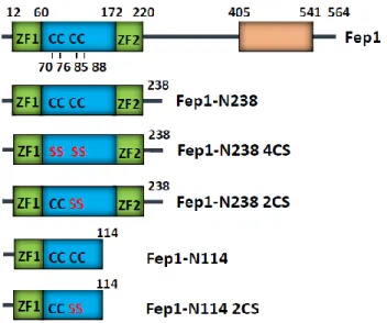
DNA as a template. The PCR products were cloned into pET15b vector (Novagen) via Nde1 and BamH1 sites, resulting in pET15b-Fep1 or pET15b-Fep1-N238 or pET15b-N114. The cysteine mutant N238 2CS (C85S/C88S), Fep1-N238 4CS (C70S/C76S/C85S/C88S) was generated by PCR mutagenesis and cloned into pET15b. All constructs were confirmed by nucleotide sequencing.
II.2. Protein expression and purification
The His-tagged recombinant proteins of full-length Fep1, N238, and Fep1-N238 4CS were expressed in E. coli BL21 (DE3) grown in LB at 37°C to OD600 of
0.5~0.6, followed by induction with 1 mM IPTG for 5 h. Harvested cells were lysed at 4°C in the binding buffer (20 mM Tris-HCl, pH 8.0, 500 mM NaCl, 5 mM imidazole) by using a high-pressure homogenizer EmulsiFlex-C3 (Avestin). Following centrifugation at 15000 g for 15 min at 4°C, the cell-free extracts were loaded onto Ni-IDA (iminodiacetic acid) column (ELPIS) pre-equilibrated with the binding buffer, and eluted with 500 mM imidazole. The partially purified proteins were then applied to HiLoad Superdex 75 column (GE Healthcare), followed by elution with 20 mM Tris-HCl buffer containing 500 mM NaCl and 1 mM DTT. For anaerobic purification, Ni-affinity chromatographies were done in the anaerobic glove box (O2 < 2 ppm; Coy Laboratory Products, Inc.).
II.3. Transformation of Escherichia coli and Yeast
For the purpose of general cloning, E. coli DH5 competent cells were used for transformation by heat shock method (42C for 1 min 30 sec.). High efficiency
34
electroporation method of E. coli (XL1-Blue) was used to amplify S. pombe genomic library plasmid of interesting target gene from the purified yeast total genomic DNA.
The S. pombe transformation was routinely done using lithium acetate (LiAc) / Polyethylene glycol (PEG) method according to Moreno et al. (1991).
II.4. Western blot analysis
Western blot analyses were performed E. coli cells were cultured and harvested at certain growth phase. Total cell extracts or fractionated extracts were resolved in SDS-PAGE (10 ~ 18%). Gel was transferred onto PROTRAN (nitrocellulose transfer membrane; Schleicher & Schuell) using TransBlot system (BioRad) at 160 ~ 180 mA for 50 min.
Filters with bound protein were washed 3 times and blocked in 10 ml of Tris-buffered saline containing 0.1% Triton-X 100 (TBS-T) with 0.5% BSA. Filters were incubated for 1 hr with proper antibodies in TBS-T containing 0.5% BSA. Excess antibodies were removed by repeated washing in TBS-T. After 1 hr incubation in TBS-T containing the secondary antibody conjugated with horseradish peroxidase, the signal was visualized with LAS3000 (Fuji) and quantified with Multi Gauge (Fuji) program.
II.5. Preparation of apoprotein
Apo-N238 was prepared by incubating the iron-bound as-isolated Fep1-N238 (100 µM) in 20 mM Tris-HCl buffer (pH 8.0) containing 500 mM NaCl, 10 mM EDTA, and 5 mM sodium dithionite (SDT) for 1 h on ice. The colorless protein solution was then buffer-exchanged into 20 mM Tris-HCl, pH 8.0, 500 mM NaCl through two consecutive PD10-desalting columns (GE Healthcare).
35
II.6. Fe-S cluster reconstitution in vitro
Reconstitution of Fe-S cluster into full-length Fep1 or Fep1-N238 was performed anaerobically (O2 < 2 ppm) in a glove box (Coy Laboratory Products, Inc.).
Following incubation of 50~100 μM apo-Fep1-N238 with 2 mM DTT for 30 min, 5-fold molar excess of ferric chloride (FeCl3) and sodium sulfide (Na2S) were added
and incubated for 2 h at room temperature. The reconstitution mixture was subjected to two consecutive chromatographies in the glove box through PD10-desalting column equilibrated with 20 mM Tris-HCl and 500 mM NaCl, to remove excess iron and sulfide.
II.7. Assays for quantification of iron, sulfide and
protein
II.7.1. Determination of iron concentration
The amount of iron in the purified protein sample was determined by using colorimetric ferrozine assay (Riemer, Hoepken et al., 2004). Aliquots (100 μL) of proteins were placed in Eppendorf tubes and mixed with 100 μL of 10 mM HCl (the solvent of the iron standard FeCl3), and 100 μL of the iron-releasing reagent (a freshly mixed solution of equal volumns of 1.4 M HCl and 4.5% (w/v) KMn04 in H2o). These mixtures were incubated for 2 h at 60°C. The HCl/ KMnO4-mediated digestion of iron-containing proteins is essential for iron quantitation of proteins. After the mixtures had cooled to room temperature, 30 μL of the iron-detection reagent (6.5 mM ferrozine, 6.5 mM neocuproine, 2.5 M ammonium acetate, and 1 M ascorbic acid dissolved in water) was added to each tube. After 30 min, 280 μL of the solution in each tube was transferred into a well of a 96-well plate and the absorbance was measured at 550 nm on a microplate reader.
36
II.7.2. Determination of sulfide concentration
Acid labile sulfide concentration was determined by using methylene blue-based color assay as described previously (Beinert, 1983). The tubes were kept tightly capped except when adding reagents. Rather than using stir bars, the tubes were closed and vortexed when mixing was called for. The procedure used was as follows. The sample volumes were brought to 100mL with pH 8 water. One at a time, each tube was opened, 300 mL 1% ZnOAc and 15 mL 12% NaOH were added simultaneously, the tube was closed tightly and vortexed. When all tubes had been treated in this way, they were allowed to sit for 15 min before addition of 75 mL DMPD (0.1% in 5 N HCl) and 2 mL FeCl3 (23 mM in 1.2 N HCl). Using Na2S z 9H2O as a standard as described by Beinert, this method proved very reproducible in our hands. In this assay, N,N-dimethyl-p-phenylenediamine (DMPD) is converted to methylene blue (MB) in the presence of sulfide and FeCl3. Precautions
were made to avoid residual amount of reducing agent such as DTT in the buffer that could hinder oxidation of DMPD by FeCl3.
II.7.3. Determination of protein concentration
Protein concentrations were determined by infrared-based peptide quantification system, using Direct Detect infrared spectrometer (Merck Millipore). Bovine serum albumin (BSA) was used as a standard.
II.8. EPR spectroscopy
Fresh Fep1-N238 samples purified anaerobically were equilibrated in 20 mM Tris-HCl buffer containing 500 mM NaCl, 1 mM DTT, and 10% (v/v) glycerol in the glove box. Fep1 proteins were either non-treated or treated with sodium dithionite (10 mM final) or ferricyanide (5 mM final), and transferred to EPR tubes. Following
37
10 min incubation at room temperature, the EPR tubes were frozen in liquid nitrogen. EPR measurements were carried out at Korea Basic Science Institute (KBSI), Western Seoul Center, for low temperature (5 K) analysis. CW X-band (9.6 GHz) EPR spectra were collected on a Bruker EMX plus 6/1 spectrometer equipped with a liquid helium cryostat (ESR900, Oxford Instrument) and ITC 503 temperature controller. All spectra were collected with the following experimental parameters: microwave frequency, 9.6 GHz; microwave power, 1 mW; modulation amplitude, 10 G; time constant, 20.48 ms; 5 scans. For measurements at liquid nitrogen temperature (123 K), samples were subjected to ESR spectrometer (JES-TE200, JEOL) at National Instrumentation Center for Environment Management (NICEM), Seoul National University.
38
CHAPTER III.
39
III. 1. Purification and UV-visible absorption
spectroscopy of full-length Fep1
III. 1. 1. Purification of full-length Fep1
Aiming at identifying biochemical characterizations of Fep1 protein,
His-tagged full length Fep1 (564 aa) was overproduced in E. coli and
anaerobically purified through Ni-IDA affinity column. Multiple protein
bands were observed on SDS-PAGE at 70 kDa and 30-35 kDa size ranges
(Fig. II-2A). LC-MS/MS analysis identified that 30-35 kDa bands are
degraded products of Fep1. A Western blot assay using 6His-probe
Antibody confirmed that bands of SDS-PAGE were 6His-tagged full length
Fep1 and 6His-tagged Fep1 degraded products (Fig. II-2B). To remove the
degraded Fep1 fragments and other possible contaminating proteins, gel
filtration chromatography was performed on Superdex 75 column,
equilibrated with 20 mM Tris-HCl, pH 8.0, 500 mM NaCl, 1 mM DTT.
Finally, we could successfully isolate full-length Fep1 only. The
single-peaked fractions centered around 128 kDa position were collected (Fig.
II-2C, red bracket) and subjected to SDS-PAGE to confirm purification of the
full-length Fep1. A prominent single band was observed, coinciding with
the calculated mass of 62.9 kDa with a hexa-histidine tag (Fig. II-2D). The
gel filtration behavior fits with the previous report that Fep1 can dimerize
through C-terminal region of Fep1 (Pelletier et al., 2005). As demonstrated
in Fig. II-2D, the purified full length protein did not undergo further
degradation, when it was concentrated through centricon (Millipore) in the
air, and kept at 4°C in the refrigerator or in the anaerobic glove box for 19 h
(lanes 2-4).
40
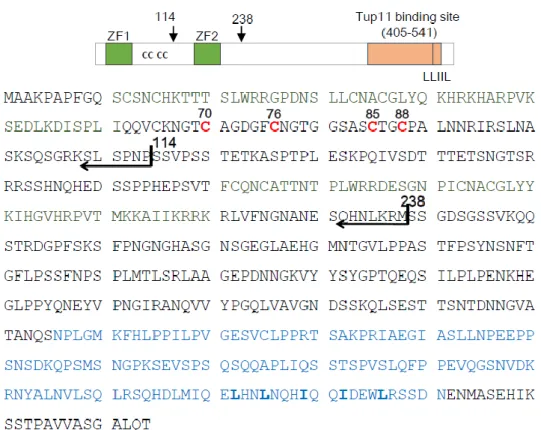
Fig II-1. Domain structure of Fep1 in S. pombe
Localization of Zinc finger (ZN) motifs and conserved cysteine residues within the sequence.
42
Fig II-2. Purification of full-length His-tagged Fep1.
A. Anaerobically purified Fep1 by Ni-agarose column.
B. LC-MS/MS analysis showed that multi-bands of SDS-PAGE were degraded
products of expressed Fep1. A Western blot assay using 6His-probe Antibody (Santa cruz, sc 804) confirmed that bands of SDS-PAGE were 6His-tagged full length Fep1 and 6His-tagged Fep1 degraded products.
C. D. A gel filtration chromatogram of Fep1 protein, following Ni-affinity column.
From Superdex 75 column, the full length Fep1 is eluted as a single peak, with the median elution volume (Ve) of 47 ml, which corresponds to 128 kDa determined by standard size marker proteins. Degraded Fep1 fragments were eluted later. The purity and stability of full-length Fep1 was monitored by SDS-PAGE. The freshly eluted Fep1 (lane 1) was concentrated by Centricon (Millipore) (lane 2), and stored either in the air (lane 3) or in the anaerobic glove box (O2 < 2 ppm; lane 4) for 19
hours.

![Fig I-6. Models for [2Fe-2S] Grx homodimers (left) and [2Fe–2S] 2+ Grx- Grx-BolA heterocomplexes (right) characterized from E](https://thumb-us.123doks.com/thumbv2/123dok_us/8543061.2302631/32.892.172.649.207.655/fig-models-grx-homodimers-bola-heterocomplexes-right-characterized.webp)