Original Research Article
Nasal septal angle deviation: effect on lateral wall in nasal obstruction
Neha Bagri
1, Kavirajan K.
1, Ranjan Chandra
1*, Yatish Agarwal
1,
Neetika Gupta
1, Shantanu Mandal
2INTRODUCTION
Diseases of the nose and paranasal sinuses are the commonly encountered condition in clinical practice. Nasal obstruction, a common presenting symptom, occurs most commonly due to septal deviation, above and beyond other causes like sinusitis, nasal polyposis, adenoid hypertrophy, atrophic rhinitis and nasal mass.
The nasal cavity is divided by a vertical partition (the “nasal septum”) into right and left side and bounded by bony sidewalls (lateral nasal walls). The nasal septum is
an essential support structure of the nose which is straight, symmetrical and meets evenly arched palate in midline.1 Septal deviation refers to convexity of the
septum to one side which may lead to deformities of the midline and lateral nasal wall structures. It has been suggested that the nasal septum is usually a midline structure until the age of seven and deviates mostly to the right thereafter.1
DNS is one of the most common causes for constant nasal obstruction. It has a variable incidence of 20-47%. 1-4 Various types of DNS are anterior dislocation, C- 1Department of Radiodiagnosis, 2Department of Otorhinolaryngology, Vardhman Mahavir Medical College &
Safdarjang Hospital, New Delhi-110029, India
Received: 01 December 2018
Accepted: 07 December 2018
*Correspondence:
Dr. Ranjan Chandra,
E-mail: drranjanchandra@gmail.com
Copyright: © the author(s), publisher and licensee Medip Academy. This is an open-access article distributed under the terms of the Creative Commons Attribution Non-Commercial License, which permits unrestricted non-commercial use, distribution, and reproduction in any medium, provided the original work is properly cited.
ABSTRACT
Background: Deviation of the nasal septum (DNS) refers to the convexity of the septum to one side disturbing the nasal physiology with obstructed nasal breathing leading to lateral nasal wall abnormalities and paranasal sinuses (PNS) mucosal disease. Knowledge of nasal morphological parameters plays an important role in planning successful nasal surgery. Our aim was to evaluate the angle of septal deviation (ASD) on CT scan and study its influence on the lateral nasal wall abnormalities and PNS mucosal disease.
Methods: A prospective cross-sectional observational study was conducted on 130 patients with clinical evidence of DNS and chronic sinusitis. The direction and severity of DNS was recorded on CT scan along with evaluation of lateral nasal wall and sinus mucosal abnormalities.
Results: Increasing ASD had statistically significant correlation with the lateral nasal wall abnormalities, most commonly, contralateral middle and inferior turbinate hypertrophy (p-value <0.0001). No significant association was found with the incidence of ipsilateral or contralateral osteomeatal complex (OMC) obstruction and sinus mucosal disease.
Conclusions: The direction and severity of septal deviation has significant impact on contralateral middle and inferior turbinate hypertrophy. The analysis of these ancillary pathologies can be of great help to the surgeon in better management of patients with nasal obstruction.
Keywords: Chronic rhinosinusitis, CT scan PNS, Lateral nasal wall, Nasal septal deviation, Septal angle
shaped deformity, S-shaped deformity, septal spur and septal thickening. C-shaped deformity is the most common type.5
DNS causes change in the airflow dynamics, alteration in the mucociliary clearance and obstruction at the osteomeatal complex resulting in nasal obstruction. Patients with increasing septal deviation are associated with higher incidence of bilateral osteomeatal complex (OMC) obstruction and bilateral chronic sinus disease.6
OMC obstruction in the ipsilateral side is attributable to nasal septal deformity and on the contralateral side due to lateral wall changes such as middle turbinate hypertrophy/pneumatisation (concha bullosa).
Clinical evaluation including Anterior Rhinoscopy and Diagnostic Nasal endoscopy continues to be gold standard before carrying out septoplasty to relieve obstructive symptoms, however CT is contemplated to assess ancillary pathologies such as obstructive middle turbinate hypertrophy, concha bullosa, chronic sinusitis, mucocele and other causes of obstruction by objective evaluation of nasal cavity which may not be visible on clinical evaluation.
Various studies have shown the relationship between the septal deviation and associated PNS pathology by CT scan, but very few studies have been reported in the literature regarding the impact of increased angle of septal deviation on lateral nasal wall, which we wish to emphasize and the outcome may help the surgeon in planning successful surgery and better management of patients with nasal obstruction.
METHODS
This study is a prospective cross-sectional observation, carried out on 130 patients in the Department of Radiodiagnosis in collaboration with the Department of Otorhinolaryngology, Vardhman Mahavir Medical College and Safdarjung Hospital, New Delhi from October 2016 to April 2018.
The study comprised of 130 adult patients with clinical evidence of nasal obstruction and chronic rhino-sinusitis (CRS) evaluated with CT scan PNS coronal view. Chronic rhinosinusitis was defined as persistent sinus disease refractory to medical treatment for minimum twelve weeks. Patients with acute sinusitis, malignancy, trauma and patients who had previous sinonasal surgery were excluded from the study.
Diagnosis and measurements
The included patients underwent detailed clinical and rhinoscopic examination and those having evidence of DNS on rhinoscopy were evaluated further for PNS pathology. Diagnostic nasal endoscopy and non contrast computed tomography were performed on these patients
image which best defined the OMC was used for calculating the degree of septal deviation.
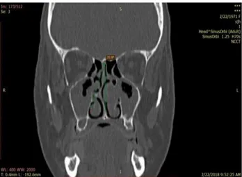
The ASD was measured as the angle between a line drawn from the crista galli to anterior nasal spine of maxilla and another line from the crista galli to the most deviated point of the nasal septum in the coronal plane (Figure 1). The patients were categorized in three groups based on the angle of septal deviation; Group I (0-7 degree), Group II (7.1- 11 degree) and Group III (>11 degree).
Figure 1: Coronal CT image showing measurement of angle of the septal deviation (ASD) -line drawn from the crista Galli to anterior nasal spine of maxilla and another line from the crista Galli to the most deviated
point of the nasal septum.
The CT scan images were studied to analyze the effect of increasing angle of septal deviation on the ipsilateral and contralateral lateral nasal wall structures, patency of OMC along with the site and pattern of mucosal abnormalities in the paranasal sinuses.
Statistical analysis
The data analysis was done using Statistical Package for Social Sciences (SPSS) version 21.0 categorical variables were presented in number and percentage (%) and continuous variables were presented as mean±SD and median.
Normality of data was tested by Kolmogorov-Smirnov test. Quantitative variables were compared using Kruskal Wallis test between three groups. Qualitative variables were correlated using Chi-Square test. P value of <0.05 was considered statistically significant.
RESULTS
Nasal septal deviation to the left (52.31%) was more prevalent as compared to the right (47.69%), however, no obvious statistical significance was noted (p value 0.196). Bony septal spurs were observed in 29% patients, among which 18% were on the left and 10% were on the right side.
On the basis of angle of septal deviation, the patients were categorized into three groups: group I (0-7 degree), group II (7.1- 11 degree), group III (>11 degree). Majority of the patients were from group II constituting of 35% patients followed by group I (33%) and group III (31%) respectively (Table 1).
The increasing angle of septal deviation was associated with increase in the incidence of contralateral middle turbinate hypertrophy and there was no evidence of ipsilateral middle turbinate hypertrophy in any of the three groups. 6.5% of patients in the group II and 17.1% in the group III showed contralateral middle turbinate hypertrophy.
Similarly, the incidence of contralateral inferior turbinate hypertrophy was also associated with increasing angles of septal deviation (Table 2). Also, there was higher incidence of bulbous type concha on the contralateral side in group II and group III patients (Table 2).
Table 1: Distribution of angles of septal deviation in groups.
Groups Male Female Total Mean Range
I 18 25 43 5.3º 2.9-7º
II 24 22 46 8.9º 7.1-10.9º
III 26 15 41 14º 11-23º
Total 68 62 130
Table 2: Middle and inferior turbinate abnormalities and their distribution as a function of degree of septal deviation.
Middle and inferior turbinate abnormalities
Group I Group II Group III
I/L C/L p-value I/L C/L p-value I/L C/L p-value
Lamellar concha 9 8 0.99 10 12 0.80 4 8 0.35 Bulbous type concha 12 11 0.99 7 13 0.20 7 10 0.58 Paradoxical middle
turbinate 2 3 0.99 1 1 0.47 1 2 0.99
Hypertrophied middle
turbinate - - - 3 0.74 - 7 0.018
Hypertrophied inferior
turbinate 5 9 0.38 5 25 <0.0001 6 28 <0.0001
Table 3: OMC obstruction and distribution of paranasal sinusitis among three groups.
OMC and paranasal sinuses
Group I Group II Group III
I/L C/L p-value I/L C/L p-value I/L C/L p-value
OMC obstruction 21 22 0.99 23 25 0.84 19 20 0.99 Frontal sinusitis 13 13 0.8 18 15 0.67 14 13 0.99 Ethmoid sinusitis 17 17 0.8 22 21 0.99 16 14 0.81 Maxillary sinusitis 23 23 0.8 30 30 0.82 22 20 0.82 Sphenoid sinusitis 8 12 0.4 14 13 0.99 9 15 0.22
Table 4: Pattern of sinusitis and their distribution among three groups.
Pattern of sinusitis
Group I
Group II Group III
I/L C/L p-value I/L C/L p-value I/L C/L p-value
Table 5: Comparison of frequency of sinus mucosal disease with other studies.
Study Maxillary
sinusitis
Anterior ethmoid sinusitis
Posterior ethmoid sinusitis
Frontal sinusitis
Sphenoid sinusitis
Mundra et al 62.3% 68.85% 13.11% 14.75% 6.56% Poorey et al 73.13% 53.73% 28.36% 43.28% 16.42%
Havas et al 24.4% 28.4% - 4.8% 11%
Kennedy and Zinrich et al 66% 78% 31% 34% 16%
Calhoun et al 43% 34% - 13% 19%
Bolger et al 77.7% 84.3% 35.4% 14.75% 25.4%
Our study 57.3% 39.6% 35% 33.1% 27.3%
The incidence of OMC obstruction was higher on the contralateral side (22.3%) as compared to the ipsilateral side (21%) among the three groups. However, there was increased incidence of OMC obstruction in group II patients compared to the group I and group III patients (Table 3). Hence, no apparent statistical significance was seen between the incidence of OMC obstruction and angle of septal deviation.
There was increased incidence of frontal, ethmoid, maxillary and sphenoid sinusitis in group II patients as compared to the group I and group III patients (Table 3), nulling the effect of angle of septal deviation on the incidence of sinus mucosal disease. Mucosal abnormalities were most frequently noted in maxillary sinus, among which 56.2% patients had right sided involvement and 58.5% had left sided involvement. No apparent correlation was seen between the side of nasal septal deviation and side of maxillary sinusitis. Sphenoid sinus was the least commonly involved. Among the various sinus mucosal disease patterns, sinonasal polyposis pattern was the most common pattern (Table 4), followed by osteomeatal unit pattern and infundibular pattern respectively.
DISCUSSION
Nasal septal deviation disturbs the nasal physiology together with conchal hypertrophy and other anatomical variations. The results in our study show that with increasing angle of septal deviation there are anatomical changes in the lateral nasal wall in the form of contralateral middle and inferior turbinate hypertrophy. However, increasing angle of septal deviation has no statistically significant effect on the OMC and mucosal pathology in the paranasal sinuses.
In our study, there was increased incidence of nasal septal deviation to the left, similar to the results of study by Madaniet al, however, in contradiction, the study results of Stallman et al, revealed that nasal septal deviation to the right was more common (51%) as compared to the left (49%).7,4 The authors in present study observed
presence of septal spur in 29% cases paralleling the study results of Shpilberg et alwho found that one third of the
patients with deviated nasal septum have associated with septal spurs.8
There was no evidence of ipsilateral middle turbinate hypertrophy in any of the three groups, rather the increasing angle of septal deviation was associated with increase in the incidence of contralateral middle turbinate hypertrophy, which is in consonance with the study results of Demir et alwho found that the measurements of the bony and mucosal structure of the contralateral middle turbinate were greater and statistically significant.9
The measurements of the bony and mucosal thickness of inferior turbinate by Orhanet al, revealed statistically significant difference between the ipsilateral and contralateral side in patients of deviated nasal septum, which is in consonance to our study results (Figure 2).10
Figure 2: Coronal CT image showing left sided deviated nasal septum with ipsilateral lamellar type
concha, contralateral bulbous type concha and contralateral inferior turbinate hypertrophy.
Our study shows higher incidence of bulbous type concha on the contralateral side (11.3%) as compared to the ipsilateral side (8.6%) among the three groups, parallel to the study results of Stallman et al.4 Elahi et al, evaluated
contralateral middle turbinate abnormalities and prominent ethmoidal bulla.6 However, in our study, this
relationship was not observed as there was increased incidence of bulbous type concha in group I patients compared to the group II and group III patients.
The incidence of OMC obstruction, in our study was higher on the contralateral side and maximum in group II patients as compared to the group I and III patients, hence, no statistically significant association was seen between increasing angle of septal deviation and the incidence of OMC obstruction, which is similar to the results of few previous studies.11,12 However, the study
results of Elahi et alstand in contradiction, who found that increasing angle of septal deviation is associated with a higher incidence of OMC obstruction.2
In this study, incidence of frontal, ethmoid, maxillary and sphenoid sinusitis was noted in group II patients compared to the group I and group III patients. Therefore, no significant association was seen between increasing angle of septal deviation and the incidence of paranasal sinus mucosal disease which is in conflict with the study conducted by Yousem et alin which they concluded that increased incidence of sinus opacification is associated with severity of nasal septal deviation.13 Calhoun et al,in
their analysis found that the septal deviation was associated with increased incidence of OMC obstruction and ethmoid sinus disease, however angle of septal deviation was not considered.14
The mucosal abnormalities in the paranasal sinuses vary from minimal mucosal thickening to total sinus opacification. In the present study, most frequently involved sinus was maxillary sinus (57.3%), while Bolger et al found most common involvement of anterior ethmoid sinuses (84.3%) which was followed by maxillary sinus involvement (77.7 %).15 On comparison
of frequency of mucosal sinus abnormalities with other studies, the results were quite variable (Table 5). Our study results matched the findings of Poorey et al showing most common involvement of maxillary sinus and sphenoid sinus involvement being the least common in frequency.12
Considering the various patterns of sinonasal inflammatory disease, we observed that the sinonasal polyposis pattern was the most common, followed by osteomeatal unit pattern and infundibular pattern respectively, which was similar to the study results of Kanwar et al.16 In contradiction, Babbel et alfound that
infundibular pattern was the most common, followed by osteomeatal unit and sinonasal polyposis pattern respectively.17 Broad spectrum antibiotic usage could be
a possible source of bias in these results and the effect of such therapies on CT scan outcome is still unknown. We assume that the antibiotic therapy would reduce the prevalence of sinus mucosal disease and the limitation that actual prevalence might be even higher still stands.
CT scan PNS stands the gold standard in evaluation of patients with chronic sinusitis with its ability to accurately map out the bony and soft tissue anatomy.18,19
CT scan in conjunction with nasal endoscopic examination is the ideal combination for evaluation of the sinonasal region. The analysis of anatomy and abnormalities of nasal septum, lateral nasal wall and paranasal sinus abnormalities is a prerequisite to safe and effective surgical treatment of sinonasal disease.
CONCLUSION
This cross-sectional observational study was conducted to evaluate the septal angle and its effect on the lateral wall of nasal cavity by CT scan, thus highlighting the ancillary pathologies which can lead to increase in the success rate of surgical procedures. The increasing angle of septal deviation was associated with increase in the incidence of lateral nasal wall abnormalities most commonly in the form of contralateral middle and inferior turbinate hypertrophy (p value <0.0001). The incidence of bulbous type concha on the contralateral side was higher (11.3%) as compared to the ipsilateral side (8.6%). The incidence of OMC obstruction and sinusitis was higher in group II as compared to group I and group III, hence there was no significant association with the increasing angle of septal deviation. Maxillary sinus was the most common sinus showing mucosal disease followed by ethmoid, frontal and sphenoidsinus. Sinonasal polyposis pattern was the most common pattern of sinonasal inflammatory disease followed by osteomeatal unit pattern, infundibular pattern and sphenoethmoidal recess pattern. Hence, CT scan PNS is recommended in patients with nasal obstruction to assess the severity of nasal septal deviation, its impact on the lateral nasal wall and paranasal sinuses which may help the surgeons in better management of the patients.
ACKNOWLEDGEMENTS
Authors would like to thank the participants of the study and the staff in the Department of Radiodiagnosis, VMMC and Safdarjung Hospital for their support and co-operation throughout the study.
Funding: No funding sources Conflict of interest: None declared
Ethical approval: The study was approved by the Institutional Ethics Committee
REFERENCES
1. Gray LP. Deviated nasal septum. Incidence and etiology. Ann Otol Rhinol Laryngol Suppl. 1978;87:3-20.
3. Mohebbi A, Ahmadi A, Etemadi M, Safdarian M, Ghourchian S. An epidemiologic study of factors associated with nasal septum deviation by computed tomography scan: a cross sectional study. BMC Ear Nose Throat Dis. 2012;12:15.
4. Stallman JS, Lobo JN, Som PM. The incidence of concha bullosa and its relationship to nasal septal deviation and paranasal sinus disease. Am J Neuroradiol. 2004;25:1613-8.
5. Moorthy P, Kolloju S, Madhira S, and Jowkar A. Clinical Study on Deviated Nasal Septum and Its Associated Pathology. Inter J Otolaryngol Head Neck Surg. 2014;3:75-81.
6. Elahi MM, Frenkiel S, Fageeh N. Paraseptal structural changes and chronic sinus disease in relation to the deviated septum. J Otolaryngol. 1997;26:236-240.
7. Madani SA, Hashemi SA, Modanluo M. The incidence of nasal septal deviation and its relation with chronic rhinosinusitis in patients undergoing functional endoscopic sinus surgery. J Pak Med Assoc. 2015;65:612-4.
8. Shpilberg KA, Daniel SC, Doshi AH, Lawson W, Som PM. CT of anatomic variants of the paranasal sinuses and nasal cavity: poor correlation with radiologically significant rhinosinusitis but importance in surgical planning. AJR Am J Roentgenol. 2015;204:1255-60.
9. Demir D, Asil K, Guven M, Kayabaşoglu G, Yılmaz MS. Assessment of the correlation between nasal septal deviation and compensatory hypertrophy of the middle turbinate. Euro Arch Otorhinolaryngol. 2015;272:2847-51.
10. Orhan I, Aydın S, Ormeci T, Yılmaz F. A radiological analysis of inferior turbinate in patients with deviated nasal septum by using computed tomography. Am J Rhinol Allergy. 2014;28:68-72. 11. Mundra RK, Gupta Y, Sinha R, Gupta A. CT Scan
Study of Influence of Septal Angle Deviation on Lateral Nasal Wall in Patients of Chronic Rhinosinusitis. Indian J Otolaryngol Head Neck Surg. 2014;66:187-90.
12. Poorey VK, Gupta N. Endoscopic and computed tomographic evaluation of influence of nasal septal deviation on lateral wall of nose and its relation to sinus diseases. Ind J Otolaryngol Head Neck Surg. 2014;66:330-5.
13. Yousem DM, Kennedy DW, Rosenberg S. Ostiomeatal complex risk factors for sinusitis: CT evaluation. J Otolaryngol. 1991;20:419-24.
14. Calhoun KH, Waggenspack GA, Simpson CB, Hokanson JA, Bailey BJ. CT evaluation of the paranasal sinuses in symptomatic and asymptomatic populations. Otolaryngol Head Neck Surg. 1991;104:480-3.
15. Bolger WE, Butzin CA, Parsons DS. Paranasal sinus bony anatomic variations and mucosal abnormalities: CT analysis for endoscopic sinus surgery. Laryngoscope. 1991;101:56-64.
16. Kanwar SS, Mital M, Gupta PK, Saran S, Parashar N, Singh A. Evaluation of paranasal sinus diseases by computed tomography and its histopathological correlation. J Oral Maxillofac Radiol. 2017;5:46-52. 17. Babbel RW, Harnsberger HR, Sonkens J, Hunt S. Recurring patterns of inflammatory sinonasal disease demonstrated on screening sinus CT. AJNR Am J Neuroradiol. 1992;13:903-12.
18. Mamatha H, Shamasundar NM, Bharathi MB, Prasanna LC. Variations of ostiomeatal complex and its applied anatomy: a CT scan study. Ind J Sci Technol. 2010 Aug 1;3(8):904-7.
19. Milczuk HA, Dalley RW, Wessbacher FW, Richardson MA. Nasal and PNS anomalies in children with chronic sinusitis. Laryngoscope. 1993;103(3):247-52.