Thinless Gyalpo et al JMSCR Volume 06 Issue 12 December 2018 Page 436 Original Article
A Comparative Study of T-tube versus Ante grade Stenting of CBD after
Open CBD Exploration for Choledocholithiasis
Authors
Thinless Gyalpo
1, Mubashar Akram
1*, Kunzes Dolma
2, Tariq P Azad
11
Department of Surgery, Government Medical College Jammu
2
Department of ENT, Government Medical College Jammu *Corresponding Author
Mubashar Akram
Email: [email protected], Tel: +91-9419129818
Abstract
Background: CBD stones once confirmed have to be extracted in order to prevent the complications related
to them. Conventionally T-tube is used for decompression of CBD after open choledocholithotomy which has its own share of complications
Aim: To compare clinical outcome in patients of choledocholithiasis undergoing CBD exploration using
either a biliary stent or T-tube as a decompression procedure.
Design and Place:This was a prospective randomized comparative study where 40 patients after being admitted in Post Graduate Department of Surgery Government Medical College, Jammu over a period of one year w.e.f. November 2015 to October 2016 with diagnosis of choledocholithiasis were divided into two
groups (Group A and Group B),each having 20 patients.
Method: 40 patients selected in study were subjected to open CBD exploration for choledocholithiasis. Out of these,20 patients in Group A underwent primary closure of the choledochotomy over a biliary stent after open CBD exploration and 20 patients in Group B underwent closure of choledochotomy over a T-tube after open CBD exploration.
Result: Primary closure of the CBD over biliary stent is a safe alternative to T-tube drainage with benefits of shorter operative time, hospital stay and lesser morbidity.
Conclusion: Closure of CBD over endobiliary stent is a modality for management of CBD stones with proven
safety and good results.
Keywords: Choledocholithiasis, Biliary Stenting, T-Tube drainage, CBD.
Introduction
Common Bile Duct (CBD) stones are identified in 10 to 15 percent of patients undergoing surgery for symptomatic gall stone disease.[37] Of these patients, it is estimated that approximately one third may spontaneously pass down the common
bile duct within 2 months without any intervention.[3] The remaining will require an endoscopic or surgical intervention to relieve the obstruction.
CBD stones may be classified as primary or secondary. Primary stones arises de novo in bile
www.jmscr.igmpublication.org Impact Factor (SJIF): 6.379
Index Copernicus Value: 79.54 ISSN (e)-2347-176x ISSN (p) 2455-0450
Thinless Gyalpo et al JMSCR Volume 06 Issue 12 December 2018 Page 437 duct (15%) and secondary stones occur by
migration along the biliary system from the gallbladder and make up to 85% of stones[.20] Choledocholithiasis may be silent and symptomless or may cause acute cholangitis with jaundice, pain and fever especially in the elderly. Traditionally, CBD stones were diagnosed with intra-operative cholangiography and were treated with open CBD exploration. Although more liberal or routine use of choledochotomy would minimize the incidence of retained stones, exploration of CBD may lead to stricture at later stage. Mc Sherry reports 0.5 percent mortality for cholecystectomy alone, 2.4 percent mortality for negative choledochotomy and 3.9 percent mortality for choledocholithotomy.[16] Advances in preoperative imaging technology such as ultrasonography, Magnetic Resonance Cholangiopancreaticography
(MRCP), Endoscopic retrograde
Cholangiopancreaticography (ERCP) and Endoscopic Ultrasound have allowed less invasive and more accurate methods of identifying and treating CBD stones[.37]
Once CBD stones are confirmed, they have to be extracted to prevent the complications such as Biliary colic, suppurative Cholangitis, Obstructive jaundice, Hepatic Abscess and Pancreatitis. Nowadays with advanced laparoscopic and endoscopic techniques available for removal of CBD stones, open exploration has become less common. Open CBD exploration is performed in situations like injury to CHD/CBD during cholecystectomy, failed endoscopic or laparoscopic removal of CBD stones, ERCP facility is limited or unavailable, patients undergoing open cholecystectomy with CBD stones or absence of advance laparoscopic equipment and expertise.
Choledochotomy for stones in the CBD was first suggested by Langenbach in 1884. The first open common bile duct exploration was performed in 1889 by Robert Abbe, a New York surgeon. He opened the duct of a 36-year-old woman with severe jaundice, removed a stone, sewed the duct with fine silk, and returned her to perfect health.
Other sources give credit for the first exploration to Londoner J.K. Thorton 1889[26], Swiss surgeon Ludwig Courvoisier 1890[1,2] or Herman Kimmell of Hamburg, Germany[.19]
Standard management of CBD stone includes choledochotomy in the supra-duodenal part[29] followed by stone extraction with confirmation of CBD clearance by passing soft catheter or dilator proximally and distally.[28] Clearance of the CBD is also confirmed by completion cholangiography or choledochoscopy. After choledochotomy it can be further managed by either primary closure of CBD with or without antegrade stenting or T-tube drainage or by bilio-enteric bypass.
The traditional practice of T-tube drainage after CBD exploration was first described by Deaver in 1904[6].In T-tube drainage there is risk of retained stones, so it is obligatory to perform a postoperative cholangiography which confirms that the CBD is clear. Later on if residual stones are present, T-tube allows access for percutaneous manipulations and extraction of stones. The T-tube has been the method of choice for CBD decompression following choledochotomy[36].
Indications for T-tube drainage
1) When significant trauma is inflicted upon the duct wall during stone removal.
2) Extensive manipulation and trauma to the head of pancreas or ampulla while removing an impacted stone.
3) Pancreatitis exists at the time of operation. 4) Transduodenal canalization of pancreatic
duct is performed.
5) There is demonstrable narrowing of lower end of CBD due to fibrotic or spastic sphincter of Oddi.
T-tube drainage allows post operative cholangiography to be performed and Residual stones can be removed from the T-tube tract avoiding a re-exploration. Use of T-tube to remove retained stone in the biliary duct was first described in 1978 by Burhenne HJ.
Thinless Gyalpo et al JMSCR Volume 06 Issue 12 December 2018 Page 438 William Halsted had described the use of primary
closure after exploration of the CBD[7]. Primary closure of CBD (choledochorraphy) was apparently used at the beginning of this century by German and French surgeons P. Duval, 1924; P. Walzel, 1933. In 1942, Mirizzi[17] cited good results in a series of 31 patients undergoing primary closure of CBD. He felt that whenever possible, placing of a tube in a delicate contractile structure like CBD, should be avoided. He has given certain requirements which should be fulfilled before choledochorraphy is contemplated:- Patency of the papilla of Vater, Complete removal of intraductal calculi, Normal pancreas and Meticulous suturing of the duct. Indications for closure of CBD over internal biliary stent
1. When stones are removed from the CBD without undue manipulation or traumatisation of walls or lumen of the CBD.
2. When the duct wall is slightly or moderately thickened but not oedematous or acutely inflamed.
3. When stones are not found otherwise normal appearing CBD or even a CBD that is dilated.
Through this study we wanted to compare T-tube versus biliary stent decompression of CBD following open choledocholithotomy.
Material and Method
This prospective randomized comparative study was conducted on 40 patients undergoing open CBD exploration in the Department of Surgery, Government Medical College Jammu from 1st November 2015 to 31st October 2016.Patients were divided into two groups:
Group A: included 20 patients who underwent
primary closure of the choledochotomy over a biliary stent after open CBD exploration for CBD stone.
Group B: included 20 patients who underwent
closure of choledochotomy over a T-tube after open CBD exploration for CBD stone.
Inclusion Criteria Exclusion Criteria
All patients
undergoing elective open
choledocholithotomy
ASA I
ASA II
Age > 80 years
Previous history of choledocholithoto my
Deranged
coagulation profile
Preoperative evaluation
The patients were assessed pre-operatively with clinical history and physical examination. Biochemical tests plus radiological evaluations like Ultrasound Abdomen and Magnetic Resonance Cholangiopancreatography (MRCP) for confirmation of the ultrasound findings were done pre-operatively.
Patients with jaundice or deranged PT received Injection Vitamin K preoperatively. Prophylactic antibiotics were administered at the time of induction. The anaesthetist in all cases noted the operation time from skin incision to the application of the last stitch.
Surgical procedure
After obtaining consent, patients underwent surgical procedure as per the study design and group allocation. All the patients underwent standard CBD exploration through choledochotomy in supraduodenal part of CBD. Before exploration of CBD, the cystic duct was ligated to obviate the risk that manipulation of gall bladder would force small stones down the cystic duct and into the common duct after the latter has been explored. After stone retrieval and completion cholangiogram or choledochoscopy, patient either underwent T-tube or biliary stent placement as per group allocation.
Technique of biliary stent placement
Thinless Gyalpo et al JMSCR Volume 06 Issue 12 December 2018 Page 439 closed with continuous 3-0 vicryl sutures over this
stent.
Technique of T-tube drainage
After the common bile duct was opened between two stay sutures above the duodenum, the stones were extracted. The choledochotomy was then closed after inserting a No.12 F/14 F gauge T-tube with the “T” lying along the length of the duct, shortening the limbs to 2.5cm in either direction and splitting the horizontal limb along its length opposite the vertical tube.
Following this, cholecystectomy was performed and a size 32 Fr drain was placed in all the patients which were brought out to the exterior through a separate stab wound and left in situ till drain output becomes nil.
Postoperative evaluation
All patients were kept nil per oral and on parenteral fluids till their bowel activity recovered. Oral intake was allowed from 12-36 hours postoperatively in the absence of vomiting and ileus. The patients were observed for complications, if any including acute pancreatitis or severe dehydration due to electrolytes imbalance or increased T-tube output. Serum amylase was done on the first postoperative day and LFT on second postoperative day. The subhepatic drain was removed once its drainage had reduced to a negligible amount. The stitches were removed on the 10th to 12th post-operative day.
Follow Up
At the time of discharge, an ultrasound of the abdomen was done in patients to rule out biliary leak, subphrenic collection, residual stones or any other complication.
1) T-tube: - T- tube was removed on or after 14th post operative day depending on T- tube cholangiogram.
2) Biliary stent placement:- Biliary stent was removed after 4 weeks by upper gastrointestinal endoscopy.
Observation
40 patients were randomized into groups A and B. The patients belonging to group A(20) underwent open CBD exploration followed by primary closure over biliary stent. Patients of group B(20) underwent open CBD exploration followed by closure over T-tube.
Sex Distribution
There were 20% (4) males and 80% (16) females in Group A and there were 15% (3) males and 85%(17) females in Group B. On analysis by fisher exact test it showed that the sex distribution between the two groups was statistically insignificant with a p value of >0.999. Thus both the groups were comparable with respect to the sex distribution of the patients.
Table 1: Sex distribution of the patients in both groups
Groups Female Percentage within group Males Percentage within group
Group A 16 80% 4 20%
Group B 17 85% 3 15%
Total 33 82.5% 7 17.5%
Age Distribution
In group A, age of the patients varied from 24-70 years with a mean age of 51.10 years. In group B, the age varied from 26-74 years with a mean age of 50.6 years. On analysis by Paired students T-test it showed that the age variation between the two groups was statistically insignificant with a p value
Thinless Gyalpo et al JMSCR Volume 06 Issue 12 December 2018 Page 440 Table 2: Age distribution of the patients in both groups
Group Minimum
Age (years)
Maximum Age (years)
Mean age (years)
Total patients in group
Standard deviation
Group A 24 70 51.10 20 11.15
Group B 26 74 50.6 20 11.07
Total patients 24 74 50.85 40 10.969
Duration of Surgery
The operating time for patients in Group A ranged from110 to 180 minutes with a mean of 121.6 minutes. The operating time for patients in Group B ranged from 112 to 210 minutes with a mean of 136.2 minutes. Analysis by the Students t test
showed the p value to be 0.0415 which means that the difference in operating times between the two groups was statistically significant. This implies that the duration of surgery was shorter in case of biliary stent group as compared to the T-tube group.
Table 3: Distribution of patients in two groups according to duration of surgery
Group N Mean Standard deviation minimum Maximum
Group A 20 121.6 17.860 110 180
Group B 20 136.2 25.281 112 210
Post Operative Hospital Stay
The post operative stay in hospital for patients in Group A ranged from 4 to 12 days with a mean of 7.05 days. The post operative stay in hospital for patients in Group B ranged from 6 to 16 days with a mean of 9.5 days. Analysis by thestudent t Test
showed the p value to be 0.002 meaning that the difference in the post operative stay in hospital between the two groups was statistically significant. This implies that the post operative stay in hospital was shorter in case of biliary stent group as compared to T-tube group.
Table 4: Distribution of patients in two groups according to duration of post operative stay in hospital
Group N Mean Std. deviation Minimum Maximum
Group A 20 7.05 1.76 4 12
Group B 20 9.50 2.74 6 16
Total 40 8.27 2.25 5 14
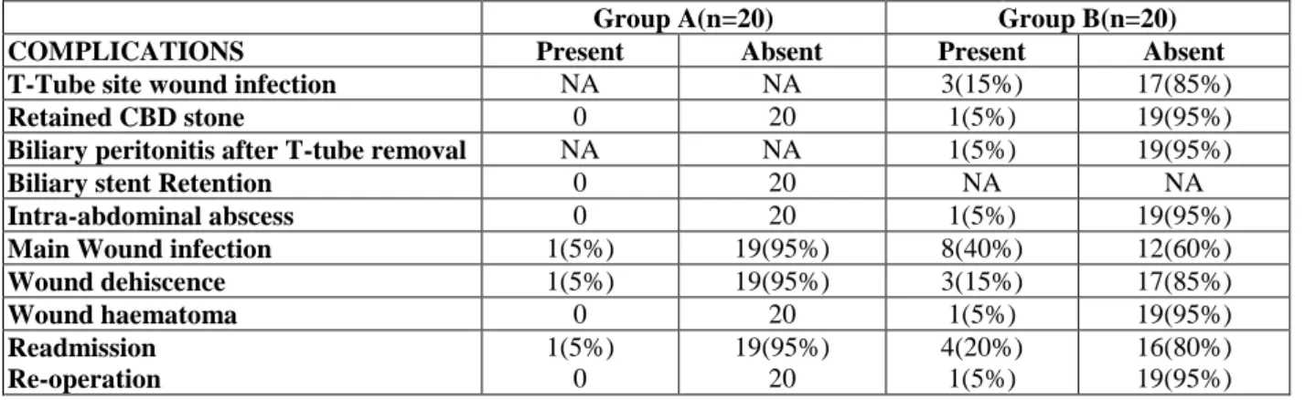
Complications
Mean duration of abdominal drain removal postoperatively was 3.90 days in group A and 3.95 days in group B. 3(15%) patients in group A and 4(20%) patients in Group B had history of Diabetes mellitus. However when the two groups were statistically compared in terms of factors like Diabetes mellitus and abdominal drain removal, there was no significant difference.
Main wound infection in postoperative period was seen in 1(5%) patient of group A and 8(40%) patients of group B. Analysis of this data by Fisher exact Test, revealed a p value of 0.0196 which was statistically significant implying that incidence of main wound infection in postoperative period is high in T-tube group as compared to biliary stent
group.
Thinless Gyalpo et al JMSCR Volume 06 Issue 12 December 2018 Page 441 Table 5 Distribution of patients in two groups according to the complications seen
Group A(n=20) Group B(n=20)
COMPLICATIONS Present Absent Present Absent
T-Tube site wound infection NA NA 3(15%) 17(85%)
Retained CBD stone 0 20 1(5%) 19(95%)
Biliary peritonitis after T-tube removal NA NA 1(5%) 19(95%)
Biliary stent Retention 0 20 NA NA
Intra-abdominal abscess 0 20 1(5%) 19(95%)
Main Wound infection 1(5%) 19(95%) 8(40%) 12(60%)
Wound dehiscence 1(5%) 19(95%) 3(15%) 17(85%)
Wound haematoma 0 20 1(5%) 19(95%)
Readmission Re-operation
1(5%) 0
19(95%) 20
4(20%) 1(5%)
16(80%) 19(95%)
Stent/T-tube removal
T-tube was removed in1 patient of group B on 14th POD while in 19 patients, it was removed after 14th POD. Biliary stentwas removed in14 patients after 4 weeks while 6 patients had biliary stent removal after 6 weeks.
Discussion
Ever since the first successful removal of CBD stones by Robert Abbe, operative exploration of the CBD at the time of cholecystectomy has been considered the benchmark to which all other treatment modalities are compared. Since its description, the T-tube has been the method of choice for CBD decompression following choledochotomy for years. Although it is true that the T-tube has been used and proven to be a safe and effective method for postoperative biliary decompression, it is not exempted from complications, which are present in upto 10% of patients.[18]
William Halstead and John Finneywere among the first to challenge the necessity of routine CBD drainage[7].To eliminate T-tube related complications after choledochotomy, endobiliary stent placement followed by primary closure of CBD has been proposed as a safe alternative.[9] This study was conducted to evaluate the safety and feasibility of primary closure of CBD with biliary stent after open CBD exploration as compared to T-tube drainage. A total of 40 patients were included in this study, 20 underwent primary closure of choledochotomy over a biliary stent
(Group A) and 20 underwent closure over a T-tube (Group B). As per our study primary closure of CBD over endobiliary stent leads to shorter convalescence with less postoperative complications when residual stones were ruled out by irrigating the CBD and hepatic ducts and by completion choledochoscopy as compared to T-tube drainage.
There were 82.5% females and 17.5% males in this study. This sex distribution is similar to that study by Parez et al,[13] the incidence of CBD stone being higher in females. The age of the patients in this study varied from 24 to 74 years with mean age of 50.85 years.
Closure of choledochotomy in a CBD of diameter less than 5mm has been associated with stricture formation. In our study, the CBD diameter varied from 9 to 15 mm (mean 11.9mm) in the stent group and from 9 to 16 mm (mean12.05mm) in the T-tube group. The minimum diameter of CBD in the present study was at least 9 mm justifying the safety of choledochotomy as the approach for CBDE.
Thinless Gyalpo et al JMSCR Volume 06 Issue 12 December 2018 Page 442 (mean 174 minutes) and Kim and Lee ( mean 188.3
minutes).[11,13]
All the patients in the CBD Exploration with biliary stent group underwent primary closure of the choledochotomy over a 7Fr 10cm biliary stent. The advantage of this stent is that the proximal flap gets engaged at the confluence of the right and left hepatic ducts and the distal flap in the duodenum. The stent was inserted into the CBD through the choledochotomy directly without fluoroscopic or choledochoscopic guidance.
It was found in our study that in cases of primary closure over stent, the patients’ stay in hospital was 7.5 days on an average. This was shorter than the stay if T-tube drainage was used, which was 9.5 days. This finding is in agreement with Parez et al (T-tube 6.8+4.7 days and stent 5.2+3.3 days).[21] CBD drainage cases had a prolonged stay of more than 2 days as compared to biliary stent cases. The benefits of a shorter postoperative stay to the patient as well as the hospital are well documented. Isla et al
(2 to 5 days; mean 3 days), Kim et al (4.8 1.5 days) and Ha et al (mean 5 days) have reported almost similar results with biliary stent placement after CBD Exploration.
Although insertion of a drain in the subhepatic space was not absolutely essential, it was preferred by most of the surgeons performing CBD Exploration. In our study, subhepatic drain was inserted in all patients and kept till the drain output reduced to a negligible amount.
Postoperative morbidity is directly related to the infective complications which range from wound and T-tube site infection to intra-abdominal abscess and life threatening acute pancreatitis. CBD Exploration, per se, significantly increases the morbidity and mortality because of infective complications. According to some surgeons[12], T-tube adds on to these complications.
15%(3) cases of T-tube drainage in our study had post-operative T-Tube wound infection and 40%(8) cases had main wound infection, which are comparable to studies by Lygidakis[14] (reported as 77%), Keighley (reported as 73%) and Parez et
al (reported 11%). It appears reasonable that T-tube drainage, requiring the introduction of a foreign body, provokes exogenous acquisition of environmental microorganisms. Apparently ascending cholangitis leads to bacteremia and distant infective complications. In evaluating the relative morbidity between the two groups, the postoperative complications were significant and this did constitute the primary cause of increased morbidity among cases undergoing T- tube drainage.
Residual calculi were not found in any of the stent placement cases as against one case (5%) of T-tube drainage detected on post-operative T-tube cholangiography. The incidence of residual stones detected on T-tube cholangiography in our study was comparable to studies by Way et al (7%), Sawyers et al (1.6%), Gillatt et al (5%), Herrington et al (5.5%), Chande et al (4.7%)[4,10,14,30]
T-tube removal lead to minor reactions in the form of pain and discomfort in most of the cases in our study. Biliary peritonitis at this time was reported in one case. The patient was diagnosed by symptoms of abdominal pain, tenderness and guarding. On USG abdomen minimal free fluid was present in the peritoneal cavity. Patient was managed conservatively. Severe reaction at the time of T-tube removal has been reported by Lygidakis et al.[14]
Readmission (T-tube 20%, stent 5%) and reoperation rate (T-tube 5%, stent 0%) in this study were comparable to the study conducted by Parez et al.[21]
Thinless Gyalpo et al JMSCR Volume 06 Issue 12 December 2018 Page 443 Conclusion
T-tube drainage is the standard practice to decompress the biliary tree[31] and prevent bile leakage due to oedema and spasm of the sphincter of Oddi.[32] It has the advantages of easy postoperative X-ray visualization of the CBD, and the potential for T-tube tract extraction of missed stones.Patient with T-tube drainage remains absent from work for 3-4 weeks with tube in place[33] and also, the associated bacteraemia necessitates antibiotic cover. T-tube has its own share of complications.[5] These include: patient discomfort, longer hospital stay, mechanical problems (dislodgement of T-tube, etc), duct stenosis after T-tube removal, risk of cholangitis from an external source via the T-tube and the possibility of bile leakage following extraction of T-tube. The entero-hepatic circulation of bile salts ceases for a week or more until the T- tube is clamped off or removed. The absence of bile in the alimentary canal postoperatively may result in slow wound healing, anorexia and constipation. The irritant foreign body reaction of a T- tube in the CBD and the infection associated with its presence may in some cases tend to increase bile drainage and may lead to severe electrolyte loss and to a persistent biliary fistula when the tube is removed. The T-tube may break off within the CBD or may be accidentally pulled out before sufficient time has elapsed for the T-tube tract to be sealed off from the peritoneal cavity or matured. Occasionally a secondary haemorrhage may arise from the CBD due to intraductal drainage.
Intra-operative deployment of biliary stent is done via the choledochotomy incision before its closure. It eliminates the complications of T-tube and allows the patient to return to unrestricted activity quickly, as the median post operative hospitalization is two days.[23] It is safe, effective, time sparing and cost effective. The stent is removed endoscopicaly after 1 month and 6-30 months follow up demonstrates no complications.[25] However, biliary stent carries some complications as clogging may occur in 10-30% cases[34] by bacterial infection and other
components as calcium bilirubinate and calcium palmitate with protein.[35] There is also the risk of pancreatitis due to ductal obstruction migration proximally or distal cholangitis and perforation. The argument that T-tube drainage in case of open CBD Exploraion would help in carrying out cholangiography postoperatively and that this tract could be utilized for removal of any residual stone does not carry much weight. This study indicates that primary closure of the CBD over biliary stent is a safe alternative to T-tube insertion.CBD Exploration followed by closure of CBD over endobiliary stent is not yet the method of choice for CBD decompression in cases of choledocholithiasis. But it has become a modality for management of CBD stones with proven safety and good results.
References
1. Beal JM. Historical perspective of gallstone disease. Surg Gynecol Obstet 1984; 158:181-9.
2. Boraschi P, Gigoni R, Braccini G, Lamacchia M, Rossi M, F. Detection of common bile duct stones before laparoscopic cholecystectomy. Evaluation with MR cholangiography. Acta Radiol 2002; 43:593-8.
3. Collins C, Maguire D, Ireland A. A prospective study of common bile duct calculi in patients undergoing laparoscopic cholecystectomy. Natural history of choledocholithiasis revisited. Ann Surg 2004; 239:28-33.
4. Corlette MB, Schatzki S, Ackroyd F. Operative cholangiography and overlooked stones. Arch Surg 1978; 113:729-34. 5. Cronan JJ. US diagnosis of
choledocholithiasis: a reappraisal. Radiol 1986; 161:133-4.
6. Deaver JB. The surgery of jaundice. Ann Surg 1925; 81:287-98.
Thinless Gyalpo et al JMSCR Volume 06 Issue 12 December 2018 Page 444 8. Halsted WS. Surgical papers. Vol 2
Baltimore; Johns Hopkins University Press,1924:427-72.
9. Hazey JW, McCreary M, Guy G, Melvin WS. Efficacy of percutaneous treatment of biliary tract calculi using the Holmium: YAG laser. Surg Endosc 2007; 21:1180-3. 10.Herrington JL, Dawson RE, Edwards WH,
Edwards LW. Further considerations in the evaluation of primary closure of the common bile duct following its exploration. Ann Surg 1957; 145:153-61. 11.Isla AM, Griniatsos J, Wan A. A technique
for safe placement of a biliary endoprosthesis after laparoscopic choledochotomy. J Laparoendosc Adv
SurgTech A 2002; 12:207-11.
12.Keighley MRB, Graham NG. Infective complications of choledochotomy with T tube drainage. Br J Surg. 1971; 58:764–9. 13.Kim EK, Lee SK. Laparoscopic treatment
of choledocholithiasis using modified biliary stents. Surg Endosc 2004;18:303-6. 14.Lygidakis NJ. Choledochotomy for biliary
lithiasis: T-tube drainage or primary closure: Effects on postoperative bacteremia and T-tube bile infection. Am J Surg 1983; 254-6.
15.Lygidakis NJ. Hazards following T tube removal after choledochotomy. Surg
Gynecol Obstet 1986;163:153–5
16.Mcsherry CK, Glenn F. The incidence and causes of death following surgery for non malignant biliary tract diseases. Ann Surg 1980; 191:271-5.
17.Mirrizi PL. Primary suture of the common bile duct in choledocholithiasis. Arch Surg 1942; 44:44-54.
18.Moesch C, Sauterreau D, Cessot F. Physicochemical and bacteriological analysis of the contents
19.Morgenstern L. A history of choledochotomy. In: Berci G, Cuschieri A, ed. Bile ducts and bile duct stones. Philadelphia: WB Saunders;1997.p 3-8.
20.Neuhaus H, Feussner H, Ungeheuer A. Prospective evaluation of the use of endoscopic retrograde cholangiography prior to laparoscopic cholecystectomy.
Endoscopy 1992; 24:745-9.
21.Perez G, Escalona A, Jarufe N. Randomized study of T-tube versus biliary stent for common bile duct decompression after open choledocholithotomy. World J Surg 2005; 29:869-72.
22.Sawyers JL, Herrington JL, Edwards WH. Primary closure of common bile duct. Am J Surg 1965; 109:107-12.
23.Saypol GM. Indications for choledochotomy in operations for cholelithiasis:an analysis of 525 cases. Ann Surg 1961;153: 567-74.
24.Teh CH, Chew SP, Teoh TA, Chua CL. Use of a biliary stent in laparoscopic choledochotomy for removal of duct stones. Br J Surg 1997; 84:1233-4.
25.Thompson MH, Tranter SE. All comers policy for laparoscopic exploration of the common bile duct. Br J Surg 2002; 89:1608-12.
26.Thornton JR, Lobo AJ, Lintott DJ. Value of ultrasound and liver function tests in determining the need for endoscopic retrograde cholangiopancreatography in unexplained abdominal pain. Gut 1992; 33:1559-61.
27.Thorton JK. Observations on additional cases illustrating hepatic surgery. Lancet 1881; 1:763.
28.Tompkins RK. Surgical management of bile duct stones. Surg Clin North Am 1990; 70:1329-39.
29.Walsh RM, Hermann RE. The conventional management of common bile duct stones prior to cholecystectomy.
Semin Laprosc Surg 1997; 4:2-8
30.Way LW, Amirand WH, Dunphy JE. Management of choledocholithiasis. Ann Surg 1972; 176:347-59.
Thinless Gyalpo et al JMSCR Volume 06 Issue 12 December 2018 Page 445 Selective preoperative endoscopic
retrograde cholangiography with sphincterotomy avoids bile duct exploration during laproscopic cholecystectomy. Gut 1995; 37:576-9. 32.White TT, Waisman H, Hopton D, Kavlie
H. Radiomanometry flow rates and cholangiography in evaluation of common bile duct disease. A study of 220 cases. Am
J Surg 1972; 123:73-9.
33.Williams EJ, Green J, Beckingham I. Guidelines on the management of common bile duct stones. Gut 2008; 57:1004-21. 34.Williams JAR. Primary duct closure versus
T-tube drainage following exploration of the common bile duct. Aust N Z J Surg 1994; 64:823-6.
35.Williams RD, Fish JC, Williams DD. The significance of biliary pressure. Arch Surg 1967; 95:374-9.
36.Williamson RCN, Usatoff V. Open biliary operations. In: R.M.Kirk.(ed). General Surgical operations. Harcourt Brace, London. vol 1ch.20 4th Ed.2000. p.375-96.