Original Research Article
Stapedotomy post-operative results and complications
Hanumant S. Giri
1, Ram C. Bishnoi
2, Pooja D. Nayak
1*, Ninad S. Gaikwad
3INTRODUCTION
Hearing empowers us and enriches our lives. Hearing enables us to socialize, communicate and even relax. Good hearing also helps to keep us safe as well as alerts us to others distress. Decreased hearing might lead to feelings of being isolated and even depression.
Otosclerosis is a hereditary localized disease of the bone derived from the otic capsule. It is characterized by alternating phases of bone formation and resorption. Mature lamellar bone is removed by osteoclastic activity and replaced by woven bone of greater thickness,
cellularity and vascularity.1 The otosclerotic focus may
be asymptomatic or lead to conductive deafness. The characteristic lesion is the deposition of woven bone at certain sites in the temporal bone such as fissula -ante-fenestrum, area of round window, anterior wall of internal acoustic canal, within footplate of stapes, around horizontal semicircular canal, cochleariform process, malleus and incus.2
Otosclerosis is diagnosed by clinical symptom of progressive conductive hearing loss, a normal tympanic membrane, and no evidence of middle ear inflammation. Treatment of otosclerosis can be of two kinds i.e. hearing
ABSTRACT
Background: Otosclerosis is a hereditary localized disease of the bone derived from the otic capsule. It is characterized by alternating phases of bone formation and resorption and patient presents with conductive hearing loss. Treatment of otosclerosis can be of two kinds: hearing aids and surgery. Stapedectomy and stapedotomy are the two surgical procedures done for treatment of otosclerosis. Present study was conducted on 30 patients with otosclerosis who underwent stapedotomy to assess the hearing results post-surgery by serial Audiometric studies and to study the complications of stapedotomy surgery.
Methods: This prospective observational study conducted on 30 patients of otosclerosis who fulfilled the inclusion and exclusion criteria.
Results: In this study of thirty cases of otosclerosis which were operated for small fenestra stapedotomy, we conclude that Hearing gain post-surgery was remarkable especially for patients with a pure conductive hearing loss. There was no deterioration in hearing after two years of follow-up. In our study on 30 patients we encountered minor complication in 4 patients (13.33%) and 1 major complication of profound sensorineural hearing loss 3.33%.
Conclusions: We conclude that stapedotomy is a relatively safe procedure with significant post-surgery hearing benefit.
Keywords: Stapedotomy, Teflon piston, Micro fenestrum, Conductive hearing loss, Otosclerosis
1Department of ENT, SMBT Institute of Medical Sciences and Research Centre, Nashik, Maharashtra, India 2Department of ENT, Sardar Patel Medical College and Associated Group of Hospital, Bikaner, Rajasthan, India 3Department of ENT, HBT Medical College and Dr R N Cooper Municipal General Hospital, Mumbai, Maharashtra,
India
Received: 26 May 2020
Revised: 06 July 2020
Accepted: 08 July 2020
*Correspondence:
Dr. Pooja D. Nayak,
E-mail: poomahe2007@gmail.com
Copyright: © the author(s), publisher and licensee Medip Academy. This is an open-access article distributed under the terms of the Creative Commons Attribution Non-Commercial License, which permits unrestricted non-commercial use, distribution, and reproduction in any medium, provided the original work is properly cited.
aids and surgery. Hearing aids are usually very effective early in the course of the disease, but eventually surgery may be required for definitive treatment. Stapedectomy and stapedotomy are the two surgical procedures done for treatment of otosclerosis. A stapedectomy consists of removing a portion of the sclerotic stapes footplate and replacing it with an implant that is anchored to the incus restoring ossicular continuity and facilitating transmission of sound waves from the tympanic membrane to the inner ear. A variant of this surgery is called a stapedotomy, performed by drilling a small hole in the stapes footplate, and the insertion of piston. The success rate of either surgery depends greatly on the skill and the familiarity of the surgeon with the procedure. However, stapedotomy is shown to have fewer complications, and thus stapedotomy is preferred under normal circumstances.
Aims and objectives
Aims and objectives were to study the hearing results in post stapedotomy patients by serial Audiometric studies on follow up at 1 month, 3 month and 6 months; to study the complications of stapedotomy surgery and to compare the results with established studies.
METHODS
Study place
Prospective observational study was conducted at BYL Nair Charitable Hospital and TNMC Mumbai.
Study period
The duration of this study was on August 2011 to April 2014.
Selection criteria for patients
30 patients with clinical features suggestive of otosclerosis who presented to ENT OPD of BYL Nair charitable hospital and TNMC Mumbai.
Inclusion criteria
Patients with impedance audiometry suggestive of ossicular fixation and patient willing to give written informed consent for surgery and to be part of the study were included.
Exclusion criteria
Patient with other causes of hearing loss and patients with external ear and other middle ear pathologies were excluded.
All 30 patients underwent detailed history taking and ENT clinical examination after obtaining Institutional Ethical Committee Clearance and Consent. Pure tone
audiometry and impedance audiometry was done to confirm the diagnosis. Routine blood investigation and pre anesthetic evaluation was done, written informed consent for the study and surgery was taken. Under all aseptic precautions patient underwent small fenestra- microscopic stapedotomy with Teflon piston insertion under local anesthesia with sedation. Post-operative follow-up with pure tone audiometry was done at the end of 1st, 3rd and 6th month following surgery. Data was
collected in a pre-structured, pretested proforma.
Statistical analysis was done using SPSS Statistical Software version 10.0.
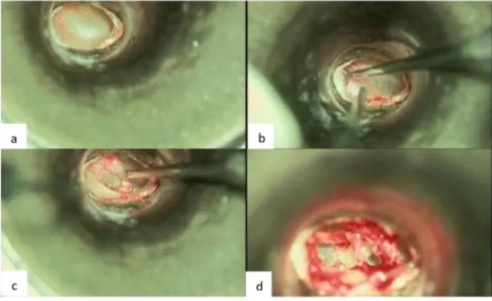
Figure 1: (a) Normal tympanic membrane, (b) incision taken 6 mm from annulus, (c) elevation of
tympano-meatal flap and (d) incudostapedial joint.
Figure 2: (a) Cutting of stapedius tendon, (b) dislocation of incudostapedial joint, (c) fenestra in
region of footplate of stapes, (d) teflon piston in situ.
RESULTS
The study consists of thirty cases of otosclerosis who underwent stapedotomy over 2.5 years. A detailed history, clinical examination and investigations were done in every patient and observations recorded.
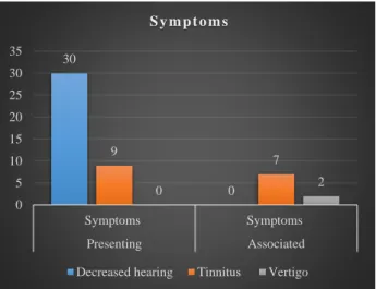
Figure 1: Clinical features.
In 9 cases (30%) tinnitus was presenting symptom with deafness while in other 7 cases (23.33%) it was an associated symptom.
The classical picture of labyrinthine vertigo was not present in any case in this study but in 2 cases (6.67%) an intermittent mild dizziness was seen.
Figure 2: Laterality of disease.
In our study about 26 patients (86.67%) had bilateral otosclerosis and 4 cases (13.33%) had unilateral otosclerosis.
All thirty cases underwent stapedotomy under local anesthesia and sedation and were followed up over a period of 3 years. In all the cases teflon piston was used as a prosthesis. Five cases had complications. The complications were managed by conservative line of treatment.
Post operative hearing
In our study patients with pure conductive loss had better air bone gap closure than patients with mixed hearing loss.
Also, nine cases (30%) with conductive hearing loss showed post-operative air bone gap of within 10 dB, while in the mixed group no case had a post-operative air bone gap of within 10 dB.
Table 1: Post-operative air conduction.
Group Post-operative air
conduction
No. of cases (%)
A Within 10 dB of bone
conduction 9 (30)
B Within 10-20 dB of bone
conduction 15 (50)
C Within 20-30 dB of bone
conduction 4 (13.33)
D Within 30-35 dB of bone
conduction 1 (3.33)
Table 2: Average air bone gap.
Type of hearing loss
Average air bone gap Average
hearing gain (dB) Pre. op
(dB)
Post. op (dB)
Mixed 37.33 19.82 17.51
Conductive 46.23 12.62 33.61
Total 43.16 15.10 28.06
Six cases (20%) in the mixed group and nine cases (30%) in the conductive group showed a post -operative air bone gap of 10-20 dB.
Table 2: Post-operative complications
One patient had tympanic membrane perforation. Temporalis fascia graft was used by underlay technique. perforation healed in this case.
The chorda tympani nerve was cut in one case (3.33%) to achieve good exposure. The patient had loss of one half of anterior two thirds of the tongue in postoperative period. No active management was done. Symptoms improved in this case.
One patient (3.33%) had profound sensorineural hearing loss on follow up 1 year after surgery, conservative treatment was given but hearing did not improve.
30
0 9
7
0 2
0 5 10 15 20 25 30 35
Symptoms Symptoms
Presenting Associated
S y m p t o m s
Decreased hearing Tinnitus Vertigo
26 4
unilateral bilateral
S. no. Complications No. of cases
(%)
1 Tympanic membrane residual
perforation 1 (3.33)
2 Chordatympani nerve palsy 1 (3.33)
3 Vertigo 1 (3.33)
4 Delayed conductive loss 1 (3.33)
5 Profound sensorineural
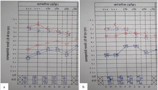
Figure 1: (a) Preoperative PTA, (b) post-operative PTA.
(a) Right ear-mild to moderately severe conductive hearing loss, left ear- moderately severe to severe conductive hearing loss. (b) Right ear (operated): Hearing sensitivity within normal limit; left ear- moderately severe to severe conductive hearing loss.
Vertigo was seen in one (3.33%) case. conservative treatment was given with bed rest. Symptoms improved in eight days.
Delayed conductive hearing loss was seen in one case (3.33%). The case was reexplored and a displaced prosthesis was noted. The new prosthesis was positioned but complete closure of air bone gap could not be attained.
DISCUSSION
We studied 30 patients with clinical diagnosis of otosclerosis for hearing outcome and complications following stapedotomy. In the majority of patients, signs of hearing loss are seen between the ages of 20 and 30 years1. In our series the maximum number of cases were
seen in the second and third decade (50%). The youngest patient being 19 years and the oldest 56 years old. From majority of earlier studies, it was reported that in clinical otosclerosis females are affected twice as often as males.1 Different studies have shown a predominance in
women.3 However, in our series males out number
females in the proportion of 1.7: 1.
The incidence of unilateral otosclerosis is 10%-15% which is consistent with our study where 86.67% of our patients had bilateral otosclerosis and 13.33% had unilateral otosclerosis.1 Pregnancy is known to precipitate
otosclerosis repeated pregnancy worsens the deafness in otosclerotic patients. This has been attributed to the role of estrogens and Progesterone. Shambaugh has reported
that in 42% of his 475 female patients there was worsening in deafness associated with pregnancy.1,3 In
our series of 30 cases out of 9 females who had a pregnancy, 5 cases (55.55% of the pregnant females) gave history of worsening of symptoms with pregnancy. Deafness was one of the presenting symptoms in all 30 cases (100%). Tinnitus was the presenting symptom in 9 cases (30%). Vertigo was an associated symptom in 2 cases (6.67%) however, it was not the presenting symptom in any. Donaldson found vertigo in 3.9% patients.4 Paracusis Willisi, defined as the ability to hear
better in noisy surroundings was absent in our series. In the present series 26 (86.67%) cases showed negative Rinne’s test on both sides and in 4 (13.33%) cases it was negative in one side. The Weber’s test was lateralized to the worse ear in all 30 cases. The side to be operated was decided on the basis of pure tone audiogram and Weber’s test. In cases where the difference was less than ten decibels in the two ears importance was given to the Weber’s test as to where it was lateralized. The absolute bone conduction test was normal in all cases in our study. Pure tone audiometry and tympanometry were done in all cases. Audiogram revealed an average of 43 dB air-bone gap in all patients. Tympanometry in all cases showed as type of curve which is suggestive of otosclerosis.
All cases were operated under local anesthesia using sedation (1 cc pentazocine and 1 cc promethazine diluted in 20 cc of normal saline injected slowly intravenously and lignocaine 2% with 1:1,00,000 adrenaline for local infiltration). The skin incision (endomeatal) was taken
under low magnification using standard incision from 6 O’clock to 12 O’clock position. According to our experience the standard incision gives adequate exposure with minimum chance of flap necrosis.
In our series we had one case (3.33%) in which the temporalis fascia graft was used for repair of the tear in the tympanomeatal flap. The perforation healed in this case. Damage to the tympnomeatal flap has been reported in 1.9% of cases by and does not preclude completion of the operation.5
Damage to the chorda tympani nerve might occur in up to 30% of cases and is more common if postero-superior bone resection is performed using a micro-drill rather than a gouge or curette.5 In our series we had to cut the
nerve in one case (3.33%). Portmann sacrificed the nerve in 12% to 15% cases.6 While dislocating the
incudostapedial joint, the force applied to dislocate the joint should be directed anteriorly, that is, away from the facial nerve and while fracturing the crura the force should be directed towards the promontory. This is to prevent injury to facial nerve in case it is exposed or there is any dehiscence in the fallopian canal. Shea has noted an incidence of idiopathic facial palsy in 0.1% of cases.7
None of our patients had any facial paresis or palsy. All stapedotomies in our series were done using the small fenestrum technique. Marquet has suggested various advantages of this technique like the fine instruments used in this technique decreases risk of tearing the membranous structure in the vestibule.8 The small hole
made on the foot plate prevents rupture of the annular ligament and so the vestibular endothelium is not breached and the contents of vestibule are not damaged. The prosthesis does not go more than 0.1 mm in the vestibule and hence prevents the chances of tearing or irritation to underlying structure. The growth of endothelium beneath the lower end of prosthesis is quick and is guided by the meniscus of perilymph; also the small curvature of the meniscus checks the entry of waste particles of bone. The inner ear is sealed rapidly from the middle ear as an extremely small opening has been made, which is almost closed by the end of the piston. The piston is firmly held in position at both ends. No pendular movement can take place and the tip remains in the center of the foot plate perpendicular to it. The main disadvantage of this technique is that, it is difficult to perform and when there is minimal fixation of foot plate, the small opening is difficult to make without mobilizing the whole foot plate and carrying out total foot plate removal. It has been shown that hearing results for the lower frequencies are not so good with this method by Smyth et al.9 Various studies have shown better short
term and long-term results with small fenestra technique compared to large fenestra by Mc-Gee, Somers, et al.10,11
Controversy still exists regarding the type and material of the prosthesis used and the use of sealing material. It is a matter of personal experience but ideally, the method
which gives better mechanical stability with good results and a minimal rate of complications is performed and preferred. Each technique has its pros and cons, but Shea’s Teflon piston is widely accepted and practiced throughout the world. Shea et al reports a success rate of 98% with air bone conduction gap of 10 decibel without any complications.12
In our study we have used the Teflon piston with a success rate of 80% with air bone conduction gap closure within 20 decibels. A series of 150 stapedectomjes without oval window sealing, using stainless steel piston was carried out by Romero-Diaz where then success rate reported was 88%, with no complication despite not sealing the oval window.13 Dawes et al used the Teflon
Piston prosthesis and stated that it is a simple procedure with a success rate of 97.5%.14 Bailey reported a success
rate of 94.5% with Teflon piston.15 Causse et al have
stated many advantages of vein graft Teflon piston interposition operation.16 Whichever be the technique
used and precautions taken complications are known to occur with the best of surgeons. We had five complications out of total of thirty cases which were profound sensorineural hearing loss, vertigo, delayed conductive loss, tympanic membrane tears, chorda tympani nerve palsy.
A number of troubles can occur in relation to the prosthesis and oval window. These include displaced prosthesis, short prosthesis, oval window fibrosis and high membrane formation.5 Crabtree et al found
displaced prosthesis to be the most common cause of failure (46%) as did Derlacki et al (82%).17,18 Ginsberg et
al found an incidence of 1.5% of slipped prosthesis in their patients operated for otosclerosis.19 We had one case
(3.33%) of delayed conductive hearing loss due to a slipped prosthesis.
One of the most dreaded complications of stapedectomy is the development of a partial or total postoperative sensorineural hearing loss. Sensorineural hearing loss may occur in the immediate postoperative period, weeks or months after operation or be delayed for months or years after the operation is performed. Quite often the cause is unknown. The causes for sensorineural hearing loss can be attributed to various factors like inappropriate instrumentation around the foot plate, floating foot-plate, acoustic trauma from drilling, rupture of the membranous inner ear, rapid loss of perilymph, foot-plate fragments or bone dust in the vestibule and serous labyrinthitis.1,5 In
general, the incidence of postoperative permanent hearing loss has ranged from 0.6 to 3% in large series.20 In our
study one patient had a dead ear. Thus, the incidence of profound hearing loss in our study is 3.33% which is high as compared to other series.
Post-operative giddiness was found in one case (3.33%) in our study in which the foot-plate was extruded while attempting a fenestrum. The footplate was removed, graft was placed over oval window, piston placed and gelfoam was placed around the piston. Patient had vertigo in the immediate post-operative period but no hearing loss at follow-up. Ginsberg et al found an incidence of 5.49% of postoperative vertigo in their study.19
Perilymph fistula formation as reported by other author are Shea (teflon & no. seal) is 2.4%; Shea (teflon and seal) is 0%; Smyth et al is 0.5%; Bellucci is 1.4%.2,9,21,22
Perilymph fistula is a serious complication as it is potentially dangerous due to the risk of meningitis and it gives rise to hearing loss which will progress if the fistula does not close either by spontaneous healing or as the result of revision surgery.1 Perilymph fistula can occur
after all operations on the stapes foot-plate. The incidence of perilymph fistula can be minimized by using a vein graft over the oval window or by using small fenestra stapedectomy or interposition operation.18 The treatment
consists of urgent re-exploration and sealing of the fistula with a soft tissue graft and a piston to keep it in place. Earlier the treatment of fistula of oval window, better are the chances of restoration of hearing otherwise a troublesome vertigo may persist with an irreversible sensorineural hearing loss.1 Fistulas are a cause of failure
in 9%-I0% of failed stapedectomy cases reported in most studies by Crabtree et al, Derlacki et al.7,17,18
Harris et al found a 5% incidence of reparative granuloma in 119 cases while reported an incidence of 1.3% in 780 stapedectomies.24 The patients present with symptoms of
hearing loss, tinnitus, and vertigo. The typical clinical presentation is that of an initial improvement in hearing after surgery with either gradual or sudden deterioration 1 to 6 weeks postoperatively. Different etiologies are put forward like infection, foreign body reaction teflon particles by Dawes et al.14,24 The treatment consists of
early re-exploration and removal of granuloma.5
One of the most alarming complication during routine stapedectomy is a “gusher- a rapid, profuse flow of fluid immediately upon opening the vestibule.5 This profuse
flow is that of cerebrospinal fluid (CSF). It is a rare complication reported in only 0.03% of cases by Causse et al over 18 years. most reported cases have been associated with congenital foot-plate fixation with sensorineural component in the pediatric population, rather than in adults with otosclerosis.19 None of our
patients had a CSF leak or a reparative granuloma.
CONCLUSION
In this study of thirty cases of otosclerosis which were operated for small fenestra stapedotomy, we conclude that hearing gain post-surgery was remarkable especially for patients with a pure conductive hearing loss. There was no deterioration in hearing after two years of follow-up. In our study on 30 patients we encountered minor
complication in 4 patients (13.33%) and 1 major complication of profound sensorineural hearing loss 3.33%. Hence, we conclude that stapedotomy is a relatively safe procedure with significant post-surgery hearing benefit.
Funding: No funding sources Conflict of interest: None declared
Ethical approval: The study was approved by the Institutional Ethics Committee
REFERENCES
1. Merchant SN, McKenna MJ, Kelly G, Swan RCI, Canter R, McKerrow WS. Otosclerosis. In: Gleeson M, Browning GG, Burton MJ, Clarke R, Hibbart J, Jones NS, et al eds. Scott-Brown’s Otorhinolaryngology, Head and Neck Surgery. 7th ed. Lon. Hodder-Arnold, 2008: 3453-3485.
2. Shea J. LXVIII Fenestration of the Oval Window. Ann Otol Rhinol Laryngol. 1958;67(4):932-951. 3. Phillip HB. Otosclerosis. Bristol [Avon]: Wright;
1981.
4. Donaldson I. Stapedectomy. A non-personal series. J Laryngol Otol. 1976;90(10):915‐8.
5. Wiet RJ, Harvey SA, Bauer GP. Complications in stapes surgery. Options for prevention and management. Otolaryngol Clin North Am. 1993;26(3):471‐90.
6. Portmann M. Procedure of "interposition" for otosclerotic deafness. Laryngoscope. 1960;70:166‐ 74.
7. Shea JJ. Thirty years of stapes surgery. J Laryngol Otol. 1988;102(1):14‐9.
8. Marquet J. Otosclerosis: small hole technique. J Laryngol Otol Suppl. 1983;8:78‐80.
9. Smyth GD, Hassard TH. Eighteen years experience in stapedectomy. The case for the small fenestra operation. Ann Otol Rhinol Laryngol Suppl. 1978;87(3 Pt 2 Suppl 49):3‐36.
10. McGee TM. Comparison of small fenestra and total stapedectomy. Ann Otol Rhinol Laryngol. 1981;90(6 Pt 1):633‐6.
11. Somers T, Govaerts P, Marquet T, Offeciers E. Statistical analysis of otosclerosis surgery performed by Jean Marquet. Ann Otol Rhinol Laryngol. 1994;103(12):945‐51.
12. Shea J, Sanabria F. A Critical Appraisal of Stapes Surgery After Ten Years. J Laryngol Otol. 1963;77(2):101-14.
13. Romero-Diaz E. Stapedectomy without window sealing. Arch Otolaryngol. 1969;89(6):807‐8. 14. Dawes JD, Curry AR. Types of stapedectomy
failure and prognosis of revision operations. J Laryngol Otol. 1974;88(3):213‐26.
15. Bailey HA Jr. Natural stapedectomy. Laryngoscope. 1968;78(5):813‐28.
17. Crabtree JA, Britton BH, Powers WH. An evaluation of revision stapes surgery. Laryngoscope. 1980;90(2):224‐7.
18. Derlacki EL. Revision stapes surgery: problems with some solutions. Laryngoscope. 1985;95(9 Pt 1):1047‐53.
19. Ginsberg IA, Hoffman SR, White TP, Stinziano GD. Hearing changes following stapedectomy: a six year follow-up. Laryngoscope. 1981;91(1):87‐92. 20. Hannley MT. Audiologic characteristics of the
patient with otosclerosis. Otolaryngol Clin N Am. 1993;26(3):373‐87.
21. Shea JJ. Polyethylene Tube Technique. Arch Otolaryngol. 1969;89(2):431.
22. Bellucci RJ. Footplate extraction in stapedectomy. Laryngoscope. 1978;88(4):701‐6.
23. Pedersen CB, Felding JU. Stapes surgery: complications and airway infection. Ann Otol Rhinol Laryngol. 1991;100(8):607‐11.
24. Harris I, Weiss L. Granulomatous complications of oval window fat grafts. Laryngoscope. 1962;72:870‐ 85.