© 2019 by the Serbian Biological Society How to cite this article: Liu J, Mao Y. Eugenol attenuates concanavalin 339 A-induced hepatitis through modulation of cytokine levels and inhibition of
mitochondrial oxidative stress. Arch Biol Sci. 2019;71(2):339-46.
Eugenol attenuates concanavalin A-induced hepatitis through modulation of cytokine
levels and inhibition of mitochondrial oxidative stress
Jing Liu1,* and Yidong Mao2
1Department of Preventive Medicine, School of Health Sciences, Wuhan University, Wuhan, Hubei, China, 430071 2Department of Pathogenic Biology, School of Basic Medical Sciences, Wuhan University, Wuhan, Hubei, China, 430071
*Corresponding author: [email protected]
Received: January 21, 2019; Revised: March 6, 2019; Accepted: March 11, 2019; Published online: March 20, 2019
Abstract: Therapeutic management of hepatitis with conventional drugs alone worsens hepatic functioning in the long term because of sustained oxidative stress. Active compounds from several plant sources have been investigated to counteract this. Eugenol, a phytochemical abundant in various plants, is known for its wide range of pharmacological effects. There is a lacuna in the deeper understanding of its hepatoprotective activity at the molecular level. Our present study aimed to determine the effects of eugenol on the changes in antioxidant components, inflammatory cytokines and modulation of mitochondrial oxidative stress in immune-mediated hepatitis. We employed a model that mimics viral hepatitis using con-canavalin A (ConA) to induce T-cell-mediated acute hepatitis. Eugenol increased (P<0.01) antioxidant enzyme activities, including reduced glutathione (GSH)-regenerating enzyme, glutathione reductase, and glucose-6-phosphate dehydroge-nase. Its antiinflammatory and antifibrogenic effects were evident from the reduction (P<0.01) in interleukin and tumor necrosis factor levels. Eugenol was found to decrease mitochondrial oxidative stress, which was elevated in hepatitis. The hepatoprotective effects of eugenol were confirmed by histological findings. The current investigation shows that eugenol exerts a hepatoprotective effect through the modulation of different pathways which include restoration of mitochondrial oxidative stress. Eugenol could be a promising candidate for human hepatitis management, warranting preclinical studies. Keywords: hepatitis; ConA; eugenol; mitochondrial ROS; antioxidant
INTRODUCTION
Every year, about 5 million people worldwide die due to hepatitis [1]. Literature data indicate that viral infections are the major causative factor of hepatitis, where there are no early markers available to detect the infection [2]. Such cases attract clinical attention only after a significant amount of liver injury and liver failure. Earlier studies have reported that serum cytokines such as tumor necrosis factor α (TNF-α), interferon γ (IFN-γ), and interleukins (IL-1 and IL-6) assume pivotal functions in animal models of acute hepatitis injury [3]. During viral infection, due to the inhibition of antioxidant enzyme synthesis, an excess generation of reactive oxygen species (ROS) occurs that further affects the cell redox equilibrium [4]. He-patocytes exposed to various toxicants are responsible for increased ROS generation in liver. The increase in
ROS activates Kupffer cells, which are responsible for increased production of proinflammatory and profi-brotic cytokines, including transforming growth factor β (TGF-β) [5]. Hepatitis B and C lead to chronic liver injury that can progress to liver fibrosis, increased ROS generation and TGF-β production [6].
and hepatitis of activated and infiltrated T cells [11]. Further, it has been reported that ConA also induces hepatitis via ROS generation. Within 12 h after ad-ministration of ConA, malondialdehyde (MDA) levels were significantly increased in experimental animals, indicating active ROS production [12]. Considering these facts, a compound known to possess antiviral, antiinflammatory and antioxidant potential is an ideal drug candidate for hepatitis management. One such a compound is eugenol.
The Food and Drug Administration (FDA) and Food and Agriculture Organization (FAO) of WHO recognize eugenol as a safe chemical [13]. Eugenol, also known as 4-allyl-2-methoxy phenol, belongs to the class of phenylpropanoids. Eugenol is a natural monoterpene abundant in many essential plant oils [14]. Previous studies have established the antioxidant [15], antiinflammatory [16] and antiviral [17] effects of eugenol. Eugenol is traditionally used in the food industry as a flavoring agent and in the cosmetic in-dustry for its fragrance [18,19]. It has been used for its antiseptic, analgesic and antibacterial properties in Asian countries. It is also used in dentistry as a cavity-filling agent [19]. Our investigation was focused on evaluating the hepatoprotective effect of eugenol in ConA-induced hepatitis.
MATERIALS AND METHODS
Experimental animals
Wistar male albino rats weighing 150-180 g were main-tained in a 12 h dark/light cycle at constant tempera-ture and humidity. A standard animal diet and water were provided ad libitum. For one week, the animals were housed under observation for adaptation. The institutional Animal Ethics Committee approved the experimental protocol used in this study (Approval No. B51242/2017).
Drugs and chemicals
ConA was obtained from Sigma-Aldrich Chemicals Co. (St. Louis, MO, USA). Eugenol, glutathione (GSH), ethylenediaminetetraacetic acid (EDTA), dimethyl sulfoxide (DMSO), and acetic acid were obtained from
Macklin Biochemical Co. Ltd. Shanghai, China. All other chemicals were of extra-pure grade or analytical grade available commercially.
Experimental design
A total of 24 animals were divided equally into four groups (6 animals per group). The first group (control) received only a single injection of phosphate-buffered saline via the tail vein. The second group of animals were administered a single dose of ConA (12 mg/kg intravenously (i.v.)) via the tail vein [20]. Group III received 5 mg/kg/day eugenol by oral gavage for 5 days before ConA administration and concomitantly during ConA administration. Eugenol alone was ad-ministered to the fourth group of animals as in group III. The animals were killed after the treatment period by cervical dislocation and samples were collected for further studies. Blood was collected and the serum was separated to determine the activities of alanine amino transferase (ALT) and aspartate amino trans-ferase (AST). The levels of other parameters, such as albumin, bilirubin and different cytokines, were also examined. Immediately after the animals were killed, liver tissues were removed, washed with ice-cold phosphate-buffered saline (PBS), and one portion was kept at -80°C for biochemical analyses. The other portions of liver tissues were fixed in 10% formalin and histopathological studies were performed. Biochemical estimations
AST, ALT, albumin, bilirubin, thiobarbituric acid reactive substances (TBARS) and GSH levels were assayed as described by the kit manufacturer (Cay-man Chemical Company, USA). An Abcam, USA, kit was used to estimate the activities of the anti-oxidant parameters, superoxide dismutase (SOD), catalase (CAT), glutathione peroxidase (GPx), glutathione reeducates (GR) and glucose 6-phos-phate dehydrogenase (G6PDH).
Cytokine assay
Measurement of mitochondrial oxidative stress
The Amplex Red Hydrogen Peroxide/Peroxidase Assay kit obtained from Thermo Fisher Scientific, USA, was used to isolate liver cell mitochondria. The samples were assessed for the level of ROS. Using reverse flow of electrons, ROS generation at complexes III and I was determined. All the experiments were performed at a constant temperature (37°C).
Histological studies
Immediately after collection, the liver tissues were rinsed in 0.1 M PBS and fixed in 10% formalin for a minimum of 72 h and a maximum of 120 h. After the minimum period, tissue sections were made from the formalin-preserved liver tissues, after dehydration, clarification and embedding in paraffin wax. The sec-tions were stained for hematoxylin and eosin (H&E). Phase contrast microscope was utilized to examine the stained liver sections. Pathophysiological differences in different experimental animal liver sections were analyzed and scored as described [21].
Statistical analysis
Sigma Plot 12.0, Systat Software, Inc., (USA) was used to analyze the statistical differences between different experimental groups. Different experimental groups were compared by one-way analysis of variance (ANOVA), followed by post hoc Bonferroni’s t tests for “control vs. ConA” or “ConA vs. the ConA+eugenol group” or “ConA vs. the ConA+eugenol group”.
RESULTS
The experimental treatment is delineated in Fig. 1A. The activities of ALT and AST and the levels of albu-min and bilirubin in the serum are shown in Fig. 1B and C. The liver injury marker enzyme ALT and AST activities in the serum were found to be increased in ConA (P<0.001) and ConA+eugenol (P<0.05) groups when compared to the control animals (group I). When a comparison was made between the ConA and ConA+eugenol group, the activities of these enzymes were found to be decreased (P<0.001), indicating a hepatoprotective effect of eugenol on animals in the
ConA-induced hepatitis group. No statistically sig-nificant differences in ALT and AST activities were observed in eugenol-alone-treated animals when compared to the control group, indicating the non-toxic nature of eugenol. Next, we analyzed liver func-tion by analyzing serum albumin and bilirubin levels. The albumin content was lowered in ConA (P<0.001) and ConA+eugenol (P<0.05) rats when compared to animals from the control group. However, albumin level was found to be elevated in ConA+eugenol-treated animals (P<0.001) in comparison to ConA animals. Bilirubin was increased in ConA- (P<0.001) and ConA+eugenol-treated animals (P<0.05) when compared with group 1 animals (control group). The level of bilirubin level was found to be decreased in the Con A+eugenol-treated group (P<0.001) when compared with the group treated only with Con A. Only the eugenol-treated animals showed a statisti-cal difference when compared with control animals.
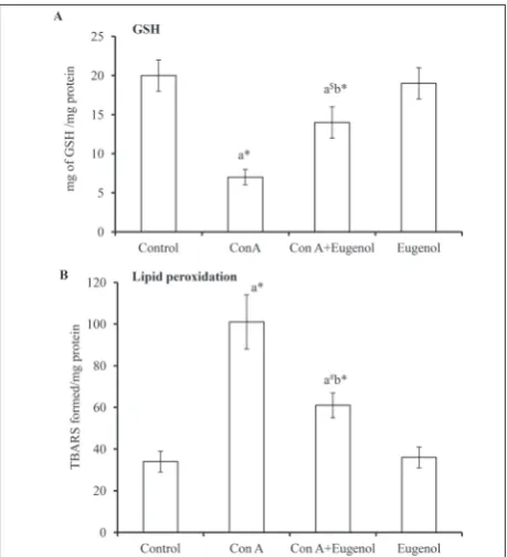
Fig. 2 shows the levels of GSH and peroxidation of lipids, in terms of TBARS, in the liver tissue of differ-ent experimdiffer-ental animals. The level of GSH (Fig. 2A) was reduced in ConA- (P<0.001) and ConA+eugenol-treated animals (P<0.05) animals when compared with the control group (group 1). The level of TBARS (Fig. 2B) was increased in the ConA- (P<0.001) and ConA+eugenol-treated (P<0.01) groups in comparison to the control group. When the ConA-alone-treated animals were compared with the ConA+eugenol group,
Fig. 1.A - The experimental regimen. B - Activities of serum
transaminases. C – Albumin and bilirubin (C) levels. Values are
stated as the mean±standard deviation (n=6). Different experi-mental groups were compared by one-way ANOVA followed by
post hoc Bonferroni’s t tests. ‘a’ vs. control; ‘b’ vs. ConA rats. *P<
the level of GSH was increased (P<0.001), whereas TBARS was decreased (P<0.001). Animals treated with eugenol alone did not show any statistically significant difference when compared to control animals.
Table 1 shows the activities of SOD, CAT, GPx, GR and G6PDH in different experimental animals. These primary antioxidant defense enzymes’ activities were decreased in ConA (P<0.001) and ConA+eugenol animals as follows: SOD (P<0.01); CAT (P<0.05); GPx (P<0.05); GR (NS) and G6PDH (NS) in comparison with control animals. In ConA+eugenol-treated animals these activities were found to be increased as follows:
SOD- (P<0.01); CAT (P<0.01); GPx (P<0.05); GR (P<0.01) and G6PDH (P<0.01) when compared with animals that received ConA alone. We did not observe any statistically different change in these parameters in animals that were treated with eugenol alone when compared to control animals.
Fig. 3 shows the inflammatory cytokines and fi-brogenic growth factors in control and experimental groups. TNF-α, IL-1β, IL-6 and fibrogenic growth fac-tors, including TGF-β, were elevated in ConA (P<0.001) and ConA+eugenol (TNF-α (NS), IL-1β (NS), IL-6 (P<0.05) and TGF-β (P<0.05)) animals when compared with control animals. When ConA+eugenol-treated animals were compared with animals administered ConA alone, these levels were decreased (P<0.001). When comparing animals treated with eugenol alone with control animals, no statistical difference was observed.
Fig. 2. Levels of GSH and TBARS in control and experimental rats. Values are stated as the mean±standard deviation (n= 6). Different experimental groups were compared by one-way ANOVA followed by post hoc Bonferroni’s t tests. ‘a’ vs. control; ‘b’ vs. ConA rats. *P< 0.001; $P< 0.05 and #P< 0.01.
Table 1. Activities of different antioxidant enzymes in control and experimental animals.
The units are as follows: for SOD units/mg protein; CAT – nmoles H2O2 utilized/mg protein; GPx – nmoles GSH utilized/mg protein; GR – nmoles
NADPH oxidized/min/mg protein; G6PD – nmoles inorganic phosphorus liberated/min/mg protein, at 37°C. Values are stated as the mean±standard deviation (n=6). Different experimental groups were compared by one-way ANOVA followed by post hoc Bonferroni’s t tests. ‘a’ vs. control; ‘b’ vs. ConA rats; ‘a’ vs. control; ‘b’ vs. ConA rats; *P< 0.001; &P< 0.01 and $P< 0.05.
Groups (n=6) SOD CAT GPx GR G6PD
Control 0.52±0.053 55.5±5.62 7.26±0.89 23±2.45 1.68±0.21
ConA 0.25±0.031a* 31.4±3.42a* 4.11±0.57a* 11±2.00a* 1.14±0.15a*
ConA+Eugenol 0.39±0.032a&b& 44.5±4.65a$b& 6.41±0.81a$b$ 19±2.16aNSb& 1.46±0.14aNSb&
Eugenol 0.56±0.64 57.5±5.45 7.56±0.78 24±3.00 1.84±0.21
Fig. 3. The effect of eugenol on different inflammatory cytokines.
TNF-α (A); IL-6 (B); IL-1β (C) and TGF- β (D). Values are stated
as the mean±standard deviation (n=6). Different experimental
groups were compared by one-way ANOVA followed by post
Mitochondrial ROS and electron flow are shown in Fig. 4. The mitochondrial ROS and reverse elec-tron flow were found to be increased in ConA and ConA+eugenol (P<0.001) animals when compared with the control group. After eugenol treatment, in the hepatitis (ConA+eugenol animals) these levels reverted to normal values (P<0.05) when compared to animals treated with ConA alone. We did not ob-serve any statistical difference in animals treated with eugenol alone.
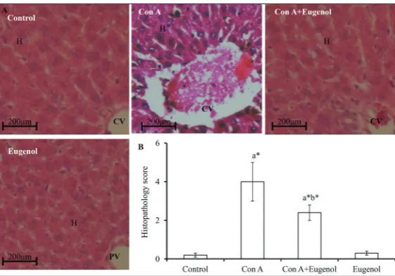
To confirm the above biochemical changes, liver histology was analyzed by a pathologist blinded to this experimental set-up. As can be seen in Fig. 5, ConA-alone-administered animals contained dead hepatocytes, exhibited dilation of sinusoids, dilation of the central vein (CV) and infiltration of lymphocytes. The above changes were minimal in ConA+eugenol-treated animals when compared to animals ConA+eugenol-treated with ConA alone. There were no histopathological differences in animals administered eugenol alone when compared to control animals. The histopatho-logical score of different experimental animals (Fig. 5B) reflected H&E observations.
DISCUSSION
As there is no technique/marker to detect liver func-tion in the early stages of liver injury [22], it is difficult to satisfactorily treat patients with acute or chronic liver injury [23]. Because of its anatomical location, the liver is prone to exposure to various chemicals and biological insults. Chemoprevention could be a means of maintaining hepatic homeostasis, and if the chemopreventive substance is from a safe natural sources such as an edible plant, it would be an ideal candidate. One such compound is eugenol, which is used in different Asian countries for treating different diseases [15-19].
In acute or chronic hepatitis, the liver is exposed to significant ROS levels. Further, it has been shown that in viral infection-induced hepatitis C or B, significant oxida-tive stress-induced damage is produced in liver tissue [24]. Similarly ConA-induced liver injury is also mediated through creased oxidative stress, observed as in-creased TBARS levels [12]. In the present investigation, we obtained similar findings. Increased serum ALT and AST activities were accompanied by increased TBARS in rats with ConA-induced hepatitis. Pretreat-ment of animals with eugenol significantly decreased serum ALT and AST activities, with a concurrent reduction in liver TBARS level. This finding showed that eugenol protected against ConA-induced hepatitis, which could be attributed to the antioxidant
Fig. 4. Mitochondrial ROS in different experimental groups. Values are stated as the mean±SEM (n=6). Different experimental groups
were compared by one-way ANOVA followed by post hoc
Bonfer-roni’s t tests. ‘a’ vs. control; ‘b’ vs. ConA rats. *P<0.001 and &P<0.01.
Fig. 5. Histological observation of the liver. A – Representative images of liver
sections of different experimental groups (600x magnification). B –
Histopatho-logical score. Values are stated as the mean±SEM (n=6). Different experimental
groups were compared by one-way ANOVA followed by post hoc Bonferroni’s t
activity of eugenol [15] that is widely reported in the literature. Eugenol pre-supplementation also increased liver metabolic function, observed as increased albumin and decreased bilirubin levels in the serum. It is worth mentioning that any molecule or compound that could restore liver function is an ideal candidate for treating hepatitis. Ravikumar et al. [20] also reported similar findings in animals with ConA-induced hepatitis and treated with plant extracts.
The liver is considered a major source for GSH. The decrease in GSH in rats with hepatitis indicated that oxidative stress mediated liver injury in ConA-induced hepatitis. In the study by Abd et al.[25], the authors showed that eugenol has the ability to stimulate the antioxidant enzyme system for xenobiotic metabolism in the liver. The decrease in liver GSH directly correlates with the reduction of SOD, CAT, GPx, GR and G6PDH activities in the liver [26]. H2O2 is primarily neutralized by GPx, which maintains a constant check on the increase in cellular peroxide levels [27]. Therefore, the decreased activities of GPx in ConA rats could be due to overproduction of H2O2 resulting in redox imbalance. Further, H2O2 and the hydroxy radical are detoxified through the heme protein CAT. We also observed reduced GR and G6PDH in ConA-administered rats, which indicated that GSH could not be replenished in these animals. The above changes were reverted in animals with eugenol pre-supplementation. Recently, Binu et al. [28] showed that eugenol restored liver function from arsenic-induced liver injury. Further, the antioxidant effects of eugenol have been highlighted in various disease models, prov-ing its versatility [29,30].
T-cell activation is not uncommon in autoimmune-mediated hepatitis. ConA activates T-cells, which are responsible for the increased levels of inflammatory cytokines, such as TNF-α, IL-6 and IL-1β. Elevated inflammatory cytokines in ConA-treated rats damage major parenchymal cells [31]. We have also observed an elevation in proinflammatory cytokines in ConA-injected rats. Our present findings agree with previous studies in which a similar elevation of inflammatory cytokines in ConA-induced hepatitis was reported [32-36]. We observed that eugenol pre- and con-comitant treatment suppressed the ConA-induced elevation of proinflammatory cytokines. This may be linked to the antioxidant and antiinflammatory
potential of eugenol. TGF-β is known to control dif-ferent basic cellular processes, such as cell adhesion, growth, migration, differentiation and extracellular matrix (ECM) accumulation [37]. TGF-β is a fibrogenic growth factor produced by different immune lineage cells. One of the fundamental mechanisms that involve the release of TGF-β from immune cells is increased ROS, as seen in different hepatitis conditions [38]. We also obtained similar findings in Con A-induced hepatitis rats. Pre- and concomitant treatment of rats with eugenol curtailed the release of this growth factor from immune cells. Similar findings were reported previously in other experimental conditions [39,40].
One sources for ROS are the mitochondria [41]. Our study is the first to report that mitochondrial ROS increase in ConA-injected animals. The increased mitochondrial ROS could be due to increased proin-flammatory cytokines and fibrogenic growth factors secreted by activated T- and Kupffer cells. Eugenol treatment could restore mitochondrial ROS by cur-tailing proinflammatory cytokines. A previous study showed that mitochondrial ROS are elevated in ste-atosis rat models, which are in turn attenuated by antioxidant treatment [42]. Similarly, we observed in our study that eugenol pre- and concomitant treat-ment shielded against ROS-induced hepatic damage. Our findings were confirmed through histological observation, which also showed that eugenol prevented ConA-induced hepatitis. In eugenol-treated rats, the normal architecture of the liver was almost restored. They also exhibited decreased central vein dilation, reduced hepatocytes death, reduced sinusoid dilation and reduction of infiltrating blood cells. Our present study indicates that eugenol, through one of these mechanisms, prevented ConA-induced hepatitis by restoring mitochondrial ROS levels, in addition to its well-known antioxidant and anti-inflammatory mechanisms.
CONCLUSIONS
profibrotic cytokine TGF-β. We showed that eugenol restores mitochondrial redox balance in ConA-induced hepatitis. Thus, eugenol could be considered as a po-tential candidate in the management of inflammatory liver injury as observed in viral infections. Further research is necessary to elucidate the molecular targets of eugenol in the hepatic system, which would aid clinicians in evaluating its therapeutic value.
Funding: This study was supported by the National Natural Sci-ence Foundation of China (81702432)
Author contributions: Both authors contributed equally to this study (study design, experimental work, data analysis, manuscript writing).
Conflict of interest disclosure: The authors declare no conflicts of interest.
REFERENCES
1. Wang T, Dai Y, Lu W, Zhou H, Chen Y, Xu X, Sun C, Cheng J. An epidemiological survey of HBV infection and low-level HBsAg in military camps in eastern China. Medicine (Balti-more). 2018;97:e12201.
2. Qiu L, Tang Q, Li G, Chen K. Long non-coding RNAs as biomarkers and therapeutic targets: Recent insights into hepatocellular carcinoma. Life Sci. 2017;191:273-82. 3. Cao Q, Batey R, Pang G, Russell A, Clancy R. IL-6, IF-γ and
TNF-α production by liver-associated T cells and acute liver injury in rats administered concanavalin A. Immunol Cell Biol. 1998;76:542-49.
4. Ruggieri A, Anticoli S, Nencioni L, Sgarbanti R, Garaci E, Palamara AT. Interplay between hepatitis C virus and redox cell signaling. Int J Mol Sci. 2013;14:4705-21.
5. Poli G. Pathogenesis of liver fibrosis: role of oxidative stress. Mol Asp Med. 2000;21:49-98.
6. Paik Y-H, Kim J, Aoyama T, De Minicis S, Bataller R, Brenner DA. Role of NADPH oxidases in liver fibrosis. Antioxid Redox Signal 2014;20:2854-72.
7. Tiegs G, Hentschel J, Wendel A. A T cell-dependent experi-mental liver injury in mice inducible by concanavalin A. J Clin Invest. 1992; 90:196-03.
8. Gantner F, Leist M, Lohse AW, Germann PG, Tiegs G. Con-canavalin A-induced T-cell-mediated hepatic injury in mice: the role of tumor necrosis factor. Hepatology. 1995;21:190-98.
9. Mizuhara H, Uno M, Seki N, Yamashita M, Yamaoka M, Ogawa T, Kaneda K, Fujii T, Senoh H, Fujiwara H. Criti-cal involvement of interferon gamma in the pathogenesis of T-cell activation associated hepatitis and regulatory mecha-nisms of interleukin-6 for the manifestations of hepatitis. Hepatology. 1996; 23:1608-15.
10. Nicoletti F, DiMarco R, Zaccone P, Salvaggio A, Magro G, Bendtzen K, Meroni P. Murine concanavalin A-induced
hepatitis is prevented by interleukin 12 (IL-12) antibody and exacerbated by exogenous IL-12 through an interferon-gamma-dependent mechanism. Hepatology. 2000;32:728-33. 11. Ballegeer M, Libert C. Different cell types involved in medi-ating concanavalin A induced liver injury: a comprehensive overview. J Gastroenterol Hepatology Res. 2016;1:1-7. 12. Shirin H, AeedH, Alin A, Matas Z, Kirchner M, Brazowski
E, Goldiner I, Bruck R. Inhibition of immune-mediated con-canavalin A induced liver damage by free-radical scavengers. Dig Dis Sci. 2010;55:268-75.
13. Hinnen C, Ranchor AV, Baas PC, Sanderman R, Hagedoorn M. Partner support and distress in women with breast can-cer: The role of patients’ awareness of support and level of mastery. Psychol Health. 2009;24:439-55.
14. Jaganathan SK, Supriyanto E. Antiproliferative and molecu-lar mechanism of eugenol-induced apoptosis in cancer cells. Molecules. 2012;17:6290-04.
15. Ogata M, Hoshi M, Urano S, Endo T. Antioxidant activity of eugenol and related monomeric and dimeric compounds. Chem Pharm Bull (Tokyo). 2000;48:1467-69.
16. Pisano M, Pagnan G, Loi M, Mura ME, Tilocca MG, Palm-ieri G, Fabbri D, Dettori MA, Delogu G, Ponzoni M, Rozzo C. Antiproliferative and pro-apoptotic activity of eugenol-related biphenyls on malignant melanoma cells. Mol Cancer. 2007;6:8-14.
17. Benencia F, Courreges M. C. In vitro and in vivo activity of
eugenol on human herpesvirus. Phytother Res. 2000;14:495-500.
18. Rastogi SC, Johansen JD, Menne T. Natural ingredients based cosmetics. Content of selected fragrance sensitizers. Contact Dermatitis 1996;34:423-26.
19. Lairungruang K, Itharat A, Panthong S. Antimicrobial activ-ity of extracts from a Thai traditional remedy called Kabpi for oral and throat infection and its plant components. J Med Assoc Thai. 2014; 97:S108-15.
20. RaviKumar S, Gnanadesigan N, Inbaneson SJ, Kalaiarasi A. Hepatoprotective and antioxidant properties of Suaeda maritima (L.) Dumort ethanolic extract on concana-valin A induced hepatotoxicity in rats. Indian J Exp Biol. 2011;49:455-60.
21. Xin HG, Zhang BB, Wu ZQ, Hang XF, Xu WS, Ni W, Zhang RQ, Miao XH. Treatment with baicalein attenuates methi-onine-choline deficient diet-induced non-alcoholic steato-hepatitis in rats. Eur J Pharmacol. 2014;738:310-18. 22. Goyal H, Zhang GM. Serum biomarkers to predict liver
fibrosis in hepatitis B. Clin Chim Acta. 2018;486:8-12. 23. Cachet X, Langrand J, Riffault-Valois L, Bouzidi C, Colas C,
Dugay A, Michel S, Boucaud-Maitre D. Clerodane furano-diterpenoids as the probable cause of toxic hepatitis induced by Tinospora crispa. Sci Rep. 2018;8:13520.
24. Ivanov AV, Valuev-Elliston VT, Tyurina DA, Ivanova ON, Kochetkov SN, Bartosch B, Isaguliants MG. Oxidative stress, a trigger of hepatitis C and B virus-induced liver carcinogen-esis. Oncotarget. 2017;8:3895-32.
26. Zhang C, Wang N, Xu Y, Tan HY, Li S, Feng Y. Molecular mechanisms involved in oxidative stress-associated liver injury induced by Chinese herbal medicine: An experimen-tal evidence-based literature review and network pharmacol-ogy study. Int J Mol Sci. 2018;19(9):E2745.
27. Tadayuki I, Akiko T, Shinya S, Ken-ichi O, Ippei H, Yuko K,
Koji T, Yoshimitsu M.A simple assay for measuring catalase
activity: A visual approach. Sci Rep. 2013;3:3081.
28. Binu P, Priya N, Abhilash S, Vineetha RC, Nair H. Protective effects of eugenol against hepatotoxicity induced by arsenic tri-oxide: An antileukemic drug. Iran J Med Sci. 2018;43:305-12. 29. Binu P, Gifty K, Vineetha RC, Abhilash S, Arathi P, Nair
RH. Eugenol, a plant-derived phenolic nutraceutical, pro-tects thiol (SH) group in myocardium from ROS-mediated oxidation under chemotherapeutic stress induced by arse-nic trioxide - a in vivo model study. Drug Chem Toxicol. 2018;4:352-57.
30. Binu P, Priya N, Abhilash S, Vineetha RC, Nair RH. Studies on curative efficacy of monoterpene eugenol on anti- leu-kemic drug arsenic trioxide induced cardiotoxicity. Biomed Pharmacother. 2017; 91:559-66.
31. Genestra M. Oxyl radicals, redox-sensitive signalling cas-cades and antioxidants. Cell Signal. 2007;19:1807-19. 32. Gantner F, Leist M, Lohse AW, Germann PG, Tiegs G,
Pag-nan G, Loi M, Mura ME, Tilocca MG, Palmieri G, Fabbri D, Dettori MA, Delogu G, Ponzoni M, Rozzo C. Concanavalin A-induced T-cell-mediated hepatic injury in mice: the role of tumor necrosis factor. Hepatology. 1995;21:190-8. 33. Bruck R, Shirin H, Hershkoviz R, Lider O, Kenet G, Aeed
H, Matas Z, Zaidel L, Halpem Z. Analysis of Arg-Gly-Asp mimetics and soluble receptor of tumour necrosis factor as therapeutic modalities for concanavalin A induced hepatitis in mice. Gut. 1997;40:133-38.
34. Klein C, Wüstefeld T, Heinrich PC, Streetz KL, Manns MP, Trautwein C ME3738 protects from concanavalin A-induced
liver failure via an IL-6-dependent mechanism. Eur J Immu-nol. 2003;33:2251-61.
35. Liang J, Zhang B, Shen RW, Liu JB, Gao MH, Li Y, Li YY, Zhang W. Preventive effect of halofuginone on concanavalin A induced liver fibrosis. PLoS One. 2013;8:82232.
36. Xia S, Han M, Li X, Cheng L, Qiang Y, Wu S, Zhang M, Xu H, Liu X, Shao Q. Dietary fish oil exacerbates concanavalin A induced hepatitis through promoting hepatocyte apop-tosis and altering immune cell populations. J Toxicol Sci. 2014;39:179-90.
37. Yoshida K, Murata M, Yamaguchi T, Matsuzaki K. TGF-β/ Smad signaling during hepatic fibro-carcinogenesis. Int J Oncol. 2014;45:1363-71.
38. Purohit V, Brenner DA. Mechanisms of alcohol-induced hepatic fibrosis: a summary of the Ron Thurman Sympo-sium. Hepatology. 2006;43:872-8.
39. Kar MS, Bhattacharjee S, Chakraborty SP, Majumdar S, Roy S. Alteration of immune functions and Th1/Th2 cytokine balance in nicotine-induced murine macrophages: immu-nomodulatory role of eugenol and N-acetylcysteine. Int Immunopharmacol. 2011;11:485-95.
40. de Araújo Lopes A, da Fonseca FN, Rocha TM, de Frei-tas LB, Araújo EVO, Wong DVT, Lima Júnior RCP, Leal LKAM. Eugenol as a promising molecule for the treat-ment of dermatitis: antioxidant and anti-inflammatory activities and its nanoformulation. Oxid Med Cell Longev. 2018;2018:8194849.
41. Mansouri A, Gattolliat CH, Asselah T. Mitochondrial dys-function and signaling in chronic liver diseases. Gastroen-terology. 2018;155:629-47.

![3,6 Di 4 pyridylpyrrolo[3,4 c]pyrrole 1,4(2H,5H) dione](data:image/gif;base64,R0lGODlhAQABAIAAAP///wAAACH5BAEAAAAALAAAAAABAAEAAAICRAEAOw==)