SIMULTANEOUS EFFECTS OF Cd(II) AND Pb(II) IONS AND
γ-IRRA-DIATION ON STABILITY OF SPIRULINA PLATENSIS
J. Monaselidze,
[a]E. Gelagutashvili,
[a]N. Bagdavadze,
[a]M. Gorgoshidze
[a]and E.
Lomidze
[a]Keywords: metal ions; γ-irradiation; Spirulina platensis; differential scanning microcalorimetry.
Effect of toxic metal ions Cd(II) and Pb(II) on cyanobacterium (blue-green algae) Spirulina platensis intact cells have been studied with optical and differential scanning microcalorimetry (DSC) methods after 7.2 kGy 137Cs gamma irradiation and without irradiation. It is shown that the addition of metal ions causes a decrease in optical absorption spectra band intensities. In the case of irradiation, the absorption band intensity decreases higher than without irradiation. The binding constant of Pb(II) with Spirulina platensis is calculated for nutrition medium with pH 9.2. DSC data show that Cd(II) and Pb(II) ions do not change the integral heat of absorption (ΔHm) that equals to 24.6 J g-1. In the case of irradiation, the DSC melting curve profile changes significantly and ΔHm decreases two times, which indicates that 50 % of proteins are denaturated. The DSC method also gives a possibility to evaluate C-phycocyanin content from deconvoluted heat absorption peak at 50 °C, which equals to 35.5 %. In case of irradiated wet mass, sub-cultured wet mass, and wet mass re-irradiated with the same dose, contents of Spirulina platensis ingredients – C-phycocyanin, chlorophyll, and carotenoids – increase as a result of the simultaneous effect of the metal ions and irradiation.
* Corresponding Authors
E-Mail: [email protected]; [email protected]
[a] I. Javakhishvili State University, E. Andronikashvili Institute of Physics. 6 M. Tamarashvili Str., Tbilisi, 0177, Georgia
Introduction
Nanotechnology gives a possibility to introduce a lot of new tools to be used in cellular and molecular biology. One of the modern trends in nanotechnology is associated with using the blue-green microalgae (cyanobacteria) Spirulina platensis that have been utilized in the food industry, pharmaceuticals, medicine and science.1 It is one of nature's first photosynthetic organisms capable of converting light directly for complex metabolic processes. One of the algae's useful qualities is its ability to protect us from radiation. Algae contain a large amount of iodine and sodium alginate that help removal of radioactive substances from the living organisms.
It was shown2 that Spirulina is a ubiquitous organism.
Spirulina platensis has attracted more attention because of its high nutritional content that includes 50–70 % protein and minerals, vitamins, amino acids, essential fatty acids, etc.3 The thermal stability of C-phycocyanin from Spirulina platensis and the compounds that additionally stabilize C-phycocyanin are crucial to food industry.4 Spirulina platensis absorbs toxic metal ions from its environment.5 It was also demonstrated that some compounds of the algal cell biomass are responsible for binding to various ions.6,7 Spirulina
platensis may be able to reduce many types of harmful stresses, including those caused by heavy metals and irradiation.8,9 In our previous works, the accumulation and biosorption of metal ions by Spirulina platensis and their components10–13 as well as Spirulina platensis usability as a matrix for production of noble metal nanoparticles14,15 have been studied.
Thermostability of Spirulina platensis cells and their component have been successfully studied with the help of differential scanning microcalorimetry (DSC).16,17
At present, some innovative technologies are focused on the metal binding capacities of various microorganisms and their components. However, the mechanism of their interaction with metal ions and gamma irradiation are unknown. In this work, we have studied the simultaneous effects of 137Cs gamma irradiation and toxic metal ions on the growth of Spirulina platensis intact cells and their constituents using UV–VIS spectrometry and DSC.
Materials and methods
Spirulina platensis IPPAS B–256 strain was cultivated in a standard Zarrouk18 alkaline saline medium at 34 ºС, illumination ∼5000 lux, at constant mixing in batch cultures.19 Cultivation of the Spirulina platensis cells was conducted for 7 days. The cell growth was evaluated by optical density by monitoring of changes in absorbance at wavelength 560 nm measured with a spectrophotometer (UV–Visible spectrometer, Cintra 10e GBC Scientific Equipment Pty Ltd, Australia). The absorption spectra from 380 to 850 nm of intact cells suspension of Spirulina platensis
(pH 9.2) in Zarrouk medium have been recorded. In all abovementioned cases, the concentration of Spirulina platensis was 1.6 mg mL-1. This was determined by instrumental measurements.20,21 The concentration of Cd(II) and Pb(II) ions was 0.5 µM.
To study the biosorption process on the Spirulina platensis
The intact cell weight content was kept constant (1.6 mg mL-1), while the initial metal concentration varied within the interval 10–3 to 10–6 M. All experiments were carried out at the ambient temperature. The dialysis was carried out in 5 mL cylindrical vessels made of organic glass. A 30µm wide cellophane membrane (Visking type, manufactures by Serva) was used as a partition. The duration of dialysis was 72 h. The metal concentration after the dialysis was measured using the atomic absorption spectrophotometer Analyst–900 (Perkin-Elmer). Each value was determined as an average of three independent estimated values with the standard deviation.
Spirulina platensis cells were exposed to 7.2 kGy -irradiation using 137Cs as a -source, at the Applied Research Center, E. Andronikashvili Institute of Physics. After the irradiation, the cells were cultivated in Zarrouk medium for 21 days. The adsorption isotherm data for metal ion binding by Spirulina cells were calculated from the Freundlich equation.22
The Spirulina platensis cell suspension and wet mass were also measured with DSC designed for diluted solutions and complex biological systems.23 The calorimeter sensitivity was 0.1 µW, the volume of measuring vessels was 0.3 cm3, the heating rate was 0.5 °C min-1, and the temperature range of measurements was from 25 to 130 °C. The accuracy of the temperature measurements was not less than 0.05 °C. The error in the determination of melting enthalpy (Hm), heat capacity dQ/dT (Cmax) was not more than 10 %.
Results and discussions
Cd(II) and Pb(II) ions effect on intact cells of Spirulina platensis was studied as a function of metal concentration at pH 9.2. The spectrum of native S. platensis biomass is illustrated in Fig. 1. Figure 1 shows the absorption characteristics of control of intact cells of Spirulina platensis. The peak at 681 nm corresponds to the absorption of chlorophyll a (Chl a). The peaks at 620.7 nm and 500 nm correspond to the absorption of phycocyanin and carotenoids, respectively. A peak at 440 nm corresponds to Soret band of Chl a.24 In Fig. 1, there are also shown the effects of Pb(II) and Cd(II) ions on the absorption of the intact cells of
Spirulina platensis.
Figure 1. Absorption spectra of intact cells of cyanobacterium
Spirulina platensis. 1 – control after incubation in nutrition medium for 7 d, 2 – same control + Pb(II), and 3 – same control + Cd(II).
Figure 1 demonstrates that the absorption intensity decreases after addition of the metal ions. The absorption is inhibited by 8, 3, 3 and promoted by 33 % at 681, 620.7, 500 and 440 nm, respectively, for Cd(II) comparing to the control. Similar results were obtained for Pb(II) – the absorption intensity was inhibited by 5, 3 and 3 % and increased by 33 % at the given wavelengths, respectively.
Figure 2. Heat absorption curve of intact Spirulina platensis cells recalculated per gram of biomass in Zarrouk medium. Conditions are the same as in Fig.1. The volume of cell suspension was 290 µl and dry biomass amount was 3.5 mg.
Figure 3. Heat absorption curve of intact Spirulina platensis cells in the presence of 0.5 µM Cd(II) recalculated per gram of biomass in Zarrouk medium. Conditions are the same as in Figure 1. The volume of cell suspension was 290 µl and dry biomass amount was 3.7 mg.
Figure 4. Heat absorption curve of intact Spirulina platensis cells in the presence of 0.5 µM Pb(II) recalculated per gram of biomass in Zarrouk medium. Conditions are the same as in Figure 1. The volume of cell suspension was 290 µl and dry biomass amount was 4.2 mg.
The curve profile changes to simpler curves in case of metal-ion treated samples contains only two separated peaks at 78 and 98 °C. As for the very weak absorption peak of chromatin at 105 °C, its thermostability does not change at the mentioned content of metal ions.23 The melting enthalpy of intact Spirulina platensis and Spirulina platensis at the presence of 0.5 µM Cd(II) and Pb(II) ions coincide within the experimental error and equals to 24.6 J g-1.
Figures 3 and 4 demonstrate that Cd(II) or Pb(II) ion concentration 0.5 µM do not damage the genetic material, but those ions change the melting curve profile, which is caused by the formation of two independent heat absorption peaks at around 78 and 97 °C. The curve deconvolution shows that the protein melting at 78 and 97 °C takes place in narrow temperature intervals, which indicate that the proteins influenced by Cd(II) and Pb(II) have high thermostability and they have highly ordered structures. It should be mentioned that we have observed a powerful heat evolution (–Q) in case of cells at presence of Cd(II) in the temperature range 30 to 50 °C, which mainly reflects respiration of cells (oxygen absorption rate), which in its turn, strongly depends on pH and heating rate.17 As far as the primary goal of this work is focused on influence of Cd(II) and Pb(II) ions on thermodynamic stability of proteomes and protein complex of Spirulina platensis that is denaturated in the temperature interval from 45 to 110 °C, we kept the samples in dark at 15 °C during 30 h in sealed DSC cells before experiments, in order to have –Q equal to about 0 J g-1. This was made to have a proper baseline for precise detection of melting parameters.
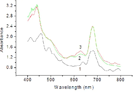
Figure 5 illustrates the absorption spectra of the intact cells of the Spirulina platensis as a control, (1) after 7.2 kGy -irradiation for 7 days, (2) control after 7.2 kGy -irradiation in the presence of Pb(II) ions, and (3) control after 7.2 kGy -irradiation in the presence of Cd(II) ions. After -irradiation, the peak intensities were decreased as follows: by 20, 11, 22 and 28 % at 681, 620.7, 500 and 440 nm, respectively, for Cd(II) ions in comparison to the irradiated control. As for Pb(II), the peak intensities were decreased by 14, 7, 18 and 20 % at the given wavelengths, respectively.
As it is seen from the abovementioned results, in both cases, cadmium(II) ions have a more significant effect on peak intensities than the lead(II) and the optical spectra positions do not change due to the effect of metals.
Figure 5. Absorption spectra of intact cells of the cyanobacterium,
Spirulina platensis. 1 – control after incubation 7 days, 2 – control after -irradiation with 7.2 kGy dose + Pb(II), and 3 – control after
-irradiation with 7.2 kGy dose + Cd(II).
The equilibrium constant was determined by the use of equilibrium dialysis and atomic absorption analysis methods for Pb(II) ions. Figure 6 presents the absorption isotherm for Pb(II) – Spirulina platensis in nutrient medium at pH 9.2, where the Freundlich adsorption model was used for the mathematical description of the biosorption of Pb(II) –
Spirulina platensis. The points presented in the figure are experimental data, and the line is derived from the Freundlich equation. The correlations between experimental data and the theoretical equation were extremely good with R2 above 0.90. Using the Freundlich isotherm, the biosorption constant (K) was determined for Pb(II)–Spirulina platensis system and were found to be equal as 1.8·10–4 M.
The Cd(II)–Spirulina platensis system sorption constant value exceeds the value given for Pb(II)–Spirulina platensis
system in the nutrient medium.10,11 The biosorption constant for Cd(II) – Spirulina platensis was dissolved in the medium at pH 8.6 was 5.1·10–4 M. We foundus that the efficiency of Cd(II) ions biosorption depends on the conditions of the uptaking processes,10,11 especially, the pH is an essential factor for Cd(II) binding of Spirulina platensis.
It was supposed25 that Cd(II), Cu(II), and Co(II) biosorption by algae biomass takes place through electrostatic interactions between the metal ions and the microbial cell walls. The results showed that carboxyl groups on algal cell biomass are the active sites for binding to various ions.26
Figure 6. Linearized Freundlich adsorption isotherms of Pb(II)–
Spirulina platensis in nutrition medium (Cb is binding metal concentration, mg g-1, and C
total is initial Pb concentration. mg L-1).
Figure 7. Absorption spectra of intact cells of cyanobacterium
Spirulina platensis. 1 – control that was sub-cultured after 7.2 kGy
-irradiation for 3 weeks, 2 – control that was sub-cultured after 7.2 kGy -irradiation for 3 weeks + Pb(II), and 3 – control that was sub-cultured after 7.2 kGy -irradiation for 3 weeks + Cd(II)
Namely, the absorption intensity decreased by 21 % at 681 nm, by 16 % at 620.7 nm, by 17 % at 500 nm, and by 25 % at 440 nm for Cd(II) ions, in comparison to the control.
Figure 8. Absorption spectra of intact cells of cyanobacterium
Spirulina platensis. 1 – irradiated mass that was sub-cultured after 7.2 kGy -irradiation for 3 weeks after repeated irradiation with same dose, 2 – irradiated mass that was sub-cultured after 7.2 kGy
-irradiation for 3 weeks after repeated irradiation with same dose + Cd(II), and 3 – irradiated mass that was sub-cultured after 7.2 kGy
-irradiation for 3 weeks after repeated irradiation with same dose + Pb(II).
As for Pb(II), the absorption intensity decreased by 10, 6, 4 and 8 % at the given wavelengths, respectively, in comparison to the irradiated wet mass.
Figure 8 demonstrates the absorption spectra of the intact cells of the irradiated Spirulina platensis mass that was sub-cultured for 3 weeks after 7.2 kGy -irradiation, and after repeated irradiation with the same dose and under effects of Cd(II) and Pb(II) ions. Curve 2 corresponds to Cd(II), and Curve 3 corresponds to Pb(II). At the presence of Cd(II) ions, the absorption intensity increased by 64, 49, 26 and 69 % at 681, 620.7, 500 and 440 nm, respectively, in comparison to the irradiated wet mass. At the presence of Pb(II), the absorption intensities are increased by 64, 59, 27 and 64 %, respectively, at the same wavelengths. Thus, the toxic metal ions promote an increase in the amount of basic components of Spirulina platensis. Namely, the presented study has demonstrated that proteins, chlorophylls and carotenoids content of Spirulina platensis significantly increases in comparison to the control as a result of the simultaneous effect of Cd(II) and Pb(II) ions and -irradiation.
Figure 9. Differential scanning microcalorimetry curve of irradiated native Spirulina platensis cells. Conditions are the same as in Figure 8. The volume of cell suspension was 290 µl and dry biomass amount was 2.1 mg.
Figure 10. Heat absorption curve of irradiated and recultivated
Figure 11. Heat absorption curve of re-irradiated Spirulina platensis
cells recalculated per gram of biomass in Zarrouk medium. Conditions are the same as in Figure 8. The volume of cell suspension was 290 µl and dry biomass amount was 1.2 mg.
The DSC measurements show that the irradiated Spirulina platensis cell suspension has a complex melting profile with weak maximums at 72 and 92 °C and a shoulder at 103 °C, the C-phycocyanin melting peak at 50 °C has been disappeared, and proteins mainly melt at 72 and 92 °C (Figure 9). We suppose that the shoulder corresponds to melting of genetic material – chromatin complex.23 The integrated heat amount is decreased to the half comparing to the non-treated sample (see Figure 2). After 3 weeks of recultivation, the same Spirulina platensis cell suspension has the curve presented in Figure 10, where the C-phycocyanin heat absorption intensity is restored and the melting temperature is shifted to higher temperatures by 6 °C, and the peak around 105 °C is very weak. For comparison, see also Figure 11.
Figure 12. Heat absorption curve as a function of the temperature of re-irradiated Spirulina platensis cells at the presence of 0.5 µM Pb(II) recalculated per gram of biomass in Zarrouk medium. Conditions are the same as in Figure 8. The volume of cell suspension was 290 µl and dry biomass quantity was 1.3 mg.
Figure 12 presents the DSC curve of re-irradiated Spirulina platensis cells at the presence of 0.5µM Pb(II). The curve shows that the C-phycocyanin peak absent, the main heat absorption occurs as a broad dominant peak around 72 °C, and a small peak appears at about 93 °C. The melting enthalpy is decreased to 1/3 compared to the intact cells. Similar results have been received for Cd(II.
The DSC data can give the value of absorption heat with high accuracy in the denaturation/melting process of
Spirulina platensis cells, therefore these peaks could be deconvoluted. Since the C-phycocyanin melts in the temperature range from 40 to 58°C (Tm = 50±1°C), from the heat calculated from the area under this peak, the C-phycocyanin content is proved to be 35±5 % of total protein amount that melts in the temperature range from 40 to 100 °C in case of Spirulina platensis in Zarrouk medium.
Acknowledgment
Paper was presented at the 5th International Conference “Nanotechnologies”, November 19–22, 2018, Tbilisi, Georgia (Nano–2018).
References
1Vonshak, A., Spirulina platensis (Arthrospira): Physiology, Cell
Biology and Biotechnology (1st Ed.), 1997, London, Taylor and Francis Ltd.
2Orio, C., Spirulina, the edible microorganism, Microbiol. Rev., 1983, 47, 551.
3Campanella, L., Crescentini, G., Avino, P., Chemical composition and nutritional evaluation of some natural and commercial food products based on Spirulina, Analusis, 1999, 27, 533.
https://doi.org/10.1051/analusis:1999130
4Martelli, G., Folli, C., Visaic, L., Daglia, M., Ferrari. D., Thermal stability improvement of blue colorant C-Phycocyanin from Spirulina platensis for food industry applications, Process Biochem., 2014, 49(1), 154.
https://doi.org/10.1016/j.procbio.2013.10.008
5Slotton, D. G., Goldman, C. R., Frank. A., Commercially grown spirulina found to contain low levels of mercury and lead,
Nutr. Rep. Int., 1989, 40(2), 1165.
6Gardea–Torresdey, J. L., Becker–Hapak, M. K., Hosea, J. M., Darnell. D. W., Effect of chemical modification of algal carboxyl groups on metal ion binding, Environ. Sci. Technol.,
1990, 19, 1372. https://doi.org/10.1021/es00079a011
7Volesky, B., Holan. Z. R., Biosorption of heavy metals, Biotechnol.
Prog., 1995, 11, 235. https://doi.org/10.1021/bp00033a001 8Sharma, K. K., Schuhmann, H., Schenk, P. M., High Lipid
Induction in Microalgae for Biodiesel Production, Energies, 2012, 5, 1532.https://doi.org/10.3390/en5051532
9Cheng, J., Huang, Y., Feng, J., Sun, J., Zhou, J., Cen, K., Mutate Chlorella sp. by nuclear irradiation to fix high concentrations of CO2, Bioresource Technol., 2013, 136, 496. https://doi.org/10.1016/j.biortech.2013.03.072
10Gelagutashvili, E., “Ch. 9. Biosorption of heavy metals by
Spirulina Platensis and their Components,” in Plants and Microbes, P. Goyal, A. Chauhan, and P. Kaushik, Eds., Mumbai, 2014, 154–174.
11Gelagutashvili, E., Comparative Study on Heavy Metals Biosorption by Different Types of Bacteria, Open J. Metal,
2013, 3, 62.https://doi.org/10.4236/ojmetal.2013.32a1008 12Gelagutashvili, E., Tsakadze, K., Effect of Hg(II) and Pb(II) Ions
on C-Phycocyanin (Spirulina platensis) , Optics Photonics J.,
2013, 3, 122. https://doi.org/10.4236/opj.2013.31020 13Gelagutashvili, E., Cyanobacteria Spirulina Platensis Basic
Protein C-Phycocyanin and Zn(II) Ions, Am. J. Nano Res.
14Kalabegishvili, T. L., Murusidze, I. G., Kirkesali, E. I., Rcheulishvili, A. N., Ginturi, E. N., Gelagutashvili, E. S., Kuchava, N. E., Bagdavadze, N. V., Janjalia, M. V., Pataraya, D. T., Gurielidze, M. A., Frontasyeva, M. V., Zinicovscaia, I. I., Pavlov, S. S., Tsertsvadze, G. I., Gabunia, V. N., Eur.
Chem. Bull., 2015, 4(1), 43. DOI:
http://dx.doi.org/10.17628/ecb.2015.4.43-49
15Kalabegishvili, T., Murusidze, I., Kirkesali, E., Rcheulishvili, A., Ginturi, E., Kuchava, N., Bagdavadze, N., Gelagutashvili, E., Frontasyeva, M. V., Zinicovscaia, I., Pavlov, S. S., Dmitriev, A. Y., Gold and silver nanoparticles in Spirulina platensis biomass for medical application, Ecol. Chem. Eng. S,2013,
20(4), 621. https://doi.org/10.2478/eces-2013-0043
16Topchishvili, L., Barbakadze, Sh., Khizanishvili, A., Majagaladze, G., Monaselidze, J., Microcalorimetric Study of Iodized and Noniodized Cells and C-Phycocyanin of Spirulina platensis,
Biomacromolecules, 2002, 3(3), 415.
17Monaselidze, J., Barbakadze, Sh., Kvirikashvili, Sh., Majagaladze, G., Khachidze, D., Topchishvili, L., Thermal Characteristics ofSpirulina platensisCells under Nongrowing Conditions at Various Values of pH Medium, Biomacromolecules, 2002,
3(4), 783.
18Zarrouk, C., Contribution al'etuded'unecyanophycee. Influence de Divers Facteurs Physiques Etchimiquessurlacroissance et Photosynthese de Spirulina maxima,PhD Thesis, 1966, Paris, University of Paris.
19Mosulishvili, L., Belokobilsky, A., Gelagutashvili, E., Rcheulishvili, A., Tsibakhashvili. N., The study of the mechanism of cadmium accumulation during the cultivation of Spirulina platensis, Proc. Georg. Acad. Sci. (Ser. Biol.),
1997, 23(1-6), 105.
20Bennet, A., Bogorad, L.,Complementary chromatic adaptation in a filamentous blue-green alga, J. Cell Biol., 1973, 58, 419.
https://doi.org/10.1083/jcb.58.2.419
21Patel, A., Mishra, S. M., Pawar, R., Ghosh, P., Purification and characterization of C-Phycocyanin from cyanobacterial species of marine and freshwater habitat, Protein Express. Purif., 2005, 40, 248.
https://doi.org/10.1016/j.pep.2004.10.028
22Freundlich. H., Over the adsorption in solution, Z. Phys. Chem., 1906, 57, 384.
23Monaselidze, J., Abuladze, M., Asatiani, N., Kiziria, E., Barbakadze, Sh., Majagaladze, G., Iobadze, M., Tabatadze, L., Holman, H.-Y., Sapojnikova, N., Characterization of chromium-induced apoptosis in cultured mammalian cells,
Thermochim. Acta, 2006, 441, 8.
https://doi.org/10.1016/j.tca.2005.11.025
24Fork, D. C., Mohanty, P., In: Light Emission by Plants and
Bacteria (Eds. A. J. Govindjee, D. C. Fork), 1986, New York, Academic Press, 451.
25Kuyucak, N., Voleskym, B., Biosorbents for recovery of metals from industrial solutions, Biotechnol. Lett., 1988, 10(2), 137.
https://doi.org/10.1007/bf01024641
26Plaza, M., Cifuentes, E., Ibanez, A., In the search of new functional food ingredients from algae, Trends Food Sci. Technol.,2008,
19, 31.
https://doi.org/10.1016/j.tifs.2007.07.012
27Privalov, P. L., In: Microcalorimetry of Macromolecules: The
Physical Basis of Biological Structures, 2012, Hoboken, Wiley, 225.