Address for correspondence
Dr. Lubna Khondker, Associate Professor Department of Dermatology & Venereology Bangabandhu Sheikh Mujib Medical University (BSMMU), Dhaka, Bangladesh.
Email: [email protected]
Original Article
Efficacy
of
combination
of
intralesional
corticosteroid injection and cryotherapy versus
intralesional corticosteroid injection alone in patients
of keloid
Introduction
A keloid may be defined as a benign growth of dense fibrous tissue, usually developing from an abnormal healing response to a cutaneous injury, extending beyond the original borders of the wound or inflammatory response.1 Clinically, they are firm nodules, which may be skin-colored, hypopigmented, or erythematous
secondary to telangiectasias.2,3 For unknown reasons, keloids occur more frequently among Blacks, Hispanics and Asians and less commonly in Caucasians.4,5 Keloids usually occur after trauma or wounding of skin.6 In addition to cosmetic disfigurement, keloids may cause protracted itch and pain resulting in a significant negative psychosocial burden for the patient.7 On histologic examination, keloids are found to have increased collagen and glycosaminoglycan deposition, both major components of the extracellular matrix.8
Clinicians always find it difficult to treat Md Qamrul Ahsan, Md Akram Ullah Sikder, Lubna Khondker
Bangabandhu Sheikh Mujib Medical University (BSMMU), Dhaka, Bangladesh
Abstract
Objective To compare the efficacy of combination of intralesional corticosteroid injection and cryotherapy with intralesional corticosteroid injection alone in patients of keloid.Methods A clinical trial was carried out in the department of Dermatology and Venereology, Bangabandhu Sheikh Mujib Medical University (BSMMU), Dhaka, Bangladesh for duration of January 2013 to August 2013. About 30 patients (group A) were treated with intralesional triamcinolone acetonide with cryotherapy and 30 patients (group B) were treated with intralesional triamcinolone acetonide.
Results Improvement rate was highest for the lesions on the chest, which was 18 (34.6%) and lowest for back 3 (5.8%). Out of all patients from group A, the mean size of the lesions were 8.17cm, 5.90 cm, 4.32 cm, and 3.57 cm at 1st visit, 2nd visit, 3rd visit and 4th visit, respectively. In group B, the mean size of the lesions was 7.50 cm, 4.92 cm, 3.00 cm, and 4.75 cm at 1st visit, 2nd visit, 3rd visit and 4th visit, respectively. Among the patients of group A and B, 27 (90%) and 25 (83.3%) cases improved, respectively.
Conclusion Each of the treatment was individually effective in the treatment of keloid but study with a larger group of patients for longer period may result in superior outcome in clinical practice through improved compliance.
Key words
keloids. Various therapeutic options have been used. Management of keloids has advanced from crude, invasive methods such as gross excision and radiation to intralesional or topical agents that act on a cellular level.9
Despite their common occurrence and the multitude of treatment modalities available, keloids remain a significant challenge for both the clinician and the patient.10 Despite the availability of many treatment modalities for the keloid, none has been found to be completely effective and satisfactory.11 Steroids are known to inhibit collagen synthesis and fibroblast growth in vitro and possess anti-inflammatory properties.12 Atrophy, one of the side effects of steroids, is utilized to achieve therapeutic effect in keloids.13 Intralesional steroid injection has a high degree of tolerability as well as effectiveness in reducing symptoms.14 It has been reported that treatment of fibroblasts with triamcinolone acetonide results in a reduction in TGF-β expression and an increase in FGF production.15
Cryotherapy is the application of extreme cold to treat or destroy keloids.13 Cryotherapy utilizes liquid nitrogen, upon evaporation, produces extreme cold, -196°. Application of liquid nitrogen to keloid tissue results in freeze destruction of keloid tissue.16 Cryotherapy has been used for smaller lesions, but its use is limited by considerable pain and sometimes prolonged healing following treatment.17 Cryotherapy has been reported to alter collagen synthesis and induce keloidal fibroblast differentiation towards a more normal phenotype.18 Some authors advocate the use of cryotherapy just prior to steroid injection in order to induce edema and thus facilitate steroid injection. Thus, in this controlled comparative study, the combined effect of intralesional corticosteroid injection with cryotherapy versus intralesional corticosteroid alone in the treatment of keloid was evaluated. Novel therapies deserve further investigation. This will likely lead to
more specific and effective treatment in the future.
Methods
A clinical trial conducted with sixty patients of keloid attended in the Department of Dermatology and Venereology, Bangabandhu Sheikh Mujib Medical University (BSMMU), Dhaka for duration of January 2013 to August 2013. Convenient type of non-probability sampling was followed. Patients were selected on the basis of inclusion and exclusion criteria. Inclusion criteria were age of the patients more than 12 years of age of either sex, having untreated keloids and patient who gave informed consent and willing to comply the study procedure. Exclusion criteria were patient with pregnancy, lactating mothers, who are under- treatment for keloids in past 12 weeks, suffering from keloid but those have contra-indications for cryotherapy, known case of corticosteroid sensitivity, patients with concomitant illnesses like renal failure, hepatic failure, peptic ulcer disease, diabetes and hypertension, and immunocompromised patients.
cryotherapy for at least 3 sessions spaced 4 weeks apart. The intralesional corticosteroid injection protocol was as follows: triamcinolone acetonide 40 mg/ml was used. The corticosteroids were injected directly into the keloid with a 30 gauge needle attached to a 1 ml insulin syringe. The volume injected differed among the patients due to varying sizes of the keloids and ranged between 0.1 ml-0.5ml.
The cryotherapy treatment protocol was as follows: EMLA cream was applied to the keloid 1 hour before treatment. The center of the keloid was sprayed with liquid nitrogen from a distance of 1 cm from the skin until the field was frozen. Patients received cryotherapy at weekly intervals using hand-held cryosurgical unit, for a maximum of six sessions or 75% flattening which ever occur in earlier. Adjacent structures were covered using adhesive putty (clay) for protection. Patients were given two freeze-thaw cycles of 15 seconds freeze time each using spot freeze technique. In case of larger lesions, the field was divided into overlapping circles of 2 cm each. Each circle was then treated separately, treating not more than 16-8 cm2 area at a time. Assessment was made one month after each treatment sessions. The parameters of the assessment included size of keloids (measured using measuring tape), and symptomatic improvement. No improvement when no flattening or <25% flattening and improvement when more than 25% flattening. Patients assessed the cosmetic appearance and the symptomatology for grading the improvement. During each visit, the size of the keloid was recorded in centimeters using the measuring tape. The clinical appearance was classified into the atrophic, hypertrophic or nodular categories. Other features like skin atrophy, erythema, ulceration, skin necrosis, hypopigmentation or hyperpigmentation, and other features were also recorded.
The strength of triamcinolone acetonide used for this study was 40 mg/ml, with up to a maximum of 2 ml per dose. The injections were scheduled at 4 weekly intervals for consecutive 3 months. Subsequently, the patients were instructed to follow up at monthly intervals for 3 months. The keloids were cleaned thoroughly with a spirit swab and the needle of the insulin syringe was inserted into the substance of keloid and the solution was pushed with adequate pressure till minimal blanching were seen. These were repeated at multiple sites on the keloid. At the end of the study, an attempt were made to analyze the effect of cryotherapy and triamcinolone acetonide in keloids and the various factors affecting the treatment outcome. Data were analyzed by computer with the help of SPSS software package. Statistical significance was set at 0.05 level and confidence interval at 95% level. Level of significance were measured by using appropriate procedures like chi square test (χ2), relative risk (RR) measurement, students t-test, ANOVA tests and others where applicable. A p value less than 0.05 were considered to be significant.
Results
In group A, 30 patients were treated with intralesional triamcinolone acetonide with cryotherapy and in group B, 30 patients were treated with intralesional triamcinolone acetonide alone.
Table 1 showed age distribution of two groups, ≤15 years and >15 years. The two groups were well matched in term of age distribution.
Table 1 Distribution of age among study groups. Age in groups
Study groups
Total Group A Group B
n % n % n %
≤ 15 Years 08 26.7 07 23.3 15 25.0
> 15 Years 22 73.3 23 76.7 45 75.0
Total 30 100.0 30 100.0 60 100.0
Χ2
value = 0.089. df = 1. P = 0.766. Not Significant (> 0.05)
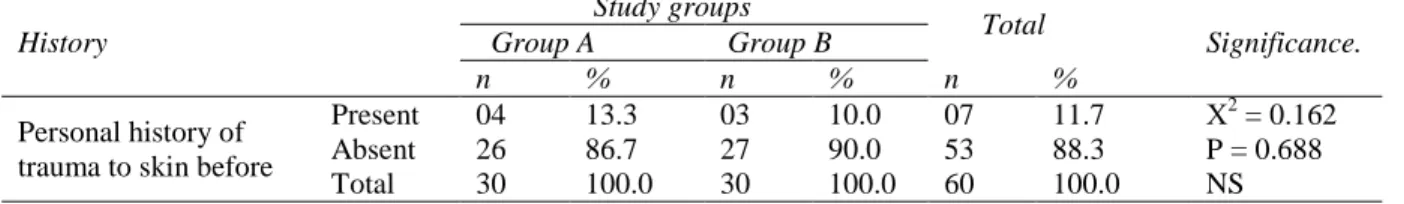
Table 2 Distribution of study groups by personal history of trauma to skin (n=60). History
Study groups
Total
Significance. Group A Group B
n % n % n %
Personal history of trauma to skin before
Present 04 13.3 03 10.0 07 11.7 Χ2 = 0.162 P = 0.688 NS
Absent 26 86.7 27 90.0 53 88.3
Total 30 100.0 30 100.0 60 100.0 Table 3 Distribution of study groups by duration of illness (n=60).
n Mean ± SD Median Range Sign.
Duration of illness (months)
Group A 30 10.00 7.24 08 2 – 36 t = 1.010
P = 0.316 NS
Group B 30 8.20 6.54 06 1 – 24
Total 60 9.10 6.90 06 1 – 36
Table 4 Distribution of study groups by number of lesion (n=60). Number of lesion
Study groups
Total Group A Group B
n % n % n %
Single 15 50.0 14 46.7 29 48.3
Multiple 15 50.0 16 53.3 31 51.7
Total 30 100.0 30 100.0 60 100.0
Χ2
value = 0.067. df = 1. P = 0.796. Not Significant (> 0.05)
Table 5 Distribution of recovery pattern among site of lesion (n=60). Site of lesion
Recovery pattern
Total Improved Not improved
n % n % n %
Chest 18 34.6 00 0.0 18 30.0
Ear 17 32.7 05 62.5 22 36.7
Extremities 10 19.2 00 0.0 10 16.6
Abdomen 04 7.7 03 37.5 07 11.7
Back 03 5.8 00 0.0 03 5.0
Total 52 100.0 08 100.0 60 100.0
Χ2
value = 8.670. df = 4. P = 0.070. Not Significant (> 0.05)
Table 6 Distribution of clinical responses among the study groups(n=60)
Size of lesion (cm) Study groups mean ± sd median range Significance
Before Treatment Group A 8.17 3.11 08 3 – 15 t = 0.779
P = 0.439
Group B 7.50 3.51 07 1 – 20
After Treatment / 2nd visit at 4th week
Group A 5.90 3.78 06 0 – 14 t = 0.104
P = 0.918
Group B 4.92 4.55 05 0 – 18
After Treatment / 3rd visit at 8th week
Group A 4.32 4.34 05 0 – 12 t = 0.104
P = 0.918
Group B 3.00 5.52 00 0 – 18
After Treatment/ 4th visit at 12th week
Group A 3.57 5.03 00 0 – 12 t = 0.104
P = 0.918
Table 7 Distribution of recovery rate among the study groups(n=60) Recovery Rate
Study groups
Total Group A Group B
n % n % n %
Improved 27 90.0 25 83.3 52 86.7
Not improved 03 10.0 05 16.7 08 13.3
Total 30 100.0 30 100.0 60 100.0
Χ2
value = 0.577. df = 1. P = 0.448. Not Significant (> 0.05)
Table 8 Distribution of duration of illness according to recovery pattern(n=60)
N mean ± sd median range Sign. Duration of Illness
(Months)
Improved 52 7.38 4.47 06 1 – 24 t = 3.733
P = 0.007 HS Not improved 08 20.25 9.59 24 6 – 36
Total 60 9.10 6.90 06 1 – 36
between two groups (p value = 0.688).
Table 3 shows that out of all patients of group A, the duration of illness ranged from 2 months to 36 months, with an average of 10 months. In group B, the duration of illness ranging from 1 month to 24 months, with an average of 8.20 months. No significant difference is observed. (p value = 0.316). Table 4 shows the distribution of number of lesions of both groups of patients. Single lesion was 15 (50%) for group A and 14 (46.7%) for group B. Multiple lesions were 15 (50%) and 16 (53.6%) for group A and group B, respectively. No significant difference is observed (p value = 0.796).
Table 5 shows the recovery pattern according to the site of distribution of lesions. Improvement rate was highest for the lesions on the chest, in 18 (34.6%) patients and lowest for back in 3 (5.8%) patients. Improvement rate for ear was 17 (32.7%), for extremities 10 (19.2%) and abdomen 4 (7.7%). No improvement was found for the lesions on the ear and abdomen, which was 5 (62.5%) and 3 (37.5%), respectively. No statistical significant difference is observed between two groups, P value = 0.070 (> 0.05)
Table 6 showed the clinical responses among the study groups. Out of all patients from group A, the mean size of the lesions was 8.17cm, 5.90 cm, 4.32 cm, and 3.57 cm at 1st visit, 2nd visit,
3rd visit and 4th visit, respectively. In group B, the mean size of the lesions was 7.50 cm, 4.92 cm, 3.00 cm, and 4.75 cm at 1st visit, 2nd visit, 3rd visit and 4th visit, respectively. No statistical significant difference is observed between two groups.
Table 7 shows the recovery rate after treatment. Among the patients of group A and B, 27 (90%) and 25 (83.3%) patients improved, respectively. The number of failure was 3 (10%) and 5 (16.7%) for group A and group B, respectively. No statistical significant difference was observed between the two groups, P value = 0.448 (>0.05).
Table 8 depicts the statistical analysis of all improved patients in both groups and duration of keloid ranging from 1 month to 24 months (mean 7.38 months). The not-improved patients had duration ranging from 6 months to 36 months (mean 20.25 months). Statistical significant difference was observed between improved and not-improved patients, P value = 0.007 (< 0.05).
Discussion
the study of Yosipovitchet al.19 and El-Tonsy et al.20 Yosipovitch et al.19 conducted a study with adolescents aged between 15 to 18 years with at least 2 keloids of at least 1 year duration were entered into the study after providing informed parental consent.19 El-Tonsy et al.20 observed that the age of the patients ranged between 2-65 years. In our study, out of all patients of group A, the duration of illness was ranged from 2 months to 36 months. In group B, the duration of illness ranged from 1 month to 24 months. Our results differed from the study of Barara et al.1 and Yosipovitch et al.19 Barara et al.1 conducted a study with 30 patients of keloids with duration of keloids under study ranged from 1-15 years. Yosipovitchet al.19 conducted a study with the mean duration of the keloids were 3.5 (range 1-4) years.
Improvement rate was highest for the lesions on the chest, 18 (34.6%) and lowest for back 3 (5.8%). No improvement was found in the lesions on the ear and abdomen, 5 (62.5%) and 3 (37.5%) cases, respectively. Barara et al.1 observed that anatomical site of keloids and etiology did not have any significant correlation with treatment outcome, either at 3 or at 6 months.
Out of all patients from group A, the mean size of the lesions reduced i.e. 8.17cm, 5.90 cm, 4.32 cm, and 3.57 cm at 1st visit, 2nd visit, 3rd visit and 4th visit, respectively. In group B, the mean size of the lesions was 7.50cm, 4.92cm, 3.00 cm, and 4.75 cm at 1st visit, 2nd visit, 3rd visit and 4th visit, respectively. Barara et al.1 showed that after 6 treatment sessions, the average flattening increased to 58.13% and average thickness reduced further to 0.213cm. The flattening of keloids varied from 12-100%. Softening was observed in all lesions except an earlobe keloid.1 Yosipovitch et al.19 showed that in terms of thickness, the keloids responded significantly better (p<0.001) to combination therapy
compared with intralesional triamcinolone alone or cryotherapy alone. Layton et al.2 conducted a study with acne keloidalis, in which intralesional triamcinolone or cryosurgery was used as treatment for keloids. They demonstrated that by treating early, and in particular vascular keloids, 85% were showed a moderate to good response in terms of flattening. Treatment with intralesional triamcinolone was also beneficial, but the response to cryosurgery was significantly better in early, vascular lesions. Prabhu et al.4 conducted a study with 29 patients. In the 15 patients who were treated with triamcinolone acetonide, the mean reduction in volume was 71.23%.
Among the patients of group A and B, 27 (90%) and 25 (83.3%) improved, respectively. El-Tonsy et al.20 observed that 8 (44%) patients showed good response with satisfactory cosmetic result. Five (28%) patients showed poor response with poor cosmetic result and many complications. In the combined cryosurgery and intralesional corticosteroids injection group, 8 (32%) patients showed excellent results. Ten (40%) patients showed good result and 7 (28%) patients showed poor response with poor cosmetic result. On the other hand in cryosurgery group, amongst idiopathic lesions, 33.3% showed good results, and 66.7% poor results. Secondary lesions showed 42.3% excellent results, 42.3% good results, and 15.4% poor results.20
effect of duration on treatment outcome, keloids were divided into 3 groups: group A: duration less than 3 years; group B: duration ranging from 3-6 years; group C: duration more than 6 years. A better percentage flattening of 51% was noted in group A lesions after 3 treatment sessions as compared to 24.5% flattening in group B and 15.8% in group C keloids. At 6 months, the percentage flattening was still highest in group A. Recent onset keloids had an early response with maximum flattening obtained in first 3 months. In contrast, older keloids with duration more than 3 years showed an initial slow response with maximum flattening being observed in the latter half of therapy.1 El-Tonsy et al.20 showed that the duration of the lesion represented an important parameter in the response of the lesion to both treatment modalities. In cryosurgery group, younger lesions (<1 year) responded well to cryosurgery followed by intralesional corticosteroid injection, as 66.7% of them showed excellent results, 33.3% showed good response, with no poor results. Older lesions (1-5 years) showed no excellent results, (1-50% showed good results and 50% showed poor response. Old lesions (>5 years) showed 16.6% excellent results, 41.7% good results and 41.7% poor response.20
Conclusion
In the light of the findings of the study we conclude that each of the treatment is individually effective but study with a larger group of patients for longer period may result in superior outcome in clinical practice through improved compliance. Further multicenter, randomized, double-blind study should be conducted with large sample size.
References
1. Barara M, Mendiratta V, Chander R. Cryotherapy in treatment of keloids:
Evaluation of factors affecting treatment outcome. J Cutan Aesthet Surg. 2012;5 :185-9.
2. Layton AM, Yip J, Cunliffe WJ. A comparison of intralesional triamcinolone and cryosurgery in the treatment of acne keloids. Br J Dermatol. 1994;130:498-501. 3. Sharma S, Bhanot A, Kaur A, Dewan SP.
Role of liquid nitrogen alone compared with combination of liquid nitrogen and intralesional triamcinolone acetonide in the treatment of small keloids. J Cosmet Dermatol. 2007;6:258-61.
4. Prabhu A, Sreekar H, Powar R, Uppin VM. A randomized controlled trial comparing the efficacy of intralesional 5-fluorouracil versus triamcinolone acetonide in the treatment of keloids. J Sci Soc. 2012;39 :19-25.
5. Mutalik S. Treatment of keloids and hypertrophic scars. Indian J Dermatol Venereol Leprol. 2005;71:3-8.
6. Shepherd JP, Dawber RP. The response of keloid scars to cryosurgery. Plast Reconstr Surg. 1982;70:677-81.
7. Zouboulis CC. Principles of cutaneous cryosurgery: An update. Dermatology. 1999;198:111-7.
8. Rusciani L, Rossi G, Bono R. Use of cryotherapy in the treatment of keloids. J Dermatol Surg Oncol. 1993;19:529-34. 9. Cruz NI, Korchin L. Inhibition of human
keloid fibroblast growth by isotretinoin and triamcinolone acetonide in vitro. Ann Plast Surg. 1994;33:401-5.
10. Robles DT, Moore E, Draznin M, Berg D. Keloids: Pathophysiology and management. Dermatol Online J. 2007;13 (3):9.
11. Pollack SV. Treatment of keloids. In: Wheeland RG, Ed. Cutaneous Surgery. Philadelphia: WB Saunders; 1994. p. 688-98.
12. Manuskiatti W, Fitzpatrick RE. Treatment response of keloids and hypertrophic sternotomy scars. Arch Dermatol. 2002;138:1149-55.
13. Berman B, Flores F. Recurrence rates of excised keloids treated with post operative triamcenolone acetonide injections or interferon α 2b injections. J Am Acad Dermatol. 1997;37:755-7.
15. English RS, Shenefelt PD. Keloids and hypertrophic scars. Dermatol Surg. 1999;25:631-8.
16. Kill J. Keloids treated with topical injection of triamcinolone acetonide (Kenalog), Immediate and long-term results. Scand J Plast Reconstr Surg. 1977;11:169-72. 17. Bock O, Schmid-Ott G, Malewski P,
Mrowietz U. Quality of life of patients with keloid and hypertrophic scarring. Arch Dermatol Res. 2006;297:433-8.
18. Kelly AP: Medical and surgical therapies for keloids. Dermatol Ther. 2004;17:212-8.
19. Yosipovitch G. A Comparative Study of Combination Intralesional Corticosteroid with Cryotherapy versus Intralesional Corticosteroid and Cryotherapy Alone in the Treatment of Keloids in Adolescents. J Dermatol Surg Oncol. 1999;5:54-6.
![Antileishmanial effect of zinc sulphate on promastigotes viability of Leishmania (L) major [MRHO/IR/75/ER]](data:image/gif;base64,R0lGODlhAQABAIAAAP///wAAACH5BAEAAAAALAAAAAABAAEAAAICRAEAOw==)