Address for correspondence
Dr. Swapna Kameti, Assistant Professor, Department of Dermatology,
Katuri Medical College & Hospital, Guntur, Andhra Pradesh, India – 500019
Original Article
Spectrum of cutaneous manifestations in diabetes
mellitus and their association with duration of
diabetes mellitus: An observational study in a tertiary
care hospital of South India
Introduction
Diabetes mellitus (DM) is the most common
endocrine disorder which exhibits a variety of
multisystem complications involving the blood
vessels, eyes, cardiovascular, renal, nervous
systems and skin. The number of people with
DM in the world increased from 108 million in
1980 to 422 million in 2014.
1The skin is
affected
by
both
the
acute
metabolic
derangements and the chronic degenerative
complications of DM.
2Chronic hyperglycemia
resulting in production of advanced glycosylated
end
products
may
be
responsible
for
pathogenesis of many diabetic complications.
3Skin manifestations can be the presenting sign of
DM but more often appear in known DM
Swapna Kameti, Senthil Kumar, Vijaya Mohan Rao*, Penchala Reddy*, C ArunaDepartment of Dermatology, Katuri Medical College & Hospital, Guntur, Andhra Pradesh, India *Department of Dermatology, Narayana Medical College & Hospital, Nellore, Andhra Pradesh, India
Abstract
Objective To study the prevalence and patterns of cutaneous manifestations among diabetic patients and their association with duration of diabetes mellitus (DM).Methods A cross-sectional study was performed on patients attending the outpatient department of
Dermatology, Venereology and Leprosy of a tertiary care hospital in South India. A total of 200 patients with dermatological manifestations having pre-existing diabetes and patients in whom diabetes was diagnosed later were included in the study. Statistical analysis was carried out using analysis of variance (ANOVA) test and SPSS 21 software.
Results Majority of patients (63%) belonged to the age group 40-59 years with female preponderance (58.5%). Cutaneous manifestations as presenting feature of diabetes were observed in 21.5% cases. The diabetic status was uncontrolled in 73% cases. Among the various cutaneous manifestations observed, cutaneous infections (52%) were most commonly observed, especially in early diabetics of upto 5 years duration and dermatoses due to chronic degenerative complications were observed in long-standing diabetics which was statistically significant (P<0.05).
Conclusion Cutaneous manifestations may be the first clue to underlying DM. Through awareness
about cutaneous manifestations of DM, dermatologist can not only take credit for detecting DM but also facilitate early diagnosis of systemic complications of DM and thereby play an important role in improvement of quality of life and management strategy of diabetic patients.
Key words
patients during the course of the disease, as
observed in 30 to 71% of diabetics.
4This work
was an attempt to study the prevalence and
patterns of cutaneous manifestations among
diabetic patients and their association with
duration of DM and help to detect and prevent
the related systemic complications of diabetes
by early institution of appropriate treatment.
Methods
The present study was a cross-sectional study
performed on patients attending the out-patient
department of Dermatology, Venereology and
Leprosy of a tertiary care hospital in South India
after obtaining institutional ethical clearance. A
total of 200 patients, including patients with
dermatological
manifestations
having
pre-existing diabetes and patients who came with
only skin disease in whom diabetes was
diagnosed later were included in the study after
taking
their
consent.
Drug-induced
and
pregnancy-induced diabetes mellitus cases were
excluded from the study. In the selected patients,
a detailed history with particular reference to
demographic details, family history of similar
complaints and of DM and duration and type of
diabetes and treatment taken, duration of various
symptoms and evolution of lesions were
enquired into. The patients were clinically
examined in good light for varied cutaneous
manifestations
and
mucous
membrane
involvement. Complete systemic examination
was done and BMI was calculated and ocular
examination was carried out by ophthalmologist.
The findings were recorded. Investigations like
random blood glucose, fasting and post prandial
blood glucose, HbA1c, oral glucose tolerance
test, urine glucose, urine ketone bodies,
complete hemogram, liver and renal function
tests, lipid profile were done. Beside laboratory
procedures like Tzanck smear, KOH mount,
Wood’s lamp examination and Gram staining
were carried out when required and skin biopsy
was done in few cases.
The ANOVA test was used to assess the
association of duration of DM with cutaneous
manifestations of DM. P<0.05 was considered
significant. Statistical analysis was carried out
using SPSS software version 21.
Results
In the present study of 200 patients with
cutaneous manifestations
(Table 1)
, majority
belonged to 5th and 6th decades with 27% and
36%, respectively with mean age of 51.84±11
years. Females constituted 58.5% of cases with
male to female ratio being 1:1.4.
Known diabetics constituted 78.5% of the study
group, whereas 21.5% patients were unaware of
their diabetic status and presented for their
dermatological problem only. The diabetic status
was uncontrolled in 146 (73%) cases. The mean
duration of DM was 5.08±6.68 years. Majority
(97.5%) of the patients belonged to type 2 DM
and 111 (55.5%) patients had a family history of
DM. In this study, 83 (41.5%) patients (51
females and 32 males) were obese.
Infections comprised the commonest group in
obese diabetics (24.5%) followed by dermatoses
more commonly associated with diabetes (20%)
such as acrochordons and acanthosis nigricans
(AN). Among the 157 known diabetics, majority
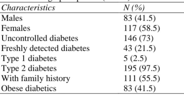
Table 1 Demographic profile (n=200).
Characteristics N (%)
Males 83 (41.5)
Females 117 (58.5)
Table 2 Duration of diabetes mellitus and type of skin manifestations (n=200). Duration Infections
Microangio-pathy
Ischemia and neuropathy
Most common dermatoses
Co-incidental dermatoses
Pruritus
Incidental 31 2 0 16 3 19
<1 year 17 0 0 9 3 7
1-5 years 24 4 1 33 8 8
6-10 years 21 10 2 11 4 5
11-15 years 7 2 2 7 3 0
16-20 years 3 7 3 2 1 1
21-25 years 1 1 2 1 0 0
>25 years 0 0 1 1 1 0
Table 3 Distribution of cutaneous manifestations of diabetes mellitus (n=200).
Characteristics N (%)
Cutaneous infections 104 (52) Fungal infections
Bacterial infections Viral infections
69 (34.5) 29 (14.5) 6 (3) Dermatoses associated with microangiopathy
Diabetic dermopathy Diabetic bullae
Disseminated granuloma annulare
20 (10) 5 (2.5) 1 (0.5) Dermatoses associated with
ischemia and neuropathy
11 ( Pruritus
Generalized pruritus Localized pruritus
40 (20) 5 (2.5) 35 (17.5) Dermatoses more commonly
associated with DM
80 (40) Acrochordons 18 (9) Acanthosis nigricans 15 (7.5) Cherry angiomas 10 (5) Lichen simplex chronicus 10 (5)
Xerosis 9 (4.5)
Vitiligo 8 (4)
Psoriasis 7 (3.5)
Lichen planus 5 (2.5)
Amyloidosis 5 (2.5)
Kyrle’s disease 2 (1) Other dermatoses coincidentally associated with DM
Seborrheic keratosis 5 (2.5) Drug reactions 4 (2) Chronic urticaria 4 (2) Hansen’s disease 2 (1) Vesiculobullous disorders 2 (1)
Scabies 2 (1)
Pyoderma gangrenosum 1 (0.5) Lichenoid polymorphous
light eruption
1 (0.5) Nevus sebaceous 1 (0.5) Marginal palmoplantar
keratoderma
1 (0.5)
(45.5%) had diabetes of less than 5 years
commonly observed especially in early diabetics
of upto 5 years duration and dermatoses due to
chronic
degenerative
complications
were
observed in long-standing diabetics
(Table 2)
,
(
P
<0.05). Among the cutaneous infections,
fungal infections (34.5%) were most frequently
observed, followed by bacterial infections
(14.5%) and viral infections (3%),
(Table 3)
.
Among
the
fungal
infections,
various
dermatophytoses (22.5%) were more commonly
noted, followed by candidal infections (12%),
(Figure 1)
. Folliculitis and furunculosis (8.5%)
were most commonly observed bacterial
infections. Generalized pruritus was found only
in 2.5% of patients as compared to 17.5% of
patients with localized pruritus. Dermatoses due
to chronic degenerative complications secondary
to microangiopathy and neuropathy were
observed in 13% and 5.5% of the cases,
respectively. Changes seen in patients due to
microangiopathy were diabetic dermopathy
(10%),
(Figure 2)
, diabetic bullae (2.5%),
(Figure 3)
and disseminated granuloma annulare
(0.5%). Various dermatoses more commonly
associated with DM observed were multiple
acrochordons (9%), followed by acanthosis
nigricans (7.5%),
(Figure 4)
and cherry
angiomas (5%),
(Table 3)
.
Figure 1 Erythema and fissuring with whitish deposits on the prepuce and the glans penis - candidal balanoposthitis
Figure 2 Multiple well-defined hyperpigmented atrophic macules over bilateral shins (shin spots) - diabetic dermopathy
Figure 3 Tense bulla on a non-inflamed base – diabetic bullae
Figure 4 Hyperpigmented velvety plaque topped by acrochordons on the neck – acanthosis nigricans and acrochordons
cataract in 11 (5.5%), retinopathy in 9 (4.5%),
chronic
renal
failure
in
7
(3.5%),
hypothyroidism in 4 (2%) and one patient each
with asthma, HIV and pulmonary tuberculosis,
respectively
.
Discussion
of being the clinical imitator with an impressive
array of signs and symptoms affecting every
organ of the body. Dermatologic disorders
associated with DM generally appear after the
primary disease or they may signal or appear
coincidentally with its onset, or even precede
diabetes by many years.
6The ratio of skin
glucose to blood is slightly higher in diabetics
(70%) than in normal people (55%).
7The
hyperglycemic state predisposes to wide variety
of skin conditions that in part are related to
microvascular
changes
(microangiopathy),
macrovascular
alterations
(atherosclerotic
cardiovascular
disease),
neuropathy,
predisposition to infections and a variety of
metabolic disturbances.
In the present study of 200 patients with
cutaneous manifestations, majority belonged to
6th (36%) and 5th (27%) decades with females
(58.5%) out numbering males (41.5%) which is
in accordance with studies carried out by
Mahajan
et al.
8and Nigam
et al.
9Studies by
Sawhney
et al.
10and Rao
et al
.
11found higher
incidence among male diabetics. Type 2 DM
(97.5%) was the most commonly observed type
which was similar to the studies by Bhat
et al.
2(97.7%), Mahajan
et al
.
8(98%), Nigam
et al.
9(82.1%)
and
Sawhney
et
al
.
10(80%),
respectively. Majority of patients 157 (78.5%)
had pre-existing DM and 43 (21.5%) presented
initially with cutaneous manifestations alone and
later were diagnosed as diabetics after
appropriate investigations. Similar frequencies
were also reported by Rao
et al.
11and Al Mutairi
et al
.
12The diabetic status was uncontrolled in
146 (73%) cases. Uncontrolled DM was seen in
55% of cases by Vahora
et al
.
13and similar
frequencies were observed by Bhat
et al
.
2,
Sawhney
et al.
10and Yosipovitch
et al
.
14The
poor control itself may be the cause or
consequence of concurrent infections.
15The
incidence of cutaneous manifestations was
commonest among early diabetics (32.5%) of
upto 5 years duration with cutaneous infections
being the most common. Similar observations
were reported by Bhat
et al.
2and Mahajan
et al.
8in their studies. The cutaneous dermatoses
associated
with
chronic
degenerative
complications
of
diabetes
such
as
microangiopathy were more common in
long-standing diabetics (6-10 years) which is in
accordance with studies by Sawhney
et al.
10, Rao
et al.
11and Al Mutairi
et al
.
12The age of onset of
diabetes in the patients with family history of
DM (55.5%) was earlier. Obese diabetics
(41.5%) had higher prevalence of acanthosis
nigricans, multiple acrochordons and fungal
infections. According to Halher,
16acanthosis
nigricans can be taken as sign of diabetes in
obese patients. In the present study, cutaneous
infections were the most common dermatoses
observed (52%), due to high number of poorly
controlled
diabetics
(73%),
which
was
statistically significant (Table 4).
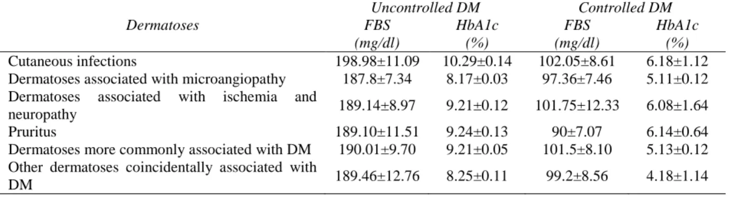
Table 4 Impact of control of DM on various dermatoses Dermatoses
Uncontrolled DM Controlled DM
FBS (mg/dl)
HbA1c (%)
FBS (mg/dl)
HbA1c (%) Cutaneous infections 198.98±11.09 10.29±0.14 102.05±8.61 6.18±1.12 Dermatoses associated with microangiopathy 187.8±7.34 8.17±0.03 97.36±7.46 5.11±0.12 Dermatoses associated with ischemia and
neuropathy 189.14±8.97 9.21±0.12 101.75±12.33 6.08±1.64
Pruritus 189.10±11.51 9.24±0.13 90±7.07 6.14±0.64
Dermatoses more commonly associated with DM 190.01±9.70 9.21±0.05 101.5±8.10 5.13±0.12 Other dermatoses coincidentally associated with
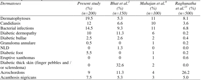
Table 5 Comparison of specific cutaneous disorders with other studies
Dermatoses Present study
(%) (n=200)
Bhat et al.2 (%) (n=150)
Mahajan et al.8 (%) (n=100)
Raghunatha et al.23 (%) (n=500)
Dermatophytoses 19.5 5.3 11 8.1
Candidiasis 12 6.6 10 3.6
Bacterial infections 14.5 9.3 11 6.8
Diabetic dermopathy 10 11.3 6 0.2
Diabetic bullae 2.5 2.6 2 0.4
Granuloma annulare 0.5 0 1 0.2
NLD 0 1.3 0 0.0
Diabetic foot 5.5 0 1 0.2
Eruptive xanthomas 0 0 1 0.6
Diabetic thick skin (finger pebbles and /
or scleredema) 0 32.6 2 0.0
Acrochordons 9 11.3 4 26.2
Acanthosis nigricans 7.5 5.3 3 5.0
This increased incidence of cutaneous infections
may be attributed to abnormalities of phagocytic
function,
cell-mediated
immunity
and
microcirculation, peripheral vascular disease,
diabetic neuropathy and hypohidrosis.
15,17Diabetic dermopathy was found in 6 (3%)
patients having peripheral neuropathy in our
study. Binkley
et al.
8found that diabetics with
peripheral neuropathy were more likely to
develop diabetic dermopathy. In the present
study, diabetic dermopathy was more common
in diabetics with duration of more than 5 years,
similar to the studies conducted by Sawhney
et
al.
10and Binkley
et al
.
18Among the various
dermatoses studied, majority had multiple
acrochordons (9%). Acrochordon has been
regarded as a sign of impaired glucose tolerance,
DM and increased cardiovascular risk.
19Studies
by Kahana
et al
.
20and Thappa
et al.
21concluded
that acrochordons may serve as markers for DM.
Acanthosis nigricans was observed in 7.5%
patients of the study group. The first major
breakthrough association of AN with insulin
resistance came from the study by Kahn.
22There
have been suggestions that insulin at high
concentrations may stimulate insulin-like growth
factor receptors on keratinocytes, thereby
promoting epidermal cell proliferation. So, every
patient of AN should be investigated for DM,
and every patient of DM should be screened for
AN. The absence of diabetic thick skin and
necrobiosis lipoidica diabeticorum (NLD) in the
present study when compared to other
studies,
2,8,23(Table 5) is mainly due to short
duration of DM in the majority of the patients.
Systemic comorbidities were present in 103
(51.5%) patients, of which hypertension (21.5%)
was most commonly observed. According to
Bhat
et al.
2, Mahajan
et al.
8and Al Mutairi
et
al
.
1246.4%, 53.1% and 44% patients were
hypertensive, respectively. Nigam
et al.
9also
reported hypertension as the most common
associated systemic disease.
Conclusion
lipoidica diabeticorum, syndromes of limited
joint mobility are extremely important to the
clinician as they are indicators of underlying
chronic degenerative complications such as
retinopathy, neuropathy and nephropathy and
hence an “alarm bell to alert the physician”.
Thus a dermatologist plays an important role in
reducing
the
dermatologic
morbidity,
improvement of quality of life and management
strategy of diabetic patients.
References
1. Worldwide trends in diabetes since 1980: a pooled analysis of 751 population-based studies with 4.4 million participants. Lancet. 2016;387:1513-30.
2. Bhat YJ, Gupta V, Kudyar RP. Cutaneous manifestations of diabetes mellitus. Int J Diab Dev Ctries. 2006;26:152-5.
3. Kalus AA, Chein AJ, Olerud JE. Diabetes mellitus and other endocrinal disorders. In: Wolff K, Goldsmith LA, Katz SI, Gilchrest BA, Paller AS, Lefell DJ, editors. Fitzpatrick’s Dermatology in General Medicine. 7th edn. New York: McGraw-Hill: 2008. p. 1461-84.
4. Al-Mutairi N. Skin diseases seen in diabetes mellitus. Bull Kuwait Inst Med Special. 2006;5:30-9.
5. Bud JL, Oledur JE. Diabetes Mellitus. In: Wolff K, Goldsmith LA, Katz SI, Gilchrest BA, Paller AS, Lefell DJ, editors. Fitzpatrick’s Dermatology in General Medicine. 7th edn. Vol. 2. New York: McGraw-Hill. p. 1651-61.
6. Jelinek JE. Cutaneous manifestations of diabetes mellitus. Int J Dermatol. 1994;33:605-17.
7. Peterka ES, Fusaro RM. Cutaneous Carbohydrate Studies IV. The skin glucose content of fasting diabetics with and without infection. J Invest Dermatol. 1966;46:459-63.
8. Mahajan S, Koranne RV, Sharma SK. Cutaneous manifestation of diabetes mellitus. Indian J Dermatol Venereol Leprol. 2003;69:105-8.
9. Nigam P K, Pande S. Pattern of dermatoses in diabetics. Indian J Dermatol Venereol Leprol. 2003;69:83-5.
10. Sawhney M, Talwar OP, Tutakne MA, Rajpathak SD. Diabetic dermoangiopathy: A clinico-pathological correlation. Indian J Dermatol Venereol Leprol. 1992;58:173-8. 11. Rao S, Pai G. Cutaneous manifestations of
diabetes mellitus. Indian J Dermatol Venereol Leprol. 1997;63:232-34.
12. Mutairi N, Zaki A, Sharma AK, Al-Sheltani M. Cutaneous manifestations of diabetes mellitus. Med Princ Pract. 2006;15:427-30.
13. Vahora R, Thakkar S, Marfatia Y. Skin, a mirror reflecting diabetes mellitus: A longitudinal study in a tertiary care hospital in Gujarat. Indian J Endocr Metab. 2013;17:659-64.
14. Yosipovitch G, Hodak E, Vardi P, Shraga I, Karp M, Sprecher E et al. The prevalence of cutaneous manifestations in IDDM patients and their association with diabetes risk factors and microvascular complications. Diabetes Care. 1998;21:506-9.
15. Feringer T, Miller FO. Cutaneous manifestation of diabetes mellitus. Dermatol Clin. 2002;20:483-92.
16. Hahler B. An overview of dermatological conditions commonly associated with the obese patient. Ostomy Wound Manage. 2006;52:34-6.
17. Sibbald RG, Schachter RK. The skin and diabetes mellitus. Int J Dermatol. 1984;23:567-84.
18. Binkley GW. Dermopathy in the diabetic syndrome. Arch Dermatol. 1965;92:625-34. 19. Crook MA. Skin tags and atherogenic lipid
profile. J Clin Pathol. 2000;53:873-4. 20. Kahana M, Grossman E, Feinstein A,
Ronnen M, Cohen M, Millet MS. Skin tags: A cutaneous marker for diabetes mellitus. Acta Dermatol Venereol. 1987;67:175-7. 21. Thappa DM. Skin tags as markers of
diabetes mellitus: An epidemiological study in India. J Dermatol. 1995;22:729-31. 22. Kahn CR, Flier JS, Bar RS, Archer JA,
Gorden P, Martin MM et al. The syndromes of insulin resistance and acanthosis nigricans: Insulin-receptor disorders in man. N Engl J Med. 1976;294:739-45.