Interleukin-15-Stimulated Natural Killer Cells Clear
HIV-1-Infected Cells following Latency Reversal
Ex Vivo
Carolina Garrido,
a,bMaria Abad-Fernandez,
a,cMarina Tuyishime,
eJustin J. Pollara,
eGuido Ferrari,
eNatalia Soriano-Sarabia,
a,bDavid M. Margolis
a,b,c,daUNC HIV Cure Center, University of North Carolina at Chapel Hill, Chapel Hill, North Carolina, USA
bDepartment of Medicine, University of North Carolina at Chapel Hill, Chapel Hill, North Carolina, USA
cDepartment of Microbiology and Immunology, University of North Carolina at Chapel Hill, Chapel Hill, North Carolina, USA
dDepartment of Epidemiology, University of North Carolina at Chapel Hill, Chapel Hill, North Carolina, USA
eDepartment of Surgery and Duke Human Vaccine Institute, Duke University Medical Center, Durham, North Carolina, USA
ABSTRACT
Current efforts toward human immunodeficiency virus (HIV) eradication
clude approaches to augment immune recognition and elimination of persistently
in-fected cells following latency reversal. Natural killer (NK) cells, the main effectors of the
innate immune system, recognize and clear targets using different mechanisms than
CD8
⫹T cells, offering an alternative or complementary approach for HIV clearance
strat-egies. We assessed the impact of interleukin 15 (IL-15) treatment on NK cell function
and the potential for stimulated NK cells to clear the HIV reservoir. We measured NK cell
receptor expression, antibody-dependent cell-mediated cytotoxicity (ADCC), cytotoxicity,
interferon gamma (IFN-
␥
) production, and antiviral activity in autologous HIV replication
systems. All NK cell functions were uniformly improved by IL-15, and, more importantly,
IL-15-treated NK cells were able to clear latently HIV-infected cells after exposure to
vori-nostat, a clinically relevant latency-reversing agent. We also demonstrate that NK cells
from HIV-infected individuals aviremic on antiretroviral therapy can be efficiently
stimu-lated with IL-15. Our work opens a promising line of investigation leading to future
im-munotherapies to clear persistent HIV infection using NK cells.
IMPORTANCE
In the search for an HIV cure, strategies to enhance immune function
to allow recognition and clearance of HIV-infected cells following latency reversal
are being evaluated. Natural killer (NK) cells possess characteristics that can be
ex-ploited for immunotherapy against persistent HIV infection. We demonstrate that NK
cells from HIV-positive donors can be strongly stimulated with IL-15, improving their
antiviral and cytotoxic potential and, more importantly, clearing HIV-infected cells
af-ter latency reversal with a clinically relevant drug. Our results encourage further
in-vestigation to design NK cell-based immunotherapies to achieve HIV eradication.
KEYWORDS
natural killer cells, HIV, latency reversal, HIV eradication,
immunotherapy, vorinostat, VOR, SAHA, immune function, IL-15, shock and kill, kick
and kill, ADCC, latency reversal, NK cell, human immunodeficiency virus, interleukins
T
he immune system is not capable of clearing human immunodeficiency virus (HIV)
infection, and thus, even in the presence of fully suppressive antiretroviral therapy
(ART), infection is chronic. However, in recent years, significant progress has been made
in strategies to disrupt the viral latent reservoir, promoting new efforts toward virus
clearance or cure (1–3). Awakening the latent reservoir of persistent infection could
represent the first step in HIV eradication therapy, but clearance of infection requires a
second step involving a potent and efficient immune response. This combined strategy
Received 12February2018 Accepted 21
March2018
Accepted manuscript posted online 28 March2018
Citation GarridoC,Abad-FernandezM,
TuyishimeM,PollaraJJ,FerrariG, Soriano-SarabiaN,MargolisDM.2018. Interleukin-15-stimulatednaturalkillercellsclear HIV-1-infectedcellsfollowinglatencyreversal ex vivo. JVirol92:e00235-18. https://doi.org/10.1128/
JVI.00235-18.
Editor FrankKirchhoff,UlmUniversityMedical
Center
faces
challenges,
such
as
insufficient
number
and
function
of
HIV-specific
CD8
⫹T
cells
(4–6),
dispersed
and
scarce
HIV
antigen
(Ag)
expression
in
latently
infected
cells,
and
selection
of
virus
populations
poorly
recognized
by
CD8
⫹T
cells
(7–9).
Therefore,
alternative
or
complementary
immune
strategies
should
be
pursued.
Here,
we
model
the
ability
to
augment
natural
killer
(NK)
cell
function
in
autologous
HIV-infected-donor
cell
systems,
demonstrating
the
potential
to
fill
this
unmet
need.
NK
cells
are
the
main
effectors
of
the
innate
immune
system.
They
are
the
first
line
of
defense
against
viral
infections
and
also
play
an
important
role
in
more
advanced
stages
of
infection
(10).
Although
controversial,
evidence
of
viral
evolution
toward
NK
cell
escape
suggests
that
these
cells
exert
specific
immunological
pressure
on
HIV
(11,
12).
NK
cells
can
eliminate
HIV-infected
cells
by
antibody-dependent
cell-mediated
cytotoxicity
(ADCC),
by
direct
lysis
via
the
release
of
cytotoxic
granules,
and
by
facilitating
priming
of
the
adaptive
immune
response.
Activation
of
NK
cells
is
governed
by
the
balance
of
positive
signals
via
the
recognition
of
cellular
stress
markers
on
the
surfaces
of
infected
or
malignant
cells
and
negative
signals
received
primarily
through
killer
cell
immunoglobulin-like
receptors
(KIRs)
engaging
the
major
histocompatibility
complex
(MHC).
It
is
especially
interesting
that
HIV
downregulates
MHC
class
I
expres-sion
on
the
surfaces
of
infected
cells,
thereby
escaping
recognition
and
lysis
by
CD8
⫹T
cells
but,
conversely,
rendering
it
more
susceptible
to
NK
cell-mediated
clearance
(13).
In
addition,
lymph
nodes
are
a
major
anatomic
HIV
reservoir
(14),
and
CD8
⫹effectors
may
be
relatively
excluded
from
lymphoid
follicles,
making
them
a
potential
sanctuary
for
persistent
infection
(15).
Recently,
it
has
been
reported
that
NK
cells
can
accumulate
in
the
follicles
of
secondary
lymphoid
organs
in
simian
immunodeficiency
virus
(SIV)-infected
African
green
monkeys
(AGMs),
natural
controllers
of
SIV
replication
(16).
These
findings
encourage
investigation
of
NK
cell-based
immunotherapies
for
HIV
eradication.
Cytokines
play
a
decisive
role
in
activating
NK
cells
by
enhancing
their
cytotoxic
potential
(17).
Interleukin
15
(IL-15)
is
a
potent
enhancer
of
NK
cytotoxic
function
and
is
under
study
in
oncology,
given
its
potential
to
improve
the
clearance
of
malignant
cells
(18).
In
the
present
work,
we
analyzed
the
potential
of
cytokine-stimulated
NK
cells
to
recognize
and
clear
latently
HIV-infected
cells
after
latency
reversal
and
performed
a
comprehensive
characterization
of
the
impact
of
cytokine
treatment
on
NK
cell
function.
We
demonstrate
that
upon
treatment
with
IL-15,
NK
cells
improve
their
capacity
to
target
and
clear
HIV-infected
cells.
RESULTS
IL-15 treatment improves antiviral activity of NK cells. HIV-infected
CD4
⫹T
cells
derived
from
8
donors
were
superinfected
with
the
laboratory
strain
JR-CSF,
and
cells
from
6
additional
donors
were
infected
with
autologous
reservoir
virus
(AR).
Infection
using
AR
is
relevant,
given
the
diversity
of
viral
isolates
within
and
between
donors,
as
well
as
the
potential
for
viral
isolates
that
have
evolved
to
escape
immune
responses.
Pooled
analysis
of
all
the
donors
showed
that
at
a
1:1
effector/target
cell
(E:T)
ratio,
autologous
NK
cells
significantly
reduced
viral
replication,
from
100%
under
the
target-alone
condition
to
31.2%
(standard
error
of
the
mean
[SEM],
4.3%;
P
⫽
0.0002),
while
IL-15
stimulation
of
NK
cells
further
decreased
viral
replication
(4.8%
[SEM,
1.3%;
P
⫽
0.0002]),
with
significant
differences
between
untreated
and
IL-15-treated
NK
cells
(
P
⫽
0.0005).
Virus
reduction
was
also
seen
at
a
1:10
E:T
ratio
for
both
untreated
NK
cells
and
IL-15-stimulated
cells
(65%
[SEM,
6.3%;
P
⫽
0.0004]
and
44.3%
[SEM,
4,8%;
P
⫽
0.0001],
respectively),
and
again,
IL-15
significantly
improved
antiviral
activity
(
P
⫽
0.008).
Finally,
at
a
1:100
ratio,
only
IL-15-stimulated
cells
exerted
a
significant
impact
on
virus
production
(79.5%
[SEM,
5.5%;
P
⫽
0.02])
(Fig.
1A).
When
the
experiments
were
analyzed
according
to
the
viral
isolate
used
for
infection
(JR-CSF
or
AR),
the
patterns
of
inhibition
were
comparable
between
the
viruses
(Fig.
1B).
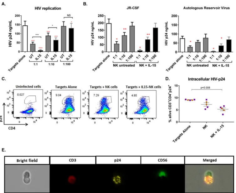
FIG 1IL-15 improves the antiviral activity of NK cells from ART-treated HIV-infected donors. (A) Viral replication measured as HIVgagp24 antigen in the
supernatants of 7-day cultures with only infected CD4⫹T cells (Targets alone) or in the presence of NK cells at different effector/target cell ratios. UT, untreated.
The red asterisks indicate statistically significant differences compared to targets alone, and black asterisks indicate differences between untreated and
IL-15-stimulated NK cells (n⫽14). (B) Viral replication in viral inhibition assays performed with JR-CSF superinfection (n⫽8) or autologous reservoir virus (n⫽
6). Wilcoxon matched-pairs signed-rank test.*,P⬍0.05;**,P⬍0.01;***,P⬍0.001. The error bars indicate standard error of the mean (SEM). (C) Representative
flow cytometry plots of intracellular p24 in cells from one donor gated on the CD3⫹population of the live fraction. (D) Proportion of live CD4⫹T cells positive
for intracellular p24 staining. Coculture of infected CD4 cells with IL-15-treated NK cells significantly reduced the proportion of live CD4⫹T cells containing p24
antigen after 5 days in culture. The orange circles correspond to cells from HIV-negative donors (n⫽2), and the purple squares correspond to cells from aviremic
HIV-positive donors (n⫽3). Mann-Whitney U test. (E) Interaction of an NK cell with an infected CD4⫹T cell visualized with ImageStreamX.
0.71%)
when
target
cells
were
cultured
with
NK
cells,
and
further,
to
5.25%
(SEM,
0.60%),
when
NK
cells
were
treated
with
IL-15
(Fig.
1D).
Finally,
we
visualized
cells
from
a
p24
intracellular-staining
experiment
using
Amnis
ImageStreamX
and
found
several
interactions
between
NK
cells
(marked
with
CD56-fluorescein
isothiocyanate
[FITC])
and
HIV-infected
target
cells
(CD3-allophycocyanin
[APC]
to
identify
targets
and
p24-phycoerythrin
[PE]
to
detect
infection)
(Fig.
1E).
Cytotoxicity
and
IFN-
␥
production
after
IL-15
stimulation.
NK
cell
cytotoxicity
FIG 2IL-15 increases the cytotoxic function of NK cells. (A) (Left and middle) Degranulation of NK cells measured by CD107a expression after coculture with
the cell line K562 or autologous HIV-superinfected CD4⫹T cells (n⫽21 andn⫽8, respectively). (Right) IFN-␥production after coculture with K562 cells
measured by intracellular staining (n⫽17). Wilcoxon matched-pairs signed-rank test. (B) (Left and middle) Comparison between degranulation and IFN-␥
production in NK cells from HIV-negative or HIV-positive donors. (Right) Fold change (IL-15/untreated) of CD107a expression and IFN-␥ production in
HIV-negative and HIV-positive donors. Mann-Whitney test. The orange circles correspond to cells from HIV-negative donors, and the purple squares correspond to cells from aviremic HIV-positive donors. The error bars indicate SEM.
significantly
improved
NK
cell
degranulation
in
the
presence
of
autologous
HIV-superinfected
CD4
⫹T
cells,
increasing
from
a
mean
of
14.5%
(SEM,
3.4%)
to
28.7%
(SEM,
4.6%)
(
P
⬍
0.01)
(Fig.
2A,
middle).
IL-15
also
enhanced
NK
cell
IFN-
␥
production
(
P
⬍
0.0001),
measured
by
intracellular
staining,
from
an
average
of
13.3%
(SEM,
2.2%)
of
NK
cells
producing
IFN-
␥
to
43%
(SEM,
4.2%)
(Fig.
2A
right).
We
also
compared
the
performance
of
NK
cells
isolated
from
HIV-negative
and
HIV-positive
donors.
NK
cells
from
HIV-negative
and
HIV-positive
individuals
degranulated
to
equal
extents,
both
without
treatment
and
after
IL-15
stimulation.
However,
NK
cell
baseline
IFN-
␥
produc-tion
was
impaired
in
aviremic
HIV-positive
donors
(
P
⫽
0.0008),
but
interestingly,
IL-15
restored
the
capacity
of
NK
cells
to
produce
IFN-
␥
to
the
same
level
as
HIV-negative
individuals
(Fig.
2B).
Impact
of
other
cytokines
on
NK
cell-mediated
cytotoxicity.
Given
that
other
FIG 3NK cell-mediated cytotoxicity after treatment with different cytokines. The graphs show the proportions of
NK cells degranulating or producing IFN-␥in the presence of K562 cells (red squares) or in the absence of target
cells (green circles) after stimulation with different cytokines or combinations, as indicated in the table below. Degranulation after culture with the cell line K562 showed a target-specific increase in cytotoxicity after stimulation
with any of the cytokines or the studied combinations (P⫽0.03). However, IL-15 showed better stimulation than
IL-12, IL-18, or IL-21 (P⫽0.03), and no difference was found between IL-15 alone and IL-15 in addition to any of
the other cytokines (P, NS). IFN-␥production upon culture with K562 cells is increased after stimulation with IL-15
or IL-12 alone or any of the cytokine combinations (P⫽0.03). The IL-15 effect was improved when IL-12 or IL-12
and IL-18 were included. However, IFN-␥production in the absence of target cells (NK alone) also increased after
IL-12 or IL-12 and IL-18 addition to IL-15, indicating nonspecific activation. Wilcoxon matched-pairs signed-rank test. The error bars indicate SEM.
statistical
significance
for
IL-15
plus
IL-12,
IL-15
plus
IL-18,
IL-15
plus
IL-21,
and
IL-15
plus
IL-12
plus
IL-21.
NK
cell
production
of
IFN-
␥
after
coculture
with
the
cell
line
K562
was
increased
when
the
cells
were
treated
with
IL-15
or
IL-12
alone,
as
well
as
with
IL-15
in
combination
with
any
of
the
other
cytokines
(
P
⫽
0.03),
but
no
effect
was
observed
upon
stimulation
with
IL-18
or
IL-21
alone.
Moreover,
addition
of
IL-12
and
IL-12
plus
IL-18
to
IL-15
significantly
increased
IFN-
␥
production
compared
to
IL-15
alone
(
P
⫽
0.03).
However,
in
the
absence
of
target
cells,
addition
of
IL-12
or
IL-12
plus
IL-18
to
IL-15
also
produced
a
significant
increase
(
P
⫽
0.03)
compared
to
IL-15
alone.
Thus,
combination
of
other
cytokines
with
IL-15,
especially
IL-12,
might
improve
NK
cell
capacity
to
produce
IFN-
␥
,
but
these
cytokine
combinations
were
associated
with
nonspecific
effects,
as
shown
by
the
significant
increase
in
IFN-
␥
production
in
the
absence
of
target
cells,
supporting
further
exploration
of
IL-15
alone.
ADCC
activity
of
NK
cells
is
enhanced
after
IL-15
stimulation.
We
evaluated
whether
IL-15
stimulation
of
NK
cells
had
an
effect
on
ADCC
activity,
using
HIV-infected
CEM.NKRCCR5
cells
as
targets
in
a
luciferase
assay.
NK
cells
were
isolated
from
6
HIV-negative
and
6
HIV-positive
ART-treated
aviremic
donors.
Untreated
or
IL-15-treated
NK
cells
were
tested
against
subtype
C
HIV-1
TV-1-infected
target
cells
in
the
presence
of
serial
dilutions
of
plasma
from
an
HIV-infected
participant.
In
both
groups,
we
observed
very
similar
statistically
significant
increases
in
target-specific
killing
when
NK
cells
were
stimulated
with
IL-15
(Fig.
4A) (
P
⫽
0.001).
Comparison
of
the
responses
from
all
the
participants
was
also
performed,
using
antibody
titers
(Fig.
4B)
and
the
area
under
the
curve
(Fig.
4C),
which
revealed
statistically
significant
differences
between
the
responses
obtained
with
untreated
and
IL-15-treated
effector
cells
(
P
⫽
0.0005).
We
did
not
observe
statistically
significant
differences
between
the
ADCC
activities
of
HIV-negative
and
HIV-positive
donors
when
we
compared
the
results
under
untreated
and
IL-15-treated
conditions.
IL-15-stimulated NK
cells
recognize
and
clear
latently
HIV-infected
cells
after
reactivation. A
modified
viral
outgrowth
assay
was
used
to
assess
the
ability
of
NK
cells
FIG4ADCCimprovesafterIL-15stimulationof NKcells.NKcellswerecultured withTV-1-infected
CEM.NKRcellsinthepresenceofHIV-negativeorA300(HIV-positive)plasmafor6h,andspecifictarget
killingwasrecordedusingdifferentplasmadilutions.(A)Meanantibody-mediatedspecifickillingwith
untreated NK cells or after overnight IL-15 stimulation using different plasma dilutions. Cytokine
treatmentimprovedADCCactivityinbothHIV-negative(n⫽ 6)andHIV-positive(n⫽ 6)donors.Means
withSEMareshown.(B)Antibodytitersobservedwithuntreated/treatedNKcells(n⫽ 12).(C)Areas
underthecurveatdifferentplasmadilutionswithuntreatedNKandIL-15-stimulatedNKcells(n⫽ 12).
TheorangecirclescorrespondtocellsfromHIV-negativedonors,andthepurplesquarescorrespondto
cellsfromART-treatedaviremicHIV-positivedonors.Wilcoxonmatched-pairssigned-ranktest.
with
the
histone
deacetylase
(HDAC)
inhibitor
vorinostat
(VOR)
(also
known
as
SAHA)
under
conditions
known
to
parallel
clinical
administration
of
the
drug
(1).
At
the
end
of
the
culture,
the
supernatants
were
harvested,
and
viral
production
was
measured
by
p24
enzyme-linked
immunosorbent
assay
(ELISA),
recording
the
number
of
wells
in
which
virus
was
recovered,
regardless
of
the
quantity
of
p24
measured.
The
underlying
assumption
was
that
each
well
would
possess
one
or
fewer
infected
cells
and
that
a
decrease
in
the
frequency
of
wells
with
detectable
p24
reflected
clearance
of
the
infected
cells.
When
resting
CD4
⫹T
cells
were
reactivated
with
PHA,
a
decrease
in
the
number
of
HIV-positive
wells
was
observed
when
NK
cells
were
added
to
the
culture
(
P
,
not
significant
[NS])
(Fig.
5A,
left
panel),
with
a
more
pronounced
reduction
when
the
NK
cells
were
first
stimulated
with
IL-15
(
P
⫽
0.031).
Of
more
clinical
relevance,
when
vorinostat
was
used
to
reverse
latency,
IL-15-stimulated
NK
cells
significantly
reduced
the
number
of
p24
HIV
⫹wells
(
P
⫽
0.015),
with
significant
differences
between
untreated
and
IL-15-treated
NK
cells
(
P
⫽
0.013)
(Fig.
5A,
right
panel).
IL-15-stimulated NK cells to confirm that nonspecific cell killing was not responsible for
the observed virus clearance (
⬍
5% annexinV-positive CD4
⫹T cells).
Impact of IL-15 exposure on activating receptor expression in NK cells.
Acti-vating NK cell receptor expression was analyzed to explore the impact of IL-15
exposure. The panel included CD16, DNAM-1, NKG2D, NKp30, and NKp46. IL-15
stim-ulation did not impact expression of CD16 or DNAM-1 but significantly increased the
expression of NKG2D and NKp30 (
P
⫽
0.03). Interestingly, IL-15 exposure also produced
a significant down-modulation of NKp46 (
P
⫽
0.03), as shown in Fig. 6.
DISCUSSION
The enhancement of the innate antiviral activity of NK cells is a viable adjunctive
clinical strategy that may be employed in efforts to clear persistent HIV infection. In the
present work, we demonstrate the ability of IL-15 stimulation to enhance NK cell
function in the context of HIV. Uniformly, IL-15 augmented NK cell function, improving
degranulation, ADCC activity, IFN-
␥
production, and direct antiviral activity in actively
replicating HIV cultures. Of most significance, IL-15-treated NK cells recognized and
cleared latently HIV-infected cells following a clinically achievable exposure to the
latency-reversing agent vorinostat.
Immunological strategies in the field of HIV eradication have focused on HIV-specific
cytotoxic T lymphocytes (CTLs) (19–21), although recently, interest in NK cells has
emerged. The capacity of NK cells to produce IFN-
␥
and upregulate NKp30 and NKp46
has been inversely associated with viral reservoir size (22), and HIV DNA decline was
FIG 5Latency clearance assay. (A) Resting CD4⫹T cells were incubated with PHA or VOR for 24 h, cocultured with or without autologous NK cells at an E:T ratio of 1:10 for another 24 h, and plated in 12 replicates in the presence of feeders for 19 days, refreshing the medium every 3 or 4 days. The supernatant was assayed for p24 ELISA to assess the presence of HIV replication. The graphs show the numbers
of wells in which p24 was detected at day 19, with a different color for each donor. (Left) Reactivation with PHA (n⫽6). (Right)
Reactivation with 335 nM VOR (n⫽8). Wilcoxon matched-pairs signed-rank test. (B) Latency clearance assays in which NK cells were
depleted after the initial 24 h of coculture with VOR-reactivated CD4⫹T cells (n⫽5). IL-15-stimulated NK cells reduced the number of
HIV⫹wells during the first 24 h of coculture with VOR-reactivated CD4⫹T cells. The error bars indicate SEM.
FIG 6Expression of activating NK cell receptors before and after IL-15 exposure. Cytokine stimulation upregulated the expression of NKG2D and NKp30, while
it decreased that of NKp46. The orange circles represent cells from healthy donors (n⫽6), and the purple squares represent cells from HIV-infected donors
Another important finding of our study is the demonstration that NK cells obtained
correlated
with
NK
cell
frequency
after
panobinostat
treatment
in
a
clinical
study
(23),
suggesting
a
role
for
NK
cells
in
the
clearance
of
HIV
infection.
Moreover,
recent
data
suggest
that
NK
cells
might
play
an
important
role
in
viral
clearance
in
the
lymph
node
(16).
However,
until
now,
the
ability
of
NK
cells
to
clear
latent
HIV
infection
and
the
effect
of
cytokine
treatment
on
this
NK
cell
antiviral
activity
have
not
been
reported.
Soluble
recombinant
human
IL-15
(rhIL-15),
the
cytokine
used
in
the
present
work,
is
sufficient
for
ex
vivo
proof-of-concept
studies.
However,
research
in
oncology
has
brought
into
clinical
testing
several
engineered
forms
of
this
potent
cytokine,
including
a
heterodimeric
form
of
the
molecule
(24)
and
a
synthetic
receptor
superagonist
(ALT-803)
(25),
that
might
have
better
profiles
for
clinical
use.
The
latter
form
of
IL-15
has
been
studied
in
the
simian
immunodeficiency
virus
model
of
HIV
infection,
ex-panding
both
NK
and
CD8
⫹T
cells
(26).
Future
directions
could
also
include
the
incorporation
of
the
IL-15
gene
into
genetically
modified
NK
cells,
so
that
the
cells
could
provide
autocrine
cytokine
support
(27,
28).
Moreover,
it
is
expected
that
additional
strategies
will
be
needed
to
further
enhance
NK
cell
function,
since
in
most
of
our
results,
despite
seeing
a
robust
antiviral
effect
of
the
stimulated
NK
cells,
total
clearance
was
not
always
observed.
Thus,
NK
cell
function
might
be
improved
by
using
other
priming
strategies,
such
as
IFN-
␣
stimulation
(29,
30)
or
therapeutic
vaccination
(31).
In
addition,
clinical
experience
gained
in
oncology
may
be
leveraged
to
design
HIV-specific
approaches,
given
the
more
advanced
stages
of
NK
cell-based
therapies
that
have
been
developed
for
cancer
(32–36).
The
most
relevant
experiment
in
this
study
is
undoubtedly
the
latency
clearance
assay
(19)
performed
on
primary
resting
CD4
⫹T
cells
from
HIV-infected
individuals
on
antiretroviral
treatment.
This
assay
is
the
most
proximate
to
the
in
vivo
situation,
eliminating
artifacts
produced
by
cell
models
or
in
vitro
infections.
As
latency
reversal
was
induced
by
VOR
at
exposures
that
have
been
safely
achieved
in
clinical
trials,
it
is
likely
that
NK
cell
targeting
and
clearance
can
be
similarly
achieved
in
vivo
.
Here,
we
proved
that
NK
cells
can
clear
infected
cells
after
VOR
exposure.
However,
a
range
of
novel
approaches
to
achieve
HIV
latency
reversal
are
under
study
(37,
38).
Thus,
in
a
similar
way,
future
work
must
address
the
effect
of
novel
latency-reversing
agents
(LRAs)
on
NK-mediated
clearance,
given
that
antigen
presentation
or
ligand
modulation
might
be
altered
by
host-targeted
LRAs
(39).
Of
interest,
there
is
evidence
that
HDAC
inhibitor
exposure
upregulates
the
surface
expression
of
ligands
for
acti-vating
receptors
in
NK
cells
(40,
41).
Nonetheless,
while
such
upregulation
is
observed
in
tumor
cells,
the
effect
of
LRA
on
latently
HIV-infected
cells
is
unknown.
For
example,
valproic
acid,
a
weak
HDAC
inhibitor,
upregulates
NKG2D
ligands
only
in
malignant
cells,
where
the
ligands
are
present
at
baseline
(42).
However,
initial
in
vitro
studies
in
HIV-infected
cell
lines
suggested
that
VOR
could
also
upregulate
certain
NKG2D
ligands
(43),
which
in
addition
may
play
an
important
role
in
facilitating
NK-mediated
ADCC
responses
(44).
Finally,
novel
latency-reversing
agents,
such
as
Toll-like
receptor
(TLR)
agonists,
may
simultaneously
reverse
HIV
latency
and
improve
NK
cell
function
(45,
46),
although
further
study
is
needed
to
definitively
determine
the
effect
of
TLR
agonists
on
NK
cells,
given
the
multiple
and
direct
effects
that
these
drugs
might
have
on
immune
cells.
In
addition
to
latency
clearance
assays,
we
conducted
virus
inhibition
assays
in
the
setting
of
active
replication
using
a
completely
autologous
system
with
NK
cells,
CD4
⫹T
cells,
and
reservoir
virus
from
the
same
donor.
The
use
of
patient-derived
viral
isolates
is
relevant,
given
that
viruses
may
differ
in
antigen
presentation.
In
our
studies,
we
observed
similar
NK
cell-mediated
viral
replication
inhibition
after
infection
with
either
JR-CSF
or
autologous
reservoir
virus.
However,
in
the
future,
should
ADCC
be
employed
to
direct
NK
cell
killing,
the
breadth
of
antibody
recognition
of
viral
species
within
the
latent
reservoir
may
be
critical.
from
HIV-positive
donors
on
ART
with
suppressed
viremia
were
sufficiently
functional
to
respond
to
IL-15
and
exerted
antiviral
activity
in
all
the
assays
performed.
Interest-ingly,
we
observed
that
IFN-
␥
production
by
HIV-positive
donors
was
impaired
com-pared
to
that
by
HIV-negative
individuals,
but
IL-15
stimulation
restored
IFN-
␥
produc-tion
in
NK
cells
from
HIV-positive
donors
to
the
level
of
HIV-negative
donors.
In
fact,
IL-15-mediated
improvement
in
the
HIV-positive
population
was
more
pronounced
than
in
the
HIV-negative
group.
The
functionality
of
NK
cells
from
HIV
⫹individuals
with
virus
suppressed
has
been
previously
described
(48,
49),
but
our
studies
further
demonstrate
their
capability
to
be
stimulated
by
IL-15
and
to
be
functional
in
an
HIV
eradication
context.
Finally,
we
observed
that
NKG2D
and
NKp30
were
upregulated
after
IL-15
treatment,
which
may
have
contributed
to
the
enhancement
of
NK
cell
function.
We
also
observed
a
downmodulation
of
NKp46
upon
IL-15
exposure.
This
is
in
accordance
with
previous
reports
(50)
and
is
troublesome,
given
the
importance
of
this
receptor
in
NK
cell
antiviral
activity
(22,
51,
52).
Therefore,
and
even
if
we
observed
an
important
improve-ment
in
NK
cell
function
despite
NKp46
downmodulation,
it
might
be
of
interest
to
consider
a
complementary
strategy
to
boost
NKp46
expression,
thus
further
increasing
NK
cell
antiviral
function.
In
summary,
the
present
study
provides
robust
evidence
that
IL-15-stimulated
NK
cells
from
virus-suppressed
HIV-positive
individuals
on
ART
can
recognize
and
clear
latently
HIV-infected
resting
CD4
⫹T
cells
after
relevant
exposure
to
the
HDAC
inhibitor
vorinostat.
This
finding
encourages
further
exploration
of
approaches
for
immunother-apies
based
on
priming
NK
cells
to
eradicate
latent
HIV
infection.
MATERIALS
AND
METHODS
Studysamples.SampleswerederivedeitherfromHIV-negativedonorsorfromHIV-infecteddonors
onARTwithsustainedplasmaviremiasuppression(⬍50copies/ml)foratleast6months.Insummary,
themeanageofdonorswas42.5(SEM,3.1)years,withsuppressedviremiaforanaverageof3.4(SEM,
0.48)years,ameantotalof743(SEM,62)CD4cells/ml,andameanof39.8%(SEM,2.1%)CD4cells.
Peripheralbloodmononuclearcells(PBMC)fromHIV-negativedonorswereobtainedfrombuffycoats
fromtheNewYorkBlood Center(LongIslandCity,NY,USA).PBMCfromHIV-infecteddonorswere
obtainedbyFicollgradientfrombuffycoatsobtainedbyleukapheresis.K562cellswereobtainedfrom
theATCC(Manassas,VA).
Ethicsstatement.Alldonorsprovidedwritteninformedconsent,andstudieswereapprovedbythe
UniversityofNorthCarolina(UNC)InstitutionalReviewBoard.Samplesusedinthestudywere
anony-mized.
Virus inhibition assays.NKcells and CD4⫹ Tcells wereisolated fromthePBMCbynegative
selection(StemCellTechnologies,Vancouver,Canada).TheNKcellenrichmentantibodycocktailincluded
CD3,CD4,CD14,CD19,CD20,CD36,CD66b,CD123,HLA-DR,andglycophorin.CD4⫹Tcellswereisolated
bynegativeselectioninparallelwithNKcellisolationfromeachdonor.TheCD4⫹Tcellenrichment
antibodycocktailincludedCD8,CD14,CD16,CD19,CD20,CD36,CD56,CD66b,CD123,Tcellreceptor␥/␦
(TCR-␥/␦),andglycophorinA.Afterisolation,NKcellswerecultured inIscove’smodifiedDulbecco’s
medium(IMDM)supplementedwith10%heat-inactivatedbovineserumand5%penicillinplus
strep-tomycin,withorwithouttheappropriatecytokines,for24h,afterwhichthecellswerewashedand
assayswereperformed.IsolatedCD4⫹Tcellswereactivatedfor24hwith2g/mlPHA(Sigma-Aldrich,
St.Louis,MO)and60U/mlIL-2(Peprotech,RockyHill,CT).Thecellsweretheninfectedbyspinoculation
for90minat2,500rpmwith50ngHIVp24per10millioncellsoftheviralstrainJR-CSForwithAR,which
isvirusisolatedfromapreviousviraloutgrowthassayperformedwithcellsfromthesamedonor(19).
Afterspinoculation,thecellswereextensivelywashedtoremovefreevirions,and50,000infectedCD4⫹
Tcellswereplatedintriplicateforeachconditionina96-wellplate.NKcells,previouslyexposedto
cytokineswhenappropriate,wereaddedtothewellsatE:Tratiosof1:1,1:10,and1:100andleftinculture
for7daysincompleteIMDMwith5U/mlIL-2;themediawererefreshedatday4.Viralproductionwas
assessedinthesupernatantbyHIVp24ELISA(ABL,Inc.,Rockville,MD),andHIVreplicationunderthe
differentNKcellconditionswascomparedtoreplicationwithonlytargetcells.Viralinhibitionwasalso
assessed bymeasuringintracellularp24 incells harvestedafter5 daysof culture.For intracellular
quantificationofp24,culturesincludedatotalof800,000cells,platedineightreplicates,whichwere
pooledtoperformtheintracellularstaining.ThecellswerefirststainedwiththeviabilitydyeZombieNIR
Fixable Viability(Biolegend,San Diego,CA),fixedwith4%paraformaldehyde/lysolecithin,andthen
resuspendedincold50%methanol,permeabilizedwith0.1%NonidetP-40,andstainedwithantibody
forp24Ag(KC-57,BeckmanCoulter,Fullerton,CA),followedbysurfacestainingwithanti-CD3,-CD4,and
-CD56.InfectionwasrecordedasthepercentageofviableCD3⫹CD4⫹cellsexpressingp24.Finally,we
visualizedthecellsfromoneoftheseintracellularp24experimentsusingtheAmnisImageStreamXMark
Cytotoxicity.NKcellcytotoxicityandIFN-␥productionwereanalyzedincoculturesofprimaryNK
cellsandK562cells(anNK-sensitivetargetcellline,duetotheirlackofMHC)withandwithoutprior
exposureoftheNKcellstocytokines.Cytotoxicitywasassessedbymeasurementofthedegranulation
markerCD107a,areliablemarkerofNKcellfunctionalactivity(53).Atotalof200,000NKcellswere
culturedwiththesamenumberofK562targetcells,alongwithPE/Cy7-CD107aantibody(Biolegend,San
Diego,CA),for2to 4 h in a 96-wellplate,adding1lofGolgiStop(BectonDickinson,FranklinLakes,NJ)
after 1h.Cells were then harvested,washed, andstained with CD56⫺-FITC and CD3-APC/Fire750
(BioLegend)instainingbufferfor20minoniceinthedark.Thecellswerethenfixedwithfixationbuffer
for 20 min at room temperature in thedark, permeabilized, andwashed withPerm/Wash buffer
(Biolegend,SanDiego,CA)twiceandstainedwithIFN-␥–PE(BioLegend)for20min.Afterwashing,the
cells were resuspended instainingbuffer and analyzedin anAttune focusing cytometer (Applied
Biosystems,FosterCity,CA).ToevaluateHIV-specificrecognition,autologousCD4⫹Tcellssuperinfected
withtheviralstrainJR-CSFwereusedastargets,insteadof thecelllineK562.Additionally, NKcell
degranulationwasassessedintheabsenceofanytargettoexaminenonspecificactivationbycytokines.
Theactivationconditionsconsistedof25ng/mlIL-15,butinsomeexperiments,25ng/mlIL-12,25ng/ml
IL-18,and/or25ng/mlIL-21(Peprotech,RockyHill,NJ)wasalsoused.
ADCCassay.WeutilizedamodifiedversionofapreviouslypublishedADCCluciferaseprocedure
(54).Briefly,CEM.NKRCCR5cells(fromA.Trkola,NIHAIDSReagentProgram,DivisionofAIDS,NIAID,NIH
[55]) were usedastargets for ADCC luciferaseassaysafterinfectionwith subtype CHIV-1IMCTV1
(GenBankaccessionno.HM215437).PurifiedNKcellswereobtainedfromsixHIV-seronegativedonors
andsixART-treatedHIV-seropositivedonors.TheNKcellswereisolatedfromcryopreservedPBMCby
negativeselectionwithmagneticbeads(MiltenyiBiotecGmbH,Germany)afterovernightincubation
withorwithout10ng/mlIL-15(MiltenyiBiotecGmbH,Germany).TheNKcellswereusedaseffectorcells
ataneffector-to-targetcellratioof5:1.Targetandeffectorcellswereplatedinopaque96-wellhalf-area
platesandcoculturedwithserialdilutionsofplasma(A300)fromanHIV-infecteddonorstartingat1:100.
The cocultureswereincubatedfor 6 h at 37°Cin5%CO2.Thefinal readoutwastheluminescence
intensitygeneratedbythepresenceofresidualintacttargetcellsthathadnotbeenlysedbytheeffector
populationinthepresenceofADCC-mediatingplasmaAbs.Thepercentageofkillingwascalculated
usingthefollowingformula:percentspecifickilling⫽ [(RLUoftargetandeffectorwell⫺ RLUoftest
well)/(RLUoftargetandeffectorwell)]⫻ 100,whereRLUisrelativelightunits.
Inthisanalysis,theRLUofthetargetpluseffectorwellsrepresentspontaneouslysisintheabsence
ofanysourceofAb.PlasmafromanHIV-seronegativedonor(CAVD002)wasusedasanegativecontrol
usingthesamedilutionscheme.ThedataarereportedaspercentspecifickillingobservedwiththeA300
plasmaaftersubtractingthebackgroundobservedinthepresenceofthenegativecontrol,andtheareas
underthecurvewerecalculatedusingPrism7(version7.0b;GraphPadSoftware).
Latencyclearanceassay.Amodifiedviraloutgrowthassay(56)wasoptimizedtoassesstheability
ofcytokine-treatedNKcellstoclearlatentlyinfectedcellsafterreactivation.RestingCD4⫹Tcellswere
negativelyisolated(StemcellTechnologies,Vancouver,Canada)fromHIV-infected,ART-treated
individ-ualswithvirussuppressedandwereculturedfor24hwithantiretroviraldrugs(10nMraltegravirand20
nMefavirenz)toavoidnewcyclesofinfection.Thecellswerethenwashedfromtheantiretroviralsand
reactivatedwithmaximalmitogenstimulation(2g/mlPHAand60U/mlIL-2)orwithvorinostat(335
nM).After24h,thecellswerewashedandplatedin12-wellplatesat1millionto2millioncellsperwell
(morecellswereusedifthedonorwasknowntohavealowfrequencyoflatentinfection).NKcellswere
addedtothecultureatanE:Tratioof1:10,and24hlater,allogeneicstimulatedCD8-depletedPBMC
(feeders)wereaddedtotheculturetopropagatetheinfection.Toconfirmthatvirusrecoveryreduction
inthepresenceofNKcellswasduetorecognitionofreactivatedCD4⫹Tcellsandnottononspecific
killingorinhibitionofviralspread,weconductedexperimentsinwhichwedepletedtheculturesof
CD56⫹cells(CD56positive-selectionkit;StemCellTechnologies)beforeaddingthefeedercells.Ineither
case,thecellswereculturedfor19days,changingthemediumevery3daysandaddingfeedercellson
day8.Thenumbersofpositivewellswerenotedbasedonp24ELISAsperformedinthesupernatantsof
theculturesatday15andday19,andconditionswithandwithouteffectorswerecompared.
Addition-ally,insomeexperiments,afterthefirst24hofcultureofthereactivatedCD4⫹Tcellswithorwithout
NKcells,celldeathwasanalyzedbyflowcytometryusingannexinV(BioLegend)staining.
Activatingreceptor expression.Apanel of NKcell activatingreceptors was analyzedbyflow
cytometrycomparinguntreatedNKcellsandcellsexposedtoIL-15.Thefollowingsurfacemonoclonal
antibodieswere used,afterstainingwiththeviabilitydye ZombieAqua: CD3-peridininchlorophyll
protein(PerCP),dsCD56-APC/Cy7,CD16-AF700,NKG2D-APC,NKp30-PE,NKp46-PE/Cy7,andDNAM-1–
FITC(Biolegend).SampleswereanalyzedontheAttunefocusingcytometer(AppliedBiosystems).The
CD3⫺CD56⫹populationwasgatedintheviablegate,andtheexpressionofeachofthereceptorswas
analyzedusingFlowJo(Ashland,OR)Xsoftware.Fluorescenceminusone(FMO)gatingcontrolsforeach
antibodywereusedtodefinethegates.
Statistical analysis. Analysis was performed with GraphPad Prism v7. Comparisons between
matchedgroupswereanalyzedusingthenonparametricWilcoxonsigned-ranktest.Statistical
signifi-cancewasassignedwhenthePvaluewas⬍0.05.ResultsareexpressedasmeansandSEM.
ACKNOWLEDGMENTS
1. Archin NM, Liberty AL, Kashuba AD, Choudhary SK, Kuruc JD, Crooks AM, Parker DC, Anderson EM, Kearney MF, Strain MC, Richman DD, Hudgens MG, Bosch RJ, Coffin JM, Eron JJ, Hazuda DJ, Margolis DM. 2012. Admin-istration of vorinostat disrupts HIV-1 latency in patients on antiretroviral
therapy. Nature 487:482– 485.https://doi.org/10.1038/nature11286.
2. Sogaard OS, Graversen ME, Leth S, Olesen R, Brinkmann CR, Nissen SK, Kjaer AS, Schleimann MH, Denton PW, Hey-Cunningham WJ, Koelsch KK, Pantaleo G, Krogsgaard K, Sommerfelt M, Fromentin R, Chomont N, Rasmussen TA, Ostergaard L, Tolstrup M. 2015. The depsipeptide ro-midepsin reverses HIV-1 latency in vivo. PLoS Pathog 11:e1005142.
https://doi.org/10.1371/journal.ppat.1005142.
3. Rasmussen TA, Tolstrup M, Brinkmann CR, Olesen R, Erikstrup C, Solomon A, Winckelmann A, Palmer S, Dinarello C, Buzon M, Lichter-feld M, Lewin SR, Ostergaard L, Sogaard OS. 2014. Panobinostat, a histone deacetylase inhibitor, for latent-virus reactivation in HIV-infected patients on suppressive antiretroviral therapy: a phase 1/2,
single group, clinical trial. Lancet HIV 1:e13– e21.https://doi.org/10
.1016/S2352-3018(14)70014-1.
4. Day CL, Kaufmann DE, Kiepiela P, Brown JA, Moodley ES, Reddy S, Mackey EW, Miller JD, Leslie AJ, DePierres C, Mncube Z, Duraiswamy J, Zhu B, Eichbaum Q, Altfeld M, Wherry EJ, Coovadia HM, Goulder PJ, Klenerman P, Ahmed R, Freeman GJ, Walker BD. 2006. PD-1 expression on HIV-specific T cells is associated with T-cell exhaustion and disease
progression. Nature 443:350 –354.https://doi.org/10.1038/nature05115.
5. Petrovas C, Casazza JP, Brenchley JM, Price DA, Gostick E, Adams WC, Precopio ML, Schacker T, Roederer M, Douek DC, Koup RA. 2006. PD-1 is
a regulator of virus-specific CD8⫹T cell survival in HIV infection. J Exp
Med 203:2281–2292.https://doi.org/10.1084/jem.20061496.
6. Trautmann L, Janbazian L, Chomont N, Said EA, Gimmig S, Bessette B, Boulassel MR, Delwart E, Sepulveda H, Balderas RS, Routy JP, Haddad EK,
Sekaly RP. 2006. Upregulation of PD-1 expression on HIV-specific CD8⫹
T cells leads to reversible immune dysfunction. Nat Med 12:1198 –1202.
https://doi.org/10.1038/nm1482.
7. Deng K, Pertea M, Rongvaux A, Wang L, Durand CM, Ghiaur G, Lai J, McHugh HL, Hao H, Zhang H, Margolick JB, Gurer C, Murphy AJ, Valen-zuela DM, Yancopoulos GD, Deeks SG, Strowig T, Kumar P, Siliciano JD, Salzberg SL, Flavell RA, Shan L, Siliciano RF. 2015. Broad CTL response is required to clear latent HIV-1 due to dominance of escape mutations.
Nature 517:381–385.https://doi.org/10.1038/nature14053.
8. Shan L, Deng K, Shroff NS, Durand CM, Rabi SA, Yang HC, Zhang H, Margolick JB, Blankson JN, Siliciano RF. 2012. Stimulation of HIV-1-specific cytolytic T lymphocytes facilitates elimination of latent viral
reservoir after virus reactivation. Immunity 36:491–501.https://doi.org/
10.1016/j.immuni.2012.01.014.
9. Borrow P, Lewicki H, Wei X, Horwitz MS, Peffer N, Meyers H, Nelson JA, Gairin JE, Hahn BH, Oldstone MB, Shaw GM. 1997. Antiviral pressure exerted by HIV-1-specific cytotoxic T lymphocytes (CTLs) during primary infection demonstrated by rapid selection of CTL escape virus. Nat Med
3:205–211.https://doi.org/10.1038/nm0297-205.
10. Lanier LL. 2008. Evolutionary struggles between NK cells and viruses. Nat
Rev Immunol 8:259 –268.https://doi.org/10.1038/nri2276.
11. Alter G, Heckerman D, Schneidewind A, Fadda L, Kadie CM, Carlson JM, Oniangue-Ndza C, Martin M, Li B, Khakoo SI, Carrington M, Allen TM,
Altfeld M. 2011. HIV-1 adaptation to NK-cell-mediated immune pressure.
Nature 476:96 –100.https://doi.org/10.1038/nature10237.
12. Elemans M, Boelen L, Rasmussen M, Buus S, Asquith B. 2017. HIV-1 adaptation to NK cell-mediated immune pressure. PLoS Pathog 13:
e1006361.https://doi.org/10.1371/journal.ppat.1006361.
13. Bonaparte MI, Barker E. 2004. Killing of human immunodeficiency virus-infected primary T-cell blasts by autologous natural killer cells is depen-dent on the ability of the virus to alter the expression of major
histo-compatibility complex class I molecules. Blood 104:2087–2094.https://
doi.org/10.1182/blood-2004-02-0696.
14. Lederman MM, Margolis L. 2008. The lymph node in HIV
pathogen-esis. Semin Immunol 20:187–195. https://doi.org/10.1016/j.smim
.2008.06.001.
15. Leong YA, Chen Y, Ong HS, Wu D, Man K, Deleage C, Minnich M, Meckiff BJ, Wei Y, Hou Z, Zotos D, Fenix KA, Atnerkar A, Preston S, Chipman JG, Beilman GJ, Allison CC, Sun L, Wang P, Xu J, Toe JG, Lu HK, Tao Y, Palendira U, Dent AL, Landay AL, Pellegrini M, Comerford I, McColl SR, Schacker TW, Long HM, Estes JD, Busslinger M, Belz GT, Lewin SR, Kallies
A, Yu D. 2016. CXCR5(⫹) follicular cytotoxic T cells control viral infection
in B cell follicles. Nat Immunol 17:1187–1196.https://doi.org/10.1038/ni
.3543.
16. Huot N, Jacquelin B, Garcia-Tellez T, Rascle P, Ploquin MJ, Madec Y, Reeves RK, Derreudre-Bosquet N, Muller-Trutwin M. 2017. Natural killer cells migrate into and control simian immunodeficiency virus replication in lymph node follicles in African green monkeys. Nat Med 23:
1277–1286.https://doi.org/10.1038/nm.4421.
17. Terme M, Ullrich E, Delahaye NF, Chaput N, Zitvogel L. 2008. Natural killer cell-directed therapies: moving from unexpected results to
suc-cessful strategies. Nat Immunol 9:486 – 494. https://doi.org/10.1038/
ni1580.
18. Guillerey C, Huntington ND, Smyth MJ. 2016. Targeting natural killer cells
in cancer immunotherapy. Nat Immunol 17:1025–1036.https://doi.org/
10.1038/ni.3518.
19. Sung JA, Lam S, Garrido C, Archin N, Rooney CM, Bollard CM, Margolis DM. 2015. Expanded cytotoxic t-cell lymphocytes target the latent HIV
reservoir. J Infect Dis 212:258 –263.https://doi.org/10.1093/infdis/jiv022.
20. Mujib S, Saiyed A, Fadel S, Bozorgzad A, Aidarus N, Yue FY, Benko E, Kovacs C, Emert-Sedlak LA, Smithgall TE, Ostrowski MA. 2017. Pharma-cologic HIV-1 Nef blockade promotes CD8 T cell-mediated elimination of
latently HIV-1-infected cells in vitro. JCI Insight 2:93684.https://doi.org/
10.1172/jci.insight.93684.
21. Liu B, Zou F, Lu L, Chen C, He D, Zhang X, Tang X, Liu C, Li L, Zhang H. 2016. Chimeric antigen receptor T cells guided by the single-chain Fv of a broadly neutralizing antibody specifically and effectively eradicate
virus reactivated from latency in CD4⫹ T lymphocytes isolated from
HIV-1-infected individuals receiving suppressive combined antiretroviral
therapy. J Virol 90:9712–9724.https://doi.org/10.1128/JVI.00852-16.
22. Marras F, Casabianca A, Bozzano F, Ascierto ML, Orlandi C, Di Biagio A, Pontali E, Dentone C, Orofino G, Nicolini L, Taramasso L, Magnani M, Marincola FM, Wang E, Moretta L, De Maria A. 2017. Control of the HIV-1 DNA reservoir is associated in vivo and in vitro with NKp46/NKp30 (CD335 CD337) inducibility and interferon gamma production by