1
Beyond organoids:
in vitro
vasculogenesis
and angiogenesis using cells from
mammals and zebrafish
Muhammad Ibrahim1,2 and Michael K. Richardson1*
1. Department of Integrative Zoology, Institute of Biology Leiden, Leiden University, The Netherlands
2. Institute of Biotechnology and Genetic Engineering, The University of Agriculture Peshawar, Pakistan
* Author for correspondence: Institute of Biology Leiden (IBL), Leiden University Sylvius Laboratory, Sylviusweg 72, 2333 BE, Leiden, The Netherlands. Tel. 0031 (0)71 527 5215, Fax: 0031 (0)71 527 4900
2
Abstract
The ability to culture complex organs is currently an important goal in biomedical research. It is possible to grow organoids (3D organ-like structures)in vitro; however, a major limitation of organoids, and other 3D culturesystems, is the lack of a vascular network. Protocols developed for establishing in vitro vascular networks typically use human or rodent cells. A major technical challenge is the culture of functional (perfused) networks. In this rapidly advancing field, some microfluidic devices are now getting close to the goal of an artificially perfused vascular network. Another development is the emergence of the zebrafish as a complementary model to mammals. In this review, we discuss the culture of endothelial cells and vascular networks from mammalian cells, and examine the prospects for using zebrafish cells for this objective. We also look into the future and consider how vascular networks in vitro might be successfully perfused using microfluidic technology.
3
1. Introduction
In multicellular animals, nutrients and oxygen are carried by the cardiovascular system, and diffuse directly into the tissues[1]. Similarly, waste products are removed from the tissues by the same system. This allows the tissues to grow and develop into functional organs[1]. The cells in a living tissue are within 100-200 µm range of a blood capillary[2]. This is important for the survival of the cells as the oxygen and nutrients cannot diffuse through the tissue beyond this range [3].A blood supply (vasculature connected to a pump) has therefore evolved to overcome the constraint on growth imposed by limited diffusion.
One area where blood vessel development is relevant is tissue engineering for regenerative medicine and organ transplantation [4]. Currently, the lack of vascularization of tissues in vitro is a major hurdle in reaching this objective [5, 6]. This is unfortunate because cultured, vascularized tissues could not only have clinical applications [4], but could also be used as an alternative to whole animal models in research [7]. There are currently great efforts directed towards growing cells and tissues from a patient’s own body (autologous transplantation), in order to overcome the potential danger of allogenic (from another individual) graft rejection, and graft-versus-host reactions[8, 9].
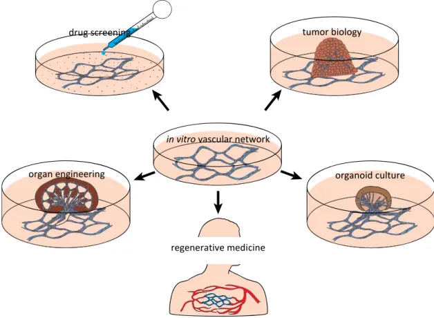
With current tissue culture techniques, tissues cannot be grown more than 100-200 µm in thickness, primarily because of the limited diffusion of nutrients and oxygen [10]. Tumor cells grown in non-adherent culture can develop into spherical masses (spheroids) up to 3mm in diameter, typically with a core of cells that are dead or dying due to diffusion limitation [11]. Similarly, masses of normal (non-malignant) cells grown in vitro are called organoids, and are currently the focus of great interest in biomedical research because they show some organization of tissues resembling in vivo organs[12]. We believe that the development of anin vitro vascular network could improve the culturing of spheroids and organoids (Figure 1) by allowing the tissues to grow and function in a way that is closer to the in vivosituation [13].
4 testing anti-angiogenic compounds in cancer research [17] or candidate drugs for their safe clinical application [18]; and studies in vascular regenerative medicine [19] (Figure 1).
Figure 1: Potential applications of a vascular network culture.
It has long been known from the field of human and animal surgery, including transplant surgery, that tissue can become re-vascularized when grafted to a suitable site[20, 21]. Similarly, developmental studies have shown that embryonic tissues can also readily become re-vascularized, and continue to grow into functional organs, when transplanted to various locations in the embryo[22].Furthermore, embryonic organ primordia can become vascularized if transplanted not only to the embryo itself, but to the vascular network in the extra-embryonic membranes. A good example of this is the chicken embryo chorioallantoic membrane (CAM) system[23, 24]. In that model, organ primordia are placed onto the highly vascular CAM, the blood vessels first having been scratched to open them up. The organ primordia can then form a vascular connection with the CAM vessels, and undergo reasonable growth and morphogenesis. The CAM, however, is highly sensitive to environmental factors [25], therefore the development of the tissue graft is not perfect, possibly because it is not submerged in a supporting volume of fluid, but rather is exposed to
in vitro vascular network
drug screening tumor biology
organ engineering
regenerative medicine
5 the air. In a sense, therefore, the CAM and other developmental systems show that the growth of organs on vascular beds is a possibility. What is needed, however, is a vascular bed ex vivo that is perfused by some kind of microfluidic system.
Most of the current research describing vasculogenesis (de novo formation of blood vessels from progenitor cells)and angiogenesis (formation of blood vessels from existing blood vessels) uses mammalian models, mainly mice. However, these models are fairly expensive, time consumingand require ethical and other permissions[26]. Endothelial cell lines such as human umbilical vein endothelial cells (HUVECs) are commonly used for developing in vitro
vascular networks[27].Other sources for developing such cultures include embryonic or adult stem cells or tissue explants. The uses and limitations of these techniques are discussed in detail in the following sections.
Zebrafish can be an alternative to mammalian models for studying vascular development[28].The zebrafish produce a large number of fertilized eggs at low cost; the embryos are externally fertilized and therefore readily accessiblefor experiments[29].In some jurisdictions, zebrafish embryos have fewer ethical restrictions. For example, in the European Union, the Directive 2010/63/EU on the protection of experimental animals allows zebrafish embryos to be used until 5 days post fertilization (dpf)without restriction[30].Finally, the zebrafish genome has been sequenced and there is a high level of conservation between zebrafish and human protein coding genes [31]. This similarity supports the use of zebrafish to model various human diseases [32, 33].Because of these advantages, the zebrafish is currently emerging as a model species to study vasculogenesis and angiogenesisin vivo[28]. Transgenic reporter lines are proving very useful in these studies [28].
6 the next section. Finally, we summarize studies that use microfluidic technology to develop perfusable vascular networks, and we discuss their potential in the field of tissue engineering.
2. Development of vasculature
in vivo
Formation of a vascular system is an essential process in embryonic development. Because multicellular tissues cannot survive without a blood supply, the cardiovascular system is one of the earliest systems formed during embryogenesis[34, 35]. The endothelial precursor cells (angioblasts) differentiate into endothelial cells and undergo the process of vasculogenesis in early embryos to form the primitive blood vessels [36].Studies on zebrafish have shown thatthe angioblasts appear in the lateral mesoderm, migrate to the midline of the embryo and form the first blood vessels [37]. In adult mice and humans,endothelial progenitor cells reside in the bone marrow asmultipotent adult progenitor cells, and contribute to the formation of new blood vessels [38].
7
3. Factors controlling vasculogenesis and angiogenesis
in vivo
3.1. Protein factors influencing vascular development
The differentiation of endothelial cells and the formation of blood vessels is mainly controlled by several protein factors[44].Important protein factors in this context are summarized inTable 1. Some of these factors are released by the endothelial cells themselves, other factors are stabilizing signals released by other cell types[44]. The differentiation of angioblasts is induced mainly by fibroblast growth factor 2 (FGF-2) and bone morphogenic protein-4 (BMP-4) [43]. FGF-2inducesthe expression of vascular endothelial growth factor (VEGF) and other important chemokines required to control vascular morphogenesis[45]. The importance of FGF-2 for vascular formation has been shown in studies on quail and zebrafish embryos [46, 47].Similarly, BMP-4 deficiency is associated with severe abnormalities in early mouse embryos, including the lack of a well-organized vasculature [48].
Among the endothelial growth factors, VEGFs play the predominant role in regulating the formation of blood vessels [49]. The VEGF family consists of several VEGF genes of which VEGF-A, which interacts with endothelial cells through VEGF receptor 2 (VEGFR2 also known as KDR or FLK1), is the main component responsible for the viability and proliferation of endothelial cells [50]. The VEGF mRNA is alternatively spliced resulting in four different isoforms of VEGF (VEGF121, VEGF165, VEGF189, VEGF206), denoted by the number of amino acids in their peptide chain[51]. These isoforms, having different rates of diffusion in the ECM due to differences in their heparin binding ability, generate a gradient, producing chemical signals for the directional migration of newly forming capillaries [52].VEGFs also have important roles in the differentiation, migration and cell-cell adhesion of endothelial cells, as well asstimulating sprouting angiogenesis and the activationof tip cells[53].
8
Table 1. Description and role of different proteins involved in vascular development.
Protein Type Name Origin Role in vascular development Ref.
Secreted
protein VEGF-A, B and C Endothelial and parenchymal cells
VEGF-A: endothelial cellsurvival factor, main regulator of vascular development; VEGF-B:coronary vascularization; VEGF-C:lymphatic system
[51]
PlGF Mesenchymal
cells Embryonic vascular development, pathological angiogenesis [54] PDGF-B Endothelial cells Vessel maturation;binds to PDGF receptor-β on
pericytes [49]
FGF-2 Multiple cell
types Differentiation of angioblasts; stimulation of VEGF expression; maintenance of vascular integrity
[43, 45]
Ang-1 Mural cells Maintenance of endothelial cell quiescence;
stabilization of vessel walls [44, 49] Ang-2 Angiogenic tip
cells Acts as antagonist of Ang-1; promotes endothelial cell proliferation [44, 49] TGF-β Endothelial and
mesenchymal cells
Embryonic vascular development; induces
vascular smooth muscle cell differentiation [57, 58]
BMP-4 Mesoderm Differentiation of angioblasts; induction of VEGF
expression [43, 59]
Egfl-7 Endothelial cells Facilitates tube formation by coordinating
endothelial cell-cell contact and migration. [60] Membrane
proteins VEGFR1, R2 and R3 Endothelial and hematopoietic cells
VEGFR1 and R2 bind to VEGF-A, B and PlGF. VEGFR2 is the main mediator of angiogenesis. R3 binds to VEGF-C
[51]
Ephrin-B2
and B4 Endothelial cells Specification of arteries and veins [43, 44]
VE-cadherin Endothelial cells Maintenance of endothelial cell-cell contact and inhibition of endothelial proliferation [61] Dll-4 Endothelial tip
cells Induces Notch signalling in response to VEGF-A in sprouting angiogenesis [62] Notch-1
and 4 Endothelial stalk cells Restricts formation of endothelial tip-cell in response to Dll-4, ensures directional sprouting [62] Abbreviations: Ang, angiopoietin; BMP, bone morphogenetic protein; Dll, delta like ligand; FGF, fibroblast growth factor; PDGF,platelet-derived growth factor; PlGF, placental growth factor; TGF-β, transforming
growth factor β; VE-cadherin, vascular endothelial cadherin; VEGF, vascular endothelial growth factor; VEGFR, vascular endothelial growth factor receptor.
9 considered to be the principle mediators of in vivo vasculogenesis and angiogenesis at all developmental stages[67].
In addition to the secreted protein factors, membrane proteins on the surface of endothelial cells also play an important role in vascular morphogenesis (Table 1). Examples of these membrane proteins include vascular endothelial cadherin (VE-cadherin), which functions to maintain endothelial cell-cell contact during VEGF-induced migration[61];and delta like ligand-4 and Notch-1, which specifythe endothelial tipvs. stalk cells during sprouting angiogenesis[62].
3.2. Role of other cell types in vascular development
In addition to the secreted and membrane-bound protein factors discussed above, cell types other than endothelial cells also contribute to the formation of blood vessels. Pericytes and smooth muscle cells promote the proliferation and survival of endothelial cells and provide structural support to the blood vessels [68, 69]. Pericytes possess a number of receptors specific for the binding of angiogenic growth factors (such as PDGF-B and TGF-β) released by
endothelial cells[70]. Similarly, the receptor for Ang-1 (a growth factor released by pericytes and other mural cells) is expressed on endothelial cells. This interaction between pericytes and endothelial cells contributes to angiogenic sprouting, vessel maturation and maintenance[70]. In the blood vessels of the central nervous system, pericytes have been reported to express tight and adherens junction proteins, thus regulating the permeability of blood-brain barrier [71].
10
3.3. Role of extracellular matrix
Extracellular matrix (ECM)contributes to the formation and diversity of blood vessels in several ways including: (i) maintaining the histological structure and elasticity of the vessels, (ii)regulating the proliferation and differentiation of endothelial cells, and (iii) transporting, modifying or blocking the angiogenic growth factors[78].The ECM is a complex network of macromolecules and its composition and properties are highly variable among different tissues, affecting the tissue-specific differentiation of stem cells [79]. The protein components of ECM interact specifically with the transmembrane protein integrinswhich activate signalling pathways inside the cells,resulting in expression of particular genes that influence cellular differentiation [80].The biophysical properties of ECM (stiffness, elasticity etc.) are also thought to induce cellular pathways (collectively known as mechanotransduction pathways) affecting cell behaviour and fate [81].
Endothelial cells synthesize their own ECM, the major components of which typically include collagen type-IV, nidogens, heparansulfate proteoglycans and laminins [82]. Research on the ECM of blood vessels have shown the presence of different ECM components at different stages of vascular development [83]. In the beginning of the process, the endothelial cells adhere to and migrate on a laminin-rich ECM which is later replaced by a collagen type-I rich ECM to support vascular tube formation [83]. Another layer of ECM in the wall of blood vessels (the medial layer or tunica media) is synthesized by smooth muscle cells and includes fibrillin microfibrils, collagens and elastin as major components [84].Large vessels have an outer adventitial layer of ECM, containing fibronectin, fibrillar collagen and other proteins [84].The ECM of actively growing blood vessels also contains higher levels of fibronectin and its distinct splice isoforms, compared to the ECM of quiescent vessels [85].At sites of injury, platelets secrete fibrinogen which coagulates into a fibrin clot providing a substrate for wound healing [86].
11 biopolymers (such as polyethylene glycol and poly-D-lysine) have also been proven to enhance vasculogenesis and angiogenesis in vitro [92, 93].
3.4. Haemodynamic factors
Shear stress generated by blood flow on the luminal surface of endothelial cells is a mechanical factor that induces intracellular biochemical pathways resulting in gene expression changes and the modulation of the structure and function of blood vessels [94]. Heparin binding EGF-like growth factor is one such factor which is expressed in response to reduced blood flow and induces vessel narrowing [95]. Other molecular pathways involved in vascular remodelling are reported to be regulated by changes in shear stress leading to the expression of PlGF [96], Notch-1 [97], and Smad6 (involved in TGB-β signalling) proteins [98].
Microfluidic culture of endothelial cells is currently an emerging technology which mimics the physiological shear stress on cultured cells to achieve the goal of culturing functional blood vessels for tissue engineering [13, 99]. A number of techniques for culturing vascular networks have been described in which endothelial growth factors, ECM components and microfluidics are combined (see following sections for detailed discussion);however, the development of fully functional blood vessels still remains a challenge [99].
4. Culture of vascular networks using endothelial cells
12
Figure 2. Schematic overview of three possible approaches to establishing cultures of vascular networks. Stem cells, depending on their source, could be embryonic stem cells, mesenchymal stem cells, or induced pluripotent cells. Endothelial cells are in blue; diverse supporting cells are represented schematically by red and yellow.
By contrast, the culture of well-defined vascular networks with a lumen requires the co-culture of multiple cell types with endothelial cells [100]. The important supporting cell types,known to induce network formation by endothelial cells, include pericytes[113],
animal or human embryos or adult tissues
stem cells organ explant
cell differentiation
pure endothelial cells
vascular sprouting
co-culture
13 mesenchymal stem cells [109], fibroblasts [107], hepatocytes [114], smooth muscle cells[115] and adipose-derived stem cells (ASCs) [106]. The importance of fibroblasts in enhancing angiogenesis has been shown in co-culture with HUVECs[107, 116]. Growth factors derived from fibroblast cultures in conditioned medium have also been shown to increase vascular morphogenesis and vessel stability from HUVECs in 3D fibrin gels[108].Furthermore, the ECM components and angiogenic growth factors secreted by fibroblasts have been found to be critical for vascular tube formation from HUVECs[117]. The culture of vascular networks is established in naturally-derived matrices (such as Matrigel® [116], collagen type-I [118] and fibrin [106]); as well as in synthetic matrices ( such as Puramatix™ [87] and polyethylene glycol hydrogel [119]) both of which types mimic native ECM. These matrices are particularly valuable for promoting successful vascular morphogenesis in vitro [92]. For a vascular network formation, the endothelial cells may be cultured on a 2D surface coated with one or a combination of these gel matrices; or in a 3D gel matrix (Table 2). In one study with HUVECs, increasing the thickness of Matrigel®was shown to increase the mean vascular cord length, and to decrease the network density [119].Combinations of different gel components can be used to mimic the complexity of natural ECM. The addition of laminin to collagen type-I scaffoldshas been shown to increase network formation and VEGFR2 expression by HUVECs in culture[90]. Similarly, increased network formation from HUVECs was observed in composite collagen type-I/fibrin matrix compared to pure collagen [110].
14 promoted vascular network formation in co-cultured mouse brain microvascular endothelial cells and ASCs in 3D collagen type-I gel [122].
Table 2. Endothelial cell cultures for vascular morphogenesis. Interacting
Cell types Culture strategy ECM Substrate Medium additives Main outcomes Possible applications Ref.
HUVEC 2D BME EGM-2 Vascular network formation. Drug screening [27]
HUVEC
HBMSC 3D Col-1 + Laminin EGM, DMEM Laminin and HBMSCs promoted vascular network formation.
Tissue
engineering [90]
HUVEC 2D Matrigel® RPMI 1640 The anti-angiogenic effect of WIF-1 was reversed by hypoxic conditions.
Cancer research [105]
HUVEC
ASC 3D Fibrin EGM-2 Vasculogenesis was more stable in co-culture with ASC.
Tissue
engineering [106]
HUVEC
HDF 2D M199, ECGS, CS HDF co-culture and bio-active silicate stimulated network formation.
Tissue
engineering [107]
HUVEC
SF 3D Fibrin EGM-2, VEGF, bFGF, Ang-1,
TGF-β
Fibroblast derived-growth factors enhancedsprouting, lumen formation and vessel stability.
Developmental
studies [108]
HUVEC
hES-MC 3D Col-1 + Fbn DMEM/F12, VEGF, bFGF, HGF
hES-MC and HGF stabilized
vascular network. Regenerative medicine, drug screening
[109]
HUVEC
MSC 3D Col-1 + fibrin EGM-2, DMEM Increasing fibrin concentration increased vascular morphogenesis.
Tissue
engineering [110]
BAEC 2D Matrigel® DMEM,
VEGF, rGAL-8
GAL-8 promoted endothelial cell migration and capillary formation.
Cancer research [111]
RAEC 2D Matrigel® DMEM,
VEGF, roxarsone
Roxarsone promoted
vascular formation in vitro. Cancer research [112]
EVC
Pericytes 3D Col-1, HA-hydrogel EGM-2 Pericytes enhanced network formation and stability. Regenerative medicine [113] CC-SMC
CJ-EC 3D Fibrin M199 Co-culture with SMCs increased length and density of vessels formed by endothelial cells.
Tissue
engineering [115]
HUVEC
HDF 2D Matrigel® EGM-2 Capillary-like network formed only in co-cultures with HDF. Tissue engineering, regenerative medicine [116] HMVEC
HDF 2D Col-1 EBM-2 Revealed Collagen binding to specific integrin on endothelial cells activating tube formation pathways.
Developmental
studies [118]
HUVEC 2D Matrigel®,
PEG Medium 200 Matrix thickness and stiffness affected vascular cord length and network density.
Tissue
engineering [119]
BEC
15
ASC requiredco-culture with
ASCs. screening
RBMVEC
ASC 3D Col-1 DMEM Stimulation of Notch 1 pathway enhanced vasculogenesis.
Regenerative
medicine [122]
Abbreviations: Ang-1, angiopoietin-1; ASC, adipose-derived stem cells; BAEC, bovine aortic endothelial cells; BEC, blood vascular endothelial cells; CC-SMC, canine carotid artery-derived smooth muscle cells; CJ-EC, canine jugular vein-derived endothelial cells; bFGF, basic fibroblast growth factor; BME, basement membrane extract (Trevigen); Col-1, collagen type-I; CS, calcium silicate; DMEM, Dulbecco’s modified Eagle’s medium; EBM, endothelial basal medium; ECGS, endothelial cell growth supplement (Promocell); ECM, extracellular matrix; EGM, endothelial growth medium; EVCs, early vascular cells;F12, Ham’s F-12 medium; Fbn, fibronectin; HA-hydrogel, hyaluronic acid based hydrogel; HBMSC, human bone marrow-derived mesenchymal stem cells; HDF, human dermal fibroblasts; hES-MC, human embryonic stem cell derived mesenchymal cells; HGF, hepatocyte growth factor; HMVEC, human dermal microvascular endothelial cells;HUVEC, human umbilical vein endothelial cells; LEC, lymphatic endothelial cells;M199, medium 199 (Lonza); MSC, mesenchymal stem cells; NHLF, human normal lung fibroblasts; PEG, polyethylene glycol hydrogel;RAEC, rat aortic endothelial cells; RBMVEC, rat brain microvascular endothelial cells;rGAL8, recombinant galectin-8; RPMI, Roswell Park Memorial Institute medium (Gibco); SF, skin fibroblasts; TGF-β, transforming growth factor beta; VEGF, vascular endothelial growth factor; WIF-1, Wnt inhibitory factor-1.
4.1. Limitations of endothelial cell culture
Endothelial cell cultures are relatively easy to maintain. However, there are certain limitations which need to be considered while carrying out endothelial culture. A blood vascular network constitutes a number of vessel types, from large vessels to micro vessels, and each vessel type has its own unique properties (including endothelial cell subtypes). Therefore it is challenging to attempt to recapitulate the formation of different vessel types using a homogeneous endothelial cell population [123]. Primary endothelial cell cultures are usually derived from terminally differentiated tissues; these cells have limited proliferative and regenerative capacity, and a short life span in vitro [124, 125].
16
5. Use of stem cells for
in vitro
vasculogenesis
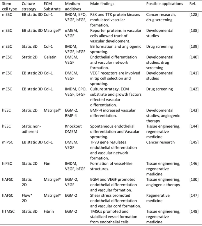
In recent years, stem cells have increasingly been used to develop vascular cultures; this is because stem cells have several advantages over terminally differentiated endothelial cells [19]. Stem cells are multipotent or pluripotent in nature, they show self-renewal, and their differentiation along various cell lineages can be manipulated by fine-tuning the culture conditions [19]. A few examples of the stem cells that can be used for endothelial, and ultimately vascular, differentiation are summarized in Table 3. Three main stem cell types used are: (i) embryonic stem cells (ESCs) [128], (ii) induced pluripotent stem cells (iPSCs) [129] and(iii) mesenchymal stem cells (MSCs) [130].
In addition, endothelial progenitor cells (EPCs), which originate in the bone marrow and contribute to the formation of new blood vessels in adults, are also useful in the study of in vitro vasculogenesis [125]. The differentiated endothelial cells arising from stem cells directly undergo vasculogenesis because of the presence of other cell types that have also differentiated from the stem cells.Alternatively, the endothelial cells can be isolated from the stem cell culture, without the unwanted additional cell types, and used for vascular morphogenesis (either in pure culture or co-culture with defined cell types) [113].
One of the advantages of using ESCs is that they can differentiate into multiple vascular cell lineages simultaneously in culture. In principle, these different lineages can contribute to the newly-formed vessels (neovessels) in a way that closely resembles the in vivo vasculogenesis in early embryos [19, 131]. Endothelial differentiation and vascular morphogenesis in ESCs is controlled by culture conditions (such as the presence of growth factors in the medium and the use of feeder layers of stromal cells, or a substratum consisting of a natural or synthetic hydrogel [131, 132]).
17 to undesired cell types such as fibroblasts [133]. By contrast, in 3D culture (suspension or gels), the proliferation of EB cells is limited, allowing greater control of the differentiation of the desired cell type [133]. Significant effects of different factors, such as culture substrate (collagen type-IV or fibronectin), cell seeding density, concentration of VEGF and FGF in medium, and culture duration, have been observed on the endothelial differentiation in human, mouse and zebrafish ESC culture [88, 136]. Similarly, TGF-β has been identified to induce vascular differentiation in human ESCs [137].
Table 3. The use of stem cell technology for endothelial differentiation and vascular development. Stem
cell type Culture strategy ECM Substrate Medium additives Main findings Possible applications Ref. mESC EB static 3D Col-1 IMDM, EPO,
VEGF, bFGF, RSK and TTK protein kinases modulated vascular formation.
Cancer research,
drug screening [128]
mESC EB static 3D Matrigel® αMEM,
VEGF Reporter proteins in vascular cells allowed track of vascular development.
Developmental
studies [138]
mESC Static 3D Col-1 IMDM,
VEGF, bFGF EB formation and angiogenic sprouting. Drug screening [139] mESC Static 2D Gelatin DMEM,
VEGF Endothelial differentiation and vascular network formation.
Developmental studies, drug screening
[140]
mESC EB static 2D Col-1 DMEM,
VEGF VEGF receptors are involved in tip cell selection and sprouting.
Developmental
studies [141]
mESC EB static 3D Col-1 IMDM, EPO,
VEGF, bFGF Culture strategy, ECM substrate and growth factors effected vascular
differentiation.
Drug screening [142]
hESC Static 2D Matrigel® EGM-2,
BMP-4 BMP-4 increased vascular differentiation. Developmental studies, angiogenic therapy
[143]
hESC Static
non-adherent Knockout DMEM Spontaneous endothelial differentiation and Vascular sprouting.
Tissue engineering, regenerative medicine
[144]
miPSC EB static 3D Col-1 DMEM,
VEGF TP73 gene regulates endothelial differentiation and vascular network formation.
Cancer research [145]
hiPSC Static 2D Fbn IMDM,
VEGF, bFGF Formation of vessel-like structures. Tissue engineering, regenerative medicine
[146]
hAFSC Static
2D Matrigel® EGM-2, VEGF EGM and VEGF promoted endothelial differentiation and vascular formation.
Tissue engineering,
angiogenic therapy [130]
hAFSC Flow*
2D Matrigel® EGM-2 Shear stress promoted endothelial differentiation and vascular cord formation.
Regenerative
medicine [147]
hTMSC Static 3D Fibrin EGM-2 TMSCs promoted and stabilized vessel formation from endothelial cells.
Tissue engineering, regenerative medicine
18
Abbreviations: αMEM, alpha-minimal essential medium (Cellgro); bFGF, basic fibroblast growth factor; BMP-4, bone morphogenetic protein-4; Col-1, collagen type-I; DMEM, Dulbecco’s modified Eagle’s medium; EB, embryoid body intermediate; ECM, extracellular matrix; EGM, endothelial growth medium (Cambrex or Clonetics); EPO, erythropoietin; Fbn, fibronectin; Flow*, on the margins of the bottom of a flask on an orbital shaker; hAFSC, human amniotic fluid-derived stem cells; hESC, human embryonic stem cells; IMDM, Iscove’s modified Dulbecco’s medium; mESC, mouse embryonic stem cells; miPSC, mouse induced pluripotent stem cells; RSK, ribosomal S6 kinase; Static, static replacement culture; TP73, tumor protein-73; TTK, threonine and tyrosine kinase; VEGF, vascular endothelial growth factor.
Another important stem cell type, similar to ESCs in pluripotency and differentiation events, is the iPSCs[149]. An advantage of iPSCs is that they can be generated by genetic reprogramming of any adult somatic cell population, and therefore raise fewer ethical concerns compared to ESCs [19]. Endothelial differentiation in iPSCs can be induced by applying similar methods used for differentiation of ESCs [150]. Furthermore, gene expression in endothelial cells derived from ESCs and iPSCs is very similar [150]. MSCs are multipotent stem cells residing in adult tissues; they have limited differentiation potential compared to ESCs and iPSCs[19]. Endothelial differentiation in human amniotic fluid derived MSCs has been shown to be inducible by VEGF [130]. MSCs derived from various tissues (bone marrow, hair follicle, adipose tissue and muscles) have been used for vascular regeneration studies reviewed in Ref. [19]. In some studies the MSCs have been reported to promote and stabilize vascular network formation from HUVECs (Table 2).
In addition to the use of pluripotent and multipotent stem cells for endothelial differentiation and in vitro vasculogenesis, the unipotent EPCs also have the ability to differentiate into mature endothelial cells and form vascular tubes in culture [125]. The advantage of EPCs for culturing vascular networks is that these cells can be easily obtained from adult tissues such as peripheral blood [19]. In vitro studies have shown that the early EPCs do not directly undergo vascularization, but release factors to stimulate angiogenesis in distantly-cultured endothelial cells in a transwell[125]. Co-culture with MSCs has been proven to enhance vascular formation from EPCs both in vitro and after implantation in vivo
[151, 152].
5.1. Issues and drawbacks with stem cell culture
19 been developed from these animals, the technique has been proven less successful for other species such as cattle, goat and dogs [149]. Furthermore, the very complexity of vascular differentiation from ESCs means that the growth factors necessary to support the generation and maintenance of multiple cell types need to be laboriously optimised [153]. Furthermore, the use of human ESCs for research raise ethical concerns [19].
ESCs and iPSCs are both pluripotent, and therefore it is challenging to direct the differentiation towards a specific lineage, and to obtain high quality pure cell cultures [149]. The iPSCs are developed by transfection of somatic cells with pluripotency genes; however, the efficiency of the process is very low (less than 1%) [154]. The iPSCs (in contrast to ESCs) are derived from adult differentiated cells by de-differentiation. Then, if re-differentiated into a specific cell type, they attain some of the characteristics of that cell type but are not identical to their normal counterparts [154]. Other issues with stem cells is that the isolation of MSCs from adult tissues requires invasive surgical procedures, and only yields small numbers of cells; the proliferation of these cells is also limited in vitro[155]. Furthermore, the MSCs isolated from different tissues or life stages are not the same, and therefore have different culture requirements and angiogenic potentials [156].
The isolation and culture methods for EPCs are only relatively recently developed (Asaharaet al., 1997 [157]). For this reason, there is no standard protocol among researchers. It should be noted that there are no specific markers for EPCs because many of the genes expressed by EPCs are also expressed in hematopoietic progenitors [158]. Similar to the iPSCs and MSCs, the number of EPCs found in isolated adult tissue cells is very low, and this greatly limits their study [19]. Finally, analysis of EPCs in long-term culture has shown that the late passage cells (45 days after the initiation of the primary culture) have changed morphology, reduced their proliferation rate, show high β-galactosidase expression and loss of vascular network formation ability, compared to the early passaged cells [159].
6. Use of tissue explants for
in vitro
angiogenesis
20 multiple cell types both in vivo and in vitro, as discussed in the previous sections. The use of tissue explants is important in this context, because these explants already contain multiple cell types, and the angiogenesis stimulated in these cultures closely represents the corresponding process in vivo[127].Furthermore, tissue explant experiments are relatively easy to perform and allow a large number of cultures to be derived from a single tissue sample[160].
Various tissue explants have been shown to have the ability to develop vascular sprouts in vitro (Table 4). Examples include cross sections of aorta called aortic rings [161]; metatarsal bones [162]; retina fragments [163]; choroid-sclera fragments [164]; and adipose tissue [165]. In most cases, the tissues for explant preparation are isolated from developing rodent embryos or neonates. Tissue explants from other species such as chick embryo aortic arch [166], rabbit aorta [167] and pig carotid artery [168] have also been adapted for sprouting angiogenesis. Furthermore, angiogenic sprouting has also been reported from human tissue explants e.g. adipose tissue [165], aortic explants from aborted embryos [169], placental explants [170, 171] and umbilical artery rings [172].
Explant cultures are usually established in a 3D gel matrix in the presence of angiogenic growth factors, and are examined for microvessel outgrowth (vascular sprouting)[127, 173]. The aortic ring model from various species is the most commonly used explant for studying
in vitro angiogenesis (Table 4). The stimulatory effect of various factors, such as angiogenic growth factors (especially VEGF) and ECM components, on the growth of vascular sprouts from aortic ring have been extensively studied, reviewed in Ref. [174]. Recentlydeveloped explant cultures, using fetal metatarsals from mice, have shown advantages over the aortic ring model, in that they do not require a 3D matrix and exogenous growth factors for vascular sprouting [162].In general, explant cultures can serve as an intermediate between the endothelial cell culture on the one hand, and in vivo models on the other. They are also thought to be more reliable for studying the mechanisms of angiogenesis andtesting the role of regulatory factors [175].
6.1. Limitations of explant cultures
21 medicine. The mouse aortic ring model shows significant variability in microvessel sprouting from explants isolated from different age and strain of animals [176]. Variability in outcome has also been reported using explants isolated from different vessel types (artery or vein) of the same individual animal [177]. The vascular sprouts in the aortic ring model regress over time in culture (with peak sprouting between days 6 and 7), and this limits the analysis time and increases variability in results with culture duration [161]. Furthermore, the aortic rings are derived from large vessels, and therefore do not truly represent in vivo angiogenesis, which is a microvascular process [127].
Table 4. Tissue explants used for sprouting angiogenesis in vitro. Tissue
explant Culture strategy ECM Substrate Medium additives Main findings Possible applications Ref. mAR Static 3D Col-1 Opti-MEM,
VEGF VEGF and collagen increased vessel sprouting. Drug screening [161] mAR Static 3D Col-1 MCDB131,
VEGF Age of the mouse inversely affected vascular sprouting from explant.
Drug screening [178]
mAR Static 3D Col-1 ESFM Endostatin inhibited vascular sprouting by modulating endothelial cell-ECM interaction.
Cancer research [179]
rAR Static 3D Matrigel® EGM-200 Ascorbate inhibited
sprouting angiogenesis. Cancer research [180] hUAR Static 3D BME EGM-2 Capillary spouting upon
VEGF stimulation. Cancer research, drug screening [172] cAA Static 2D Matrige® bFGF, VEGF Chemical compound
releasing nitric oxide inhibited angiogenesis
Cancer research,
drug screening [181]
mAT static 3D Col-1 MCDB131,
VEGF Angiogenic sprouting. Drug screening [182] hAT Static 2D Matrigel® EBM-2,
EGM-2MV Angiogenic capacity reflected donor’s physiology.
Angiogenic therapy,
drug screening [165]
mRE Static 2D PTFE DMEM,
VEGF VEGF stimulated vascular sprouting. Drug screening [183] mRE Static 3D Fibrin DMEM,
VEGF VEGF stimulated sprouting angiogenesis in a dose dependent manner.
Drug screening [184]
mMT Static 2D Gelatin OR
col-1 αMEM Vascular sprouting occurred without additional growth factors.
Developmental studies, drug screening
[162]
mMT Static 2D αMEM,
VEGF VEGF recovered the impaired vascular sprouting in endoglin deficient explants.
Diseases modelling,
drug screening [185]
mPE Static 2D Col-1 M199, VEGF,
bFGF Isoforms of VEGF differentially stimulated vascular sprouting.
Developmental
22
Abbreviations: αMEM, alpha-minimal essential medium; bFGF, basic fibroblast growth factor; BME, basement membrane extract (BD Biosciences); cAA, chick aortic arch; Col-1, collagen type-I; DMEM, Dulbecco’s modified Eagle’s medium; EBM, endothelial basal medium (Lonza); ECM, extracellular matrix; EGM-MV, endothelial growth medium microvascular; EGM, endothelial growth medium (Cascade Biologics); ESFM, endothelial serum-free medium (Life Technologies); hAT, human adipose tissue explant; hUAR, human umbilical arterial ring; M199, medium 199 (Gibco); mAR, mouse aortic ring; mAT, mouse adipose tissue explant; MCDB131, basal medium (Invitrogen); mMT, mouse metatarsal explant; mPE, mouse proepicardium explant; mRE, mouse retinal explant; Opti-MEM, minimal essential reduced-serum medium (Gibco); PTFE, polytetrafluroethylene membrane; rAR, rat aortic ring; Static, static replacement culture; VEGF, vascular endothelial growth factor.
Tissues containing microvascular networks (e.g. adipose tissue and retina) can be used for explant preparation. However, these tissues are more difficult to isolate and, like the aortic explant, show variability between experiments [163]. The high levels of capillary sprouting observed in adipose tissue explant cultures are in many senses an advantage; however they do make it difficult to identify all the sprouts individually and interpret the results [165]. Similarly, angiogenic sprouts from fetal mouse metatarsal explants present microvascular features; however, their isolation and culture procedures also require advanced technical skills, which are key to the reproducibility of the research[162]. Finally, the metatarsal and chick aortic arch explants are isolated from developing embryos and have high proliferative capacity; therefore, angiogenesis in these models does not represent the in vivo situation in adults [127].
7. Zebrafish: a new model species for studying
in vivo
vasculogenesis and angiogenesis
The zebrafish is a freshwater teleost fish [187] that is emerging as a model of choice for studying vasculogenesis and angiogenesis[188]. The embryos and larvae are often used in these studies because of their external fertilization, optical transparency at early stages, and the ease of exposure to test substances (by simply adding the compound to the swimming water) [189]. Furthermore, the genome comparison study has revealed that there is at least one orthologue in zebrafish genome for more than 70% of human protein coding genes [31]. Vascular development and function in zebrafish are relatively conserved, compared to the same processes in other vertebrates [188].
23 the living, transparent embryo [28]. Enhanced visualization of vascular development can be achieved by injecting fluorescent micro particles into the blood stream, or by using transgenic lines such as kdrl:GFP and fli:GFPthat express green fluorescent protein (GFP) in vascular cells [28].
For these and other reasons, vascular development in zebrafish — from early differentiation of angioblasts to the maturation of blood vessels—has been extensively studied [32, 37, 190, 191]. Studies have shown similar angiogenic responses to the test substance irisin, in zebrafish embryos in vivo, and in HUVECs in vitro[192]. In another example, the genetic mutation (gridlock), which causes aortic malformations and congenital heart defects in humans, showed similar phenotypic effects in zebrafish [193]. Zebrafish have been successfully utilized to model several human vascular diseases reviewed in Ref.[32].
Similarly, a zebrafish in vivo xenograft model has been developed to study human carcinomas [194]. These studies have shown successful invasion, metastasis and extravasation of various human tumorcells in zebrafish embryos and adults [194]. It has been demonstrated that the transplantation of human WM-266-4 melanoma cells and breast adenocarcinoma cells in zebrafish embryos induced angiogenesis in the host vasculature; this led to the formation and infiltration of neovessels into the tumor masses[195, 196].Other examples of human carcinomas studied in zebrafish include breast cancer bone metastasis [197], uveal melanoma [198] and retinoblastoma [199].
The zebrafish possesses remarkable regenerative capacity in several organs (including the caudal fin and heart [200, 201]) which makes it a useful model for studying regeneration [202]. The regeneration of organs also involves the regeneration of blood vessels, and therefore the regenerative capacity of zebrafish is also important for vascular regeneration studies [28].
7.1. Zebrafish transgenic reporter lines for vascular studies
24 endothelial and blood cells; that line permitted the observation of the development of the vascular system and blood flow simultaneously [203].
The most important transgenic line that we are utilizing to study vascular development in zebrafish is the kdrl:GFP line (Figure 3A). This transgenic line isalso known as Tg(kdr:eGFP) or
Tg(flk1:eGFP) [204]. In this line, GFP is specifically expressed in endothelial cells under the control of the VEGFR2 or kdr-like gene [204]. The kdrl:GFP line allows high resolution analysis of single cell migration and vascular development in living embryos [204].The utility of
kdrl:GFP zebrafish embryos has been confirmed as a high-throughput model for screening the effect of toxic compounds on vascular development[205].
Other transgenic zebrafish lines are available for vascular studies, although they have some limitations. For example, In Tg(Tie2:eGFP) the GFP expression is relatively weak in the vascular cells[206]. Similarly, in Tg(fli1:eGFP) the GFP is expressed in certain non-vascular cells which interferes with the results especially in the head region of the embryo [204]. Furthermore, studies on Tg(fli1:eGFP) have shown changes in the gene expression of a number of genes, compared to the wild type embryos, which may affect the results while using transgenic zebrafish for experiments [207].
In summary, zebrafish is a high-throughput, easily quantifiable, fast developing and relatively inexpensive in vivo model for vascular studies. However, there are some drawbacks associated with this model. For example, the relevance of zebrafish embryo model to understand human angiogenesis is questioned, as there is a large evolutionary time difference between the two species [175]. Therefore, preclinical drug screening in zebrafish should always be followed by validation in mammalian models beforegoing to clinical trials [28].
8. Future prospects for using zebrafish cells for
in vitro
vasculogenesis and angiogenesis
25 drawbacks associated with adapted cell lines can be avoided [208]. In addition to the above mentioned characteristics, zebrafish also possess specific desirable features for in vitro
applications.
8.1. Cell culture techniques in zebrafish
The external fertilization andlarge number of fast developing embryos, allow easy harvesting of large numbers of cells and quantities of tissues from different developmental stages[209]. Zebrafish cells grow at a lower temperature (26-28 °C) than chick and mouse cells and do not usually require a CO2-enriched atmosphere [209]. These properties allow zebrafish cells to be grownat room temperature, although the use of a simple incubator is recommended to help maintain sterile conditions[209]. The protective covering of the chorion, which is present until hatching at around 48 hpf, partly isolates the embryos from the environment[210]. This is important for in vitro studies because it maintains the embryos in an aseptic condition[211].
To harvest sterile cells or tissues from zebrafish embryos it is necessary to decontaminate the surface of the chorion. Using this approach, it is possible to isolate and culture sterile cells from blastula (3 hpf) or gastrula (24 hpf) stage embryos [209, 212].In a recent study, we have shown that embryos with a chorion decontaminated at 24 hpf could be further cultured to 5 dpfunder aseptic conditions [173]. The tissues and cells isolated from these embryos were successfully maintained free of contamination for eight days in culture.
8.2. Zebrafish embryonic stem cells
26
Figure 3: Confocal images of transgenic zebrafish kdrl:GFP whole embryo (A) and EB culture (B-D). (A) A 5 dpf kdrl:GFP embryo showing florescent endothelial cells forming blood vessels. Scale bar, 200 µm. (B, C and D) Developing embryoid body on subsequent days of culture (day 1, day 6 and day 8, respectively) on a mixture of collagen type-I, Geltrex™ and fibrin substratum, showing the development of vascular network-like structures from kdrl:GFP+ endothelial cells. Scale bars, 100 µm.
The induction of myogenic differentiation in zebrafish primary ESCs has been shown by culturing these cells on a laminin substratum, in medium containing insulin [216], FGF [217] or sonic hedgehog protein [218]. In another study, the seeding density (between 1 and 2 × 10⁴ cells/cm2) of zebrafish primary ESCs, co-culture with a zebrafish fibroblast-like cell line (ZF4) and medium supplementation with insulin, were found to induce cardiomyocyte differentiation [219]. Similarly, an increase in the generation of primordial germ cellresembling-cells was found in zebrafish blastocyst cell cultures after the addition of BMP-4, EGF and retinoic acid to the medium [220]. Furthermore, the use of FGF and VEGF have been shown to increase differentiation towards the endothelial cell lineage in zebrafish blastocyst cells [221].
Zebrafish blastocyst cells aggregate into EBs in culture. We have shown that the percentage of endothelial-like (kdrl:GFP+) cells in EBs is increased by culturing them in suspension (i.e.
hanging drop culture)rather than in adherent culture, and by adding endothelial growth supplements, including VEGF, to the medium [136]. We found that the kdrl:GFP+ cells in EB
culturesform vascular network-like structures on hydrogel substrates (Figure 3B-D). A
27
8.3. Cultures of specialised zebrafish cell types
Attempts have been made to develop cultures of specialised cell types using progenitor cells isolated from zebrafish embryos.In one study, neural crest cells, isolated from dissociated 14 hpf (10-somite stage) embryos, were maintained in culture [222]. These cells were shown to proliferate, migrate and differentiate into neurons, chondrocytes and glial cells in vitro[222]. Strategies have also been described for the culture of primary neurons from developing brain and spinal cord cells of zebrafish embryos [223]. We have found that the endothelial cells in dissociated hearts, isolated from 5 dpf zebrafish embryos,form colonies ona fibronectin substratum; however, these cultures could only be maintained for short periods, possibly because of low seeding-density [173].
These studies suggest that zebrafish embryonic cell culture can be an important model for studying endothelial differentiation and vascular morphogenesis in vitro. In principle, it could be possible to use zebrafish kdrl:GFP embryos for the isolation of endothelial cells using fluorescence activated cell sorting (FACS). The development of these cells into vascular networks in response to various signals could then be readily tracked in live cultures without the need to fix and stain them. Furthermore, tissues and organ explants from zebrafish embryos can be a promising model for sprouting angiogenesis, as we have shown using liver and heart explants from 5 dpf embryos [173].
In summary, the zebrafish allows easy access to large numbers of primary cells, and vascular development in cultures derived from these cells occurs in a complex environment of other cell types. In contrast, it is difficult to access primary cells and tissues in mammalian models, and the vascular culture using endothelial cell lines such as HUVECs does not reflect the complex process in vivo.
8.4. Disadvantages of zebrafish in vitro model
28 growth [213]. The lower incubation temperature for zebrafish cells may not be ideal for human cells if a co-culture has to be established.
The zebrafish embryonic cells usually have to be cultured on a feeder layer of growth-arrested cells to maintain their pluripotency [211]. The development of vascular network in primary zebrafish embryonic cells and tissues in the presence of other supporting cell types may be considered as an advantage as it closely mimic the in vivo situation. However, this provides less control over the in vitro vascular development compared to endothelial cell lines. Furthermore, because the zebrafish is a relatively new research model, the differentiation and culture conditions for its cells still needs to be optimised.
9. Organoids: un-vascularized organotypic cultures
A remarkable capacity for self-organization in vitro is shown by stem cells (embryonic or adult pluripotent stem cells) and primary tissues (organ explants or dissociated cells). This capacity allows them to develop into ‘organoids’ — 3D cell masseswith organ-like properties[224]. In an organoid, multiple organ-specific cell types are arranged together, recapitulating some properties (structural and functional) of an organ [225]. The ability to control the differentiation in pluripotent stem cells makes them a model of choice for organoid cultures[226]. Using these cells, organoids of a number of organs including brain, retina, stomach, intestine, lungs, liver and pancreas, have been developed (reviewed in Ref. [12]).The organoid culture not only allowsthe study of developmental processes,it also represents an emerging model system to study diseases, to screen toxicants, and to develop personalized and regenerative medicine[12, 226].
29 reported [228]. Finally, the simultaneous effect of growth factors and extracellular matrix in these cultures may inhibit some pathways required for organogenesis [229].
The major issue with organoids, as with other tissue engineering approaches,is the lack of vascularization. This necessarily means that organoids are diffusion-limited and cannot fully develop into mature organs in vitro[228]. One study demonstrated the development of 3D neural organoid constructs with a vascular network inside, by co-culturing neural progenitor cells, endothelial cells, MSCs and macrophage precursors [230]. A similar neurovascular spheroid model has been developed using primary cortical tissue cells isolated from 1-3 days postnatal mice and rats [231]. In another study theintestinal organoids developed from human ESCs were implanted in mice under the kidney capsule[232]. These organoids further expanded, maturated and demonstrated intestinal morphology and function after vascularization by the host vasculature[232].
Taking it a step further, the use of decellularized whole organs as scaffolds for culturing bio-artificial organs are now gaining interest. In this technique the cell population of an organ is removed by perfusion with detergents or by mechanical means, while retaining the original three dimensional shape of the extracellular matrix[233]. These organ scaffolds are then repopulated with stem cells, endothelial cells or a mixture of specific cell types (depending on which organ was used) to generate an organ culture [233]. Using this strategy Ottet al. have developed a heart construct using decellularized rat heart seeded with cardiac or endothelial cells [234]. The recellularized construct retained the physiological structure of the heart and showed contractions, pump function and response to drugs [234].Similarly, decellularized rat intestine becomes vascularized when human endothelial cells are seeded into its empty vascular channels[235].
10. Beyond organoids: microfluidics and the development of a
functional vascular network
30 microfluidic technologies to pump blood or some nutrient liquid through the vascular network.
Microfluidic systems give control over the biophysical and biochemical microenvironment of cells, allowing the analysis of complex interactions between different cell types and the respective signalling molecules [236].Microfluidic devices(chips) are made from various material (including glass and poly dimethyl-siloxane), using microfabrication techniques. They contain hollow channels that can be filled with ECM components and medium [13]. The devices can simply be connected to a medium reservoir to generate a passive flow through the channels; or to a pump for more constant flow rate (Table 5).Using unique combinations of materials (with different structural and mechanical properties), microfabrication techniques, ECM components, growth factors, cell types and flow rates, a controlled environment can be created to promote vascular development [236].
These properties of microfluidic systemsmight, in the future, be adaptable to the vascularisation of organoids in vitro. This, in turn, could allow more advanced development, by providing chemical cues and nutrient/waste exchange through a perfusable vascular bed. Connecting an organoid culture to a microfluidic vascular bed would also allow the required changes in gene expression in response to the shear stress generated by the flow of medium.
Microfluidicscould also make it possible to manipulate the course of organogenesis in organoid cultures. For example, the flow rate could be increased with time to fulfil the increasing nutrient requirements of the growing tissue. Designing a microfluidic device with multiple channels connected to different parts of the culture chamber, could also allow the delivery of a specific growth factor to aspecific region of the developing organ. Furthermore, a microfluidic vascularized organ culture would make it possible to visualize the core of optically dense tissues by introducing staining solutions through the vasculature.
10.1. Examples of microfluidic vascular culture systems
31 physiological responses to flow-induced shear stress [237]. The flow rate in this experiment was 5 µL/min, which generated a shear stress of 0.31 - 7.22 dyne/cm2. This value is close to the physiological level of shear stress (1 to 10 dyne/cm2) [237]. It should be noted that the shear stress generated by a particular flow rate depends on the dimensions of the microfluidic channels. In the same microfluidic device the authors showed that the presence of flow (regardless of flow direction) facilitates vasculogenesis by HUVECs, and that angiogenic sprouting occurs only in the direction opposite to the flow direction[238]. Similarly, angiogenic sprouting in 3D collagen type-I gel from HUVEC cultures in a microfluidic device showed morphological features resembling in vivo angiogenesis [239].
Table 5. Microfluidic devices for the culture of vascular networks. Device
design* Cell types used Flow conditio n
3D
matrix Medium additive s
Main outcomes Possible
applications Ref.
HMVEC Passive Col-1,
PDL EGM-2MV, VEGF
Endothelial tube-like structures regressed in PDL treated channel after 2-3 days.
Tissue
engineering [93]
HUVEC NHLF HPP Active: 5 µL/min
Fibrin EGM-2 Perfusable 3D vascular
network. Tissue engineering, drug screening
[237]
HUVEC
NHLF Passive Fibrin EGM-2 Flow stimulated angiogenic sprouting opposite to direction of flow.
Cancer
research [238]
HUVEC Passive Col-1 EGM-2 Directional neovessel growth in response to angiogenic signals.
Drug
screening [239]
HUVEC HBMSC OB Active: 2 µL/min
Fibrin EGM-2MV, VEGF, Ang1
Tumorcellextravasation into the surrounding bone-mimicking matrix. Cancer research, drug screening [240] HUVEC
NHLF Passive Fibrin EGM-2 Easy quantification of tumor cell extravasation. Cancer research [241] HMVEC Active:
1-2 µL/min
Col-1 EGM-2MV, VEGF
Metalloproteinases modulated the length and diameter of vessels in 3D ECM. Developmenta l studies, tissue engineering [242] ECFC-EC NHLF CC
Passive Fibrin,
laminin EGM-2 A vascularized tumor model responding to chemical stimuli.
Cancer
research [243]
iPSC-EC
NHLF Passive Fibrin VVM iPSC-ECs formed inter-connected capillaries with lumina
Disease modelling, drug screening
[244]
iPSC-EC Passive PEG VVM,
VEGF iPSC-ECs self-organized into tubular networks, VEGF enhanced network formation.
Tissue engineering, drug screening
32
ECFC-EC
NHLF Passive: 0-1 mm/s**
Fibrin EGM-2, VEGF, bFGF, 5% O2
Endothelial cells self-organized into a perfused interconnected capillary network. Tissue engineering, drug screening [246] ECFC-EC
NHLF Passive: 8-100 µm/s**
Fibrin EGM-2,
20% O2 Flow modulated cellular communications between different chambers by carrying cell-secreted morphogens.
Drug
screening [247]
HUVEC
HBVP Passive: 10 µL/min
Col-1 M199, ECGS, VEGF, bFGF
The vascular cells showed an inflammatory
response when stimulated.
Diseases
modelling [248]
HUVEC Passive: 0.7-8 µL/min
POMaC
EGM-2MV The pre-formed vascular network allowed the growth of tissues and showed physiological responses. Tissue engineering, drug screening [249] HPMEC
AEC Active: 0.3 µL/min
Col-1 EBM-2 Capillary side responded physiologically to alveolar infection by releasing cytokines.
Drug
screening [250]
RBME
RCC Passive Fbn, PDL Neurobasal™, bFGF
Vascular side stimulation with TNF-α activated microglia and astrocytes on the neural side.
Disease modelling, drug screening [251] HBMVEC hiPSC-CN Astrocytes Pericytes Active: 2 µL/min Lamini
n, Col-1 EBM-2 The BBB model increased permeability and activated cytokines in response to LPS stimulation.
Disease modelling, drug screening
[252]
ZBC-EB Active: 20 µL/min Col-1, Geltrex ™, fibrin LDF, VEGF, EGS, bFGF
The endothelial cells in the EBsformed longer and wider sprouts in
microfluidic culture.
Drug
screening [173]
Abbreviations and symbols:Active, flow is generated by a pump with constant flow rate; PDL, poly-D-lysine; AEC, alveolar epithelial cells; Ang-1, angiopoietin-1; bFGF, basic fibroblast growth factor; CC, cancer cells (colorectal, breast or melanoma cell line); Col-1, collagen type-I; EBM, endothelial basal medium; ECFC-EC, human endothelial colony forming cell-derived endothelial cells;ECGS, endothelial cell growth supplement;EGM, endothelial growth medium; EGM-2MV, endothelial growth medium microvascular;Fbn, fibronectin;HBMSC, human bone marrow-derived mesenchymal stem cells; HBMVEC, human brain-derived microvascular endothelial cells;HBVP, human brain vascular pericytes;hiPSC-CN, humaninduced pluripotent stem cell-derived cortical neurons; HMVEC, human dermal microvascular endothelial cells; HPMEC, human pulmonary microvascular endothelial cells;HPP, human placental pericytes; HUVEC, human umbilical vein endothelial cells; iPSC-EC, human induced pluripotent stem cells-derived endothelial cells;M199, medium 199 (Lonza); NHLF, human normal lung fibroblasts; OB, osteoblasts; Passive, the flow is generated by hydrostatic pressure of the medium in a reservoir; PEG, polyethylene glycol hydrogel;POMaC, poly(octamethylene maleate (anhydride) citrate); RBME, rat brain microvascular endothelial cells; RCC, E-18 rat cortical cells; TNF-α, tumor necrosis factor alpha; VEGF, vascular endothelial growth factor; VVM, VascuLife® VEGF medium; ZBC-EB, zebrafish blastocyst cell-derived embryoid bodies; *, see Figure 4 for further description of device design; **, flow velocity calculated by tracking the movement of fluorescent dextran particles through the network.
33 the lumen openings[241]. This allows the vascular network to be perfused by chemical compounds or cells. The device allows rapid quantification of changes in vascular networks in response to test compounds[241].Using the same design, Jeon et al. have shown the extravasation of cancer cells, introduced through the perfused vascular network, into the surrounding bone-mimicking matrix [240]. In a similar device, human colorectal or breast cancer cells co-cultured with endothelial cellsin 3D ECM in the middle channel have been shown to develop into vascularizedtumor aggregates[243]. These tumor-like structures showed reduced growth, and sometimes even regression occurred,in response to standard vascular targeting therapies infused via the microvessels[243].
Based on a similar principle, Moya et al.[246] designed a chambered central channel with each chamber connected to the two microfluidic channels on either side through a 30 µm pore (Figure 4B). In that study the authors demonstrated that the endothelial cells, co-cultured with fibroblasts in 3D fibrin matrix in the central chambers, self-assembled into an interconnected capillary network,that anastomosed on each side to the channels (Table 5). This device allows the culture of multiple, vascularized microtissues which were nearly identical between the different chambers[253]. The geometry of microfluidic channels and media reservoirs in this device, allow better control over the flow distribution in each chamber to generate near physiological shear stress [253]. Furthermore, these channels also allowed the transport of chemical signals from one culture chamber to the other [247].Using a modified version of this device, with an extended central chamber connected by multiple pores to the side channels, Hsu et al.have shown that interstitial flow and hypoxia can independently stimulate vasculogenesis [254].
34 that study, the HUVECswere seeded into the cavities of cast, which first formed a lining to the cast channels, and then formed vascular sprouts into the surrounding matrix [248].
Figure 4. Microfluidic devices commonly used for culturing vascular-like networks. (A) Three channel microfluidic device [238, 240]. The middle channel (m) is filled with extracellular matrix with embedded cells. The two side channels (s) conduct the medium flow (arrows). (B) Device with a central culture chamber connected to two channels on either side [246]. Endothelial cells embedded in 3D matrix in the central chamber self-organize into a network, allowing medium perfusion from one channel to the other. (C) Microfluidic device mimicking capillary-tissue interface with endothelial cells on one side and epithelial cells on other side of a porous membrane [250]. The medium flow (arrows) can be established on the endothelial side only, or on both sides. (D) In this device, endothelial cells line the built-in channels cut into a hydrogel with embedded supporting cells [248, 249]. (E) A microfluidic channel slide with a 3D matrix plug (P) in the middle of the channel [173]. The medium flows on either side of the plug (a small amount may penetrate the gel by diffusion). Note that (B and D) comes closest to a growing vascular bed connected to the microfluidic system. Arrows showing the direction of the medium flow. For further discussion of microfluidic devices in the field of vascular culture, see Refs. [13, 99, 256].
In a similar device, a perfused vasculature has been developed by seeding HUVECs inside moulded channels in a synthetic scaffold [249]. The network thus formed was able to vascularize cardiac and hepatic tissues cultured on the outside of the scaffold. The material used for the scaffold in that study waspoly(octamethylene maleate (anhydride) citrate) (POMaC;a biodegrade-able and biocompatible scaffold) [249]. In that setup, the micro pores
A B
C
s m s
D
E
35 incorporated into the scaffold allowed the uptake of nutrients, chemical compounds and cells from the vessels, and the release of metabolites into the vessels by the surrounding tissue [249].By increasing the flow rate (from 0.7 to 8.0 µL/min) across the lifespan of the culture, this device allowed the growth of tissues of up to 2 mm thickness.
In most of the cases discussed above, endothelial cell lines are used to culturevascular networksin a microfluidic system. The stem cells and explant cultures have been very little used in such studies. We have shown that zebrafish EBs embedded in a 3D matrix(mixed collagen type-I, Geltrex™ and fibrin gel components),formed longer and wider vascular sprouts when cultured in a microfluidic channel (Figure 4E) compared to the conventional (static) 96-well plates[173].
10.2. Technical challenges for the future
As we have discussed above, scientists are getting closer to reaching the goal of a functional vascular network perfused by microfluidics in vitro. However, there are some severe technical challengesto be overcome. Presumably, some kind of synthetic interface or connector will be needed to connect the living vessels with microfluidic system. Another major challenge will be to maintain an increasing blood flow as the tissue explant, attached to the vascular network, grows in size. Thus, the growing tissue will require vessels of increasing diameter, and this in turn will require an expanding connection to the microfluidic system. Solutions to these problems will require intensive research.
11. Conclusions
36 functional vascular network can be developed, that have the ability to transport the required factors for the growth and to remove metabolites of the tissues in vitro. The major challenges in establishing such a system,that the future studies must overcome, would be to recapitulate the key features of vascular system such as barrier function and vasoactivity, besides weather or not the vascular network is perfusable. Further research will be required to develop a microfluidic system in which there is a remodelling of the vascular-to-hardware connection, as would be essential in order to meet the increasing demands of the growing tissue.
Acknowledgements
This work was supported by the Smart Mix programme of the Netherlands Ministry of Economic Affairs and the Netherlands Scientific Research Council (NWO)[grant number SSM06010]; and the Generade programme of the Centre of Expertise Genomics in Leiden, The Netherlands[grant number 2016_004].
References
[1] J.R. Levick, The microcirculation and solute exchange, An Introduction to cardiovascular physiology, Taylor & Francis2010, pp. 166-187.
[2] R.Y. Kannan, H.J. Salacinski, K. Sales, P. Butler, A.M. Seifalian, The roles of tissue engineering and vascularisation in the development of micro-vascular networks: a review, Biomaterials 26(14) (2005) 1857-75.
[3] R.K. Jain, P. Au, J. Tam, D.G. Duda, D. Fukumura, Engineering vascularized tissue, Nat. Biotechnol. 23(7) (2005) 821-3.
[4] V. Marx, Tissue engineering: organs from the lab, Nature 522(7556) (2015) 373-7.
[5] J.J. Kim, L. Hou, N.F. Huang, Vascularization of three-dimensional engineered tissues for regenerative medicine applications, Acta Biomater. 41 (2016) 17-26.
[6] H. Bae, A.S. Puranik, R. Gauvin, F. Edalat, B. Carrillo-Conde, N.A. Peppas, A. Khademhosseini, Building vascular networks, Sci. Transl. Med. 4(160) (2012) 160ps23.
[7] F. Groeber, L. Engelhardt, J. Lange, S. Kurdyn, F.F. Schmid, C. Rucker, S. Mielke, H. Walles, J. Hansmann, A first vascularized skin equivalent for as an alternative to animal experimentation, ALTEX 33(4) (2016) 415-422.
[8] M. Rezvani, A.A. Grimm, H. Willenbring, Assessing the therapeutic potential of lab-made hepatocytes, Hepatology 64(1) (2016) 287-94.


![Figure 4. Microfluidic devices commonly used for culturing vascular-like networks. (A) Three channel microfluidic device [238, 240]](https://thumb-us.123doks.com/thumbv2/123dok_us/8126428.2155227/34.892.168.717.186.618/figure-microfluidic-devices-commonly-culturing-vascular-networks-microfluidic.webp)
