TOOLS FOR THE ADVANCEMENT OF CELL-BASED SCREENING FOR NEUROLOGICAL DISORDERS
Kent Ross Gordon
A dissertation submitted to the faculty at the University of North Carolina at Chapel Hill in partial fulfillment of the requirements for the degree of Doctor of Philosophy in
the Department of Biomedical Engineering in the School of Medicine.
Chapel Hill 2017
Approved by:
Anne Marion Taylor
Robert Dennis
J. Michael Ramsey
Donald Lo
iii ABSTRACT
Kent Ross Gordon: Tools for the Advancement of Cell-based Screening for Neurological Disorders
(Under the direction of Anne Marion Taylor)
The pharmaceutical industry has changed in multiple ways. Major
consolidation has continued with increasing mergers and acquisitions. Concurrently, there has been a decreasing trend in the number of new drugs being
commercialized. Of this reduced output, new drug discovery has increasingly
focused on treatment of neurological disorders, and R&D outsourcing has increased in the form of partnerships with academic drug discovery centers. As public
institutions, it is critical for these partnerships to have low cost solutions for their drug discovery needs. In this spirit new focus has been directed at developing technology to improve drug screening for neurological diseases. This new technology includes microfluidic devices for increasing throughput as well as the use of human induced pluripotent stem cell (iPSC) derived neurons which present an advantage over animals for modeling human diseases. Although they show great promise, human iPSC-derived neurons are still hindered by many challenges, including long
iv
using immortalized non-neuronal cell lines in neurological screening; however screening on neurons represents the possibility of better outcomes due to their phenotypic and morphological accuracy. Thus there is an increased demand for technology to expand neuron throughput for screening. This work explores the use of microraft arrays to increase throughput for neuron-based neurological disorder drug screening. Microraft arrays are culture devices consisting of an array of 1,600 releasable, paramagnetic, polystyrene microrafts (500 µm x 500 µm x 100 µm) each serving as an individual culture surface. The device is used to culture both primary rat neurons as well as human neurons derived from embryonic stem cells, and new tools are created to support this device for screening applications. Individual
microraft cultures were maintained in multi-well plates and tools were developed to isolate and transport of individual microrafts to facilitate screening studies.
v
vi
ACKNOWLEDGEMENTS
I first extend my gratitude to my advisor Anne M. Taylor for her support. Her kindness and generosity has meant so much to me over the years. I thank my wife Neina Gordon for her love, support, patience, and being the amazing woman she is. I’d like to thank all the members of my committee for their support and guidance. They are all
remarkable people and I look up to their excellence. I thank Bob Dennis for serving as chair of my committee and always having my back. I thank my family for their love and support, most of all my parents for always being there for me and staying positive. I thank the members of the Taylor lab past and present for their support, well wishes, and friendship: Tharkika Nagendran, Rebecca Bigler, Joyce Kamande, Mark Niedringhaus, and Nicholas Hallfors. I thank Nancy Allbritton and her lab, specifically Matt Disalvo, Yuli Wang, and Pete Attayek for supplying the Cellraft arrays and answering all of my questions. I give special thanks to Vilma Berg for believing in me and always being there. I give special thanks to Kathy Wood for her support, encouragement, and working tirelessly to create impactful programs for minorities at UNC. I give special thanks to Glenn Walters for his help,
vii
TABLE OF CONTENTS
LIST OF TABLES ... xii
LIST OF FIGURES ... xiii
LIST OF ABBREVIATIONS ... xvi
Ch. 1 Background ... 1
1.1 Introduction ... 1
1.2 Current State of Treatment for Neurological Diseases ... 1
1.2.1 Huntington’s Disease ... 3
1.2.2 Fragile X syndrome ... 4
1.2.3 Duchenne Muscular Dystrophy ... 5
1.2.4 Spinal Muscular Atrophy ... 7
1.2.5 Friedreich’s Ataxia ... 9
1.3 Current State of Commercial Drug Discovery R&D ...10
1.3.1 Current State of Pharmaceutical R&D and Future Directions ...10
1.3.2 Public Private Partnerships in Drug Development ...12
1.4 Current State of Technology for Screening for Neurological Conditions ...13
1.4.1 Array Based Microfluidic Systems ...14
1.4.2 Droplet Based Microfluidic Systems ...18
viii
1.5.1 History and Advantages of Human iPSCs ...20
1.5.2 Current Use of hiPSC-derived Neurons ...23
1.5.3 Challenges of hiPSC-derived Neurons ...27
1.6 Summary ...30
1.8: BIBLIOGRAPHY ...52
Chapter 2: Neuronal Cell Culture Performance of Microraft Arrays ...59
2.1 Introduction ...59
2.2 Materials and Methods ...63
2.2.1 Microraft arrays and well plates ...63
2.2.2 Rat neuron culture ...64
2.2.3 Stem Cell Culture ...65
2.2.4 Immunocytochemistry ...66
2.2.5 Microscopy ...66
2.2.6 Image processing and analysis ...67
2.2.7 Statistics ...67
2.3 Results and Discussion ...67
2.3.1 Rat Neuron Cell Culture ...67
2.3.2 hESC-Derived Neuron Cell Culture ...69
2.4 Conclusions ...69
2.5 Tables & Figures ...72
ix
Chapter 3: Transfer of Microrafts...78
3.1 Introduction ...78
3.2 Materials and Methods ...79
3.2.1 Design and Analysis ...79
3.2.2 Magnetic Wand Materials ...80
3.2.3 Fabrication of Magnetic Wand ...80
3.2.4 Microraft Array Fabrication ...80
3.2.5 Fluid Handling Materials ...81
3.3 Results and Discussion ...81
3.3.1 Design and Fabrication of Magnetic Wand ...81
3.3.2 Analysis of the Magnetic Wand ...83
3.3.3 Testing of the Magnetic Wand ...84
3.3.4 Fluidic Approach ...85
3.4 Conclusions ...87
3.5 Tables & Figures ...89
3.6 BIBLIOGRAPHY ...99
Chapter 4: Magnetic Centering of Microraft in 384-Well Microtiter Plates ... 100
4.1 Introduction ... 100
4.2 Materials and Methods ... 102
4.2.1 Design and Analysis ... 102
x
4.2.3 Magnet Array Plate Fabrication ... 103
4.2.4 Microraft Array Fabrication ... 103
4.2.5 Microraft Transport ... 104
4.2.6 Cell Culture ... 105
4.2.7 Fluorescent dye labeling ... 106
4.2.8 Imaging and Image Processing ... 106
4.2.9 Statistical Analysis ... 107
4.3 Results ... 107
4.3.1 Design and Fabrication of Magnet Array Plate ... 107
4.3.2 Magnetic Field Analysis ... 108
4.3.3 Magnetic Force Analysis ... 109
4.3.4 Centering Performance Evaluation ... 110
4.3.5 Cell Viability Evaluation ... 113
4.4 Conclusions ... 114
4.5 Tables & Figures ... 117
4.6 BIBLIOGRAPHY ... 122
Chapter 5: Protein Measurement Assay using Microrafts ... 124
5.1 Introduction ... 124
5.2 Methods ... 125
5.2.1 Rhodamine B Microraft Fabrication ... 125
xi
5.2.3 Cell Culture ... 126
5.2.4 Cell Lysis and Recombinant FMRP ... 126
5.2.5 ELISA Assay ... 127
5.2.6 Flow Cytometry ... 128
5.2.7 Immunofluorescence ... 128
5.2.8 Image Acquisition ... 129
5.2.9 Image Analysis ... 129
5.2.10 Statistical Analysis ... 129
5.3 Results ... 130
5.3.1 Quantification of Microrafts ... 130
5.3.2 Development of Screening Assay ... 131
5.3.2.1 Assay Proof of Principle ... 131
5.3.2.2 Antibody Concentration Optimization ... 132
5.3.3 Immunofluorescence FMRP Measurement ... 134
5.4 Conclusions ... 135
5.5 Tables & Figures ... 137
5.6 BIBLIOGRAPHY ... 152
Chapter 6: Conclusions ... 153
xii
LIST OF TABLES
Table 1.1. Current therapies in the pipeline to treat Huntington’s Disease ...31
Table 1.2. Current therapies in the pipeline to treat fragile X syndrome ...32
Table 1.3. Current therapies in the pipeline to treat Duchenne Muscular Dystrophy ...33
Table 1.4. Current therapies in the pipeline to treat spinal muscular atrophy ...34
Table 1.5. Current therapies in the pipeline to treat Friedreich’s ataxia ...36
Table 1.6. hiPSC-derived neuron type development ...50
xiii
LIST OF FIGURES
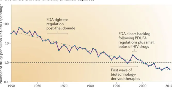
Figure 1.1. The number of new drugs approved by the US Food and Drug
Administration (FDA) per billion US dollars (inflation-adjusted) spent on
research and development (R&D) has halved roughly every 9 years ... 37
Figure 1.2. Approvals of new chemical entities by the US FDA: 1940-2010 ...38
Figure 1.3. Wlodkowic et al. microfluidic live-cell array (array cytometer). ... 39
Figure 1.4. Dimov et al. integrated microfluidic array plate (IMAP) ...40
Figure 1.5. Wang et al. multilayer microfluidic cell array ...41
Figure 1.6. Gao et al. microfluidic localized, multiple cell culture array ...42
Figure 1.7. Kim et al. programmable microfluidic cell array. ...43
Figure 1.8. Zhou et al. openly accessible microfluidic liquid handlers. ...44
Figure 1.9. Du et al. microfluidic droplet array system for drug combination screening ...45
Figure 1.10. Zhang et al. superhydrophobic microwell array chip (SMARchip). ...46
Figure 1.11. Popova et al. droplet-microarray (DMA) reverse cell screening platform. ...47
Figure 1.12. Jakiela et al. microdroplet chemostats for bacterial growth ...48
Figure 1.13. Bogojevic et al. DMF device used for multiplexed cell-based assays ...49
Figure 2.1. Microraft array design schematics ...72
Figure 2.2. Cell culture performance of Cellrafts with rat neurons ...73
Figure 2.3. Neuronal cell viability versus cell density using Cellrafts ...74
Figure 2.4. Human ESC-derived neurons grown on Cellrafts.. ...75
Figure 3.1. Magnetic wand detailed drawings and 3D assembly.. ...89
Figure 3.2. Finite element analysis of probe length.. ...90
Figure 3.3. Finite element analysis of the probe tip.. ...91
Figure 3.4. Finite element analysis of magnet probe-probe interaction. ...92
xiv
Figure 3.6. Magnetic probe encased with Hy-Mu80 magnetic shield. ...94
Figure 3.7. Magnetic wand performance testing with 2-magnet combinations.. ...95
Figure 3.8. Magnetic wand performance testing with 3-magnet combinations.. ...96
Figure 3.9. Microraft transfer performance with a sixteen-channel pipette at various draw/dispense volumes. ... 97
Figure 3.10. Success rate for each number of Cellrafts transferred using various draw/dispense volumes. ... 98
Figure 4.1. Microraft arrays and three dimensional modeling and drawings of the magnet array plate.. ... 117
Figure 4.2. Results of the magnetic field analysis of a microraft at various heights within a microtiter plate well.. ... 118
Figure 4.3. Magnetic force analysis of microrafts at different axial (Z) and radial positions.. ... 119
Figure 4.4. The magnet array plate effectively centers the microrafts.. ... 120
Figure 4.5. Cell viability was not affected by the transfer and centering process.. ... 121
Figure 5.1 384-well microplate experimental layout showing distribution of different numbers of microrafts per well... 137
Figure 5.2 Rhodamine B infused microrafts quantified using plate reader. ... 138
Figure 5.3. Fluorescence signals for rhodamine B infused microrafts.. ... 139
Figure 5.4. Bead based ELISA format. ... 140
Figure 5.5. FMRP detection using flow cytometer and a bead-based ELISA.. ... 141
Figure 5.6. 384-well microplate experimental layout showing samples in red and yellow and controls in various colors. ... 142
Figure 5.7. Flow cytometer fluorescence signals.. ... 143
Figure 5.8. 384-well microplate experimental layout showing samples in red yellow and controls in various colors. ... 144
Figure 5.9. Flow cytometer fluorescence signals.. ... 145
Figure 5.10. Flow cytometer fluorescence signals produced from the lysate of neurons grown on microrafts. ... 146
xv
Figure 5.12. Representative 20X images of rate hippocampal neurons plated at
1MM cells/mL... 148
Figure 5.13. Representative 20X images of rate hippocampal neurons plated at 500K cells/mL. ... 149
Figure 5.14. Representative 20X images of rate hippocampal neurons plated at 250K cells/mL. ... 150
Figure 5.15. Representative 20X images of rate hippocampal neurons plated at 0K cells/mL. ... 151
Figure 6.1. FMRP measurements in NSCs using TR-FRET assay by Kumari et al. ... 159
Figure A.1. Modified Cellraft array design for 384-well automation. ... 164
Figure A.2. Custom magnetic wand array design for 384-well automation ... 165
xvi
LIST OF ABBREVIATIONS
ALS Amyotrophic Lateral Sclerosis ASD Autism spectrum disorder CNS Central nervous system DMD Duchenne Muscular Dystrophy ELISA Enzyme-linked immunosorbent assay ESC Embryonic Stem Cell
FMR1 Fragile X mental retardation 1 FMRP Fragile X mental retardation protein FRDA Friedreich's ataxia
FXN Frataxin
FXS Fragile X Syndrome HD Huntington's disease
hiPSC Human induced pluripotent stem cell HTS High-throughput screening
xvii M&A Mergers and acquisitions
mRNA Messenger RNA PDMS Polydimethylsiloxane RNA Ribonucleic acid
SMA Spinal Muscular Atrophy SMN Spinal motor neuron
1
Chapter 1 Background
1.1 Introduction
Neurological disorders including neurodevelopmental and neurodegenerative diseases represent a disease segment with one of the largest unmet need. While undergoing many structural changes, the pharmaceutical industry is now focusing more intensely on this disease segment. For these reasons, new technology is needed to reduce costs. The technology to-date however has yet to make a strong impact on drug discovery and here we learn why this is the case. In this review, an assessment is made of the current state of treatment for neurological diseases followed by the current state of pharmaceutical R&D. Finally, new technological solutions including physical devices and cellular innovations are described along with their implementation.
1.2 Current State of Treatment for Neurological Diseases
2
phase II clinical trials for the treatment of Parkinson’s disease.1
Data has shown an increase in attrition rates for new pharmaceutical projects across all phases, but especially Phase II and III clinical trials.2 One of the reasons for these increased attrition rates is the discrepancies between the animal models used in preclinical trials and human subjects used in later clinical trials. Another reason is that
screening and preclinical work has not been carried out because it is not financially practical for most organizations.
Of the many neurological diseases and disorders, the ones that can benefit the most from new cost saving technology are those in which well-defined
biomarkers exist, and are simple in nature such as single gene mutations or those in which a known protein is absent or over expressed. For these reasons, some of the most attractive disorders for drug discovery include Huntington’s disease (HD), Fragile X Syndrome (FXS), Duchenne Muscular Dystrophy (DMD), Spinal Muscular Atrophy (SMA), and Friedreich’s ataxia (FRDA).
None of these diseases and disorders has a cure and a possible reason for this is the lack of preclinical testing on human cells. Currently the largest
3
(hiPSC) derived neurons present a promising alternative and will be described in a later section.
1.2.1 Huntington’s Disease
Huntington’s disease is an autosomal dominant neurodegenerative disorder caused by a CAG repeat expansion in the huntingtin gene. This mutation results in an expanded polyglutamine (polyQ) repeat in the huntingtin protein (HTT). Repeats >36-39 trigger the disorder and longer expansions lead to an early onset.3; 4 The polyQ repeat expansion leads to a buildup of misfolded HTT which is toxic to neurons in the central nervous system (CNS).
HD is an inherited disorder that typically begins between the ages of 30 to 50 and gets progressively worse after onset. This disease is characterized by
uncontrollable motor functions, known as chorea, and cognitive deficits in patients. The impairment in cognition includes attention deficit and loss of short- and long-term memory as well as reduced depth perception.5 Other symptoms include slurred speech, emotional instability, and difficulty feeding and swallowing.
There is no cure for HD and current treatments only treat symptoms. So far the most successful drug used to treat HD is Tetrabenazine (Zenazine) by
Lundbeck. Tetrabenazine, a high-affinity inhibitor of mono-amine uptake into vesicles of presynaptic neurons, was approved in 2008 for chorea in HD patients.6 Looking at the current pipeline (Table 1.1), therapeutic approaches for HD include addressing mutant HTT modification and degradation, addressing signaling
4
very early in development. The major drugs that have failed in the pipeline include Dimebon (Latrepirdine), Coenzyme Q10, PDE10A Inhibitor, Pridopidine, VX15, and PBF-999. All of these drugs failed in phase II or III trials suggesting that they were non-toxic but ineffective. It’s possible that this lack of efficacy could be due to the failure to test these compounds on human cells prior to entering the clinic. In preclinical studies, Dimebon and Coenzyme Q10 were tested on the YAC128 HD transgenic mouse while the PDE10A inhibitor was tested on the R6/2 HD mouse model. Pridopidine was tested on CHO cells expressing human D2short dopamine receptors.
1.2.2 Fragile X syndrome
Fragile X syndrome (FXS) is well known neurodevelopmental disorder and one of the leading causes of autism. FXS is caused by a CGG repeat expansion in the 5’ untranslated region of the fragile X mental retardation 1 (FMR1) gene located on the X chromosome. This mutation leads to loss of the fragile X mental
retardation protein (FMRP). FMRP is a ribonucleic acid (RNA) binding protein which serves to transport intracellular RNA and regulate the translation of target
messenger RNAs (mRNA).
5
moderate to severe mental retardation, delays in social development, attention deficit, hyperactivity, anxiety, reduced motor coordination, and an increased incidence of epilepsy.8
Currently there is no cure for FXS. Most current therapies target anxiety and other symptoms rather than addressing the root cause of the disease. Treatments in the pipeline (Table 1.2) for FXS target neuro-transmitter dependent receptors, cell signaling molecules, translation regulators, and specific targets of FMRP. In terms of these potential treatments, major clinical failures include Arbaclofen, Basimglurant (RO4917523), and Mavoglurant (AFQ056). Arbaclofen is a GABA-B agonist while Basimglurant (RO4917523) and Mavoglurant (AFQ056) are both mGlur5
antagonists. These are all neuro receptor ligands and instead of addressing the root cause of the disease, they only function to ameliorate the FXS phenotype
downstream at neural synapses. Most importantly however, all three of these therapies were tested on the fmr1 knockout mouse model in preclinical studies and not on human cells.
1.2.3 Duchenne Muscular Dystrophy
Duchenne muscular dystrophy (DMD) is the most common form of muscular dystrophy and the most common neuromuscular disease. DMD is caused by disruptions in the DMD gene which consists of 79 exons on the X chromosome. These disruptions include deletions (~65%), duplications (~10%), point mutations (~10%), and other rearrangements (15%), and lead to the loss of the protein,
6
DMD affects 1 in 3,500 boys and is first observed at the age of 3 to 5 years. From onset, the disease is characterized by rapid progression of muscle weakness and degeneration. By the age of 12 most boys lose the ability to walk and death occurs by the age of 20 or 30 usually due to respiratory failure. Girls have a 50 percent chance of inheriting and passing the defective gene to their children.11-13
There is no cure for DMD. Glucocorticoid drug therapy represents the best therapeutic option. Examples of this family of drugs include the corticosteroids, prednisone, and deflazacort. These drugs unfortunately have many side effects, but are effective in slowing the rate of muscle deterioration, reducing inflammation, and delaying the disease progression overall. Furthermore, it is not yet clear
mechanistically how these drugs function to ameliorate the symptoms of DMD.11
Current therapeutic research for DMD can be divided into two approaches – therapies that attempt to upregulate dystrophin production and those that attempt to treat specific phenotypes of the disease. Methods used to restore dystrophin
include cell therapy, gene therapy, exon skipping, and suppression of stop codons, while those used to treat the disease phenotypic side effects include
anti-inflammatory, anti-fibrotic, and antioxidant agents, myostatin pathway inhibition, neuronal nitric oxide synthase (NNOS) pathway enhancement, and Utrophin upregulation.
7
oligomer, Tadalafil is a GMP-hydrolyzing phosphodiesterase 5A (PDE5A) inhibitor, Drisapersen is a 2’O-methyl-phosphorothioate oligonucleotide (2’OMePS) which is a type of antisense oligonucleotide targeting exon 51, ACE-031 is a muscle growth factor, PTC124 (Ataluren) is a stop codon suppressor, BMN053 (Pro-053) is a 2’OMePS targeting exon 53, BMN044 (Pro-044) is a 2’OMePS targeting exon 44, and halofuginone (HT-100) is an anti-fibrotic TGF-β inhibitor. The majority of these therapies were tested on human myoblasts except for Tadalafil and halofuginon (HT-100) which were tested on the mdx mouse model.14-16
1.2.4 Spinal Muscular Atrophy
Spinal muscular atrophy (SMA) is a neuromuscular disorder caused by mutation or deletion of the survival motor neuron (SMN) 1 gene which leads to a reduction or loss of the SMN protein. The function of the SMN protein includes RNA transcription, pre-mRNA splicing, small nuclear ribonucleoprotein (snRNP)
biogenesis, axonal transport, and cytoskeletal dynamics.17 Humans also possess an SMN2 gene which is paralogous to SMN1 with the exception of a translational
silence occurring at nucleotide 840 which leads to alternative splicing. The majority of the SMN protein is therefore produced by SMN1 however SMN2 does produce low levels of the protein. Loss of the SMN protein leads to degradation of motor neurons in the spinal cord.
8
located in the trunk and distal limbs, thus causing difficulties in breathing and movement.
There is no cure for SMA. Current therapeutic research on SMA has focused on four major therapeutic approaches. These approaches include replacement or correction of the mutated SMN1 gene, modulation of the SMN2 gene,
neuroprotection of distressed motor neurons, and prevention and restoration of muscle function loss.
Currently 4-AP (Dalfampridine-ER, Ampyra) is showing the most promise in clinical trials. This drug has been approved to treat muscle fatigue in patients with multiple sclerosis and is sold and produced by Acorda Therapeutics.19 Although the studies on SMA have been completed successfully, Chiriboga et al. from Columbia University found no positive improvement in motor function or ambulation in adults ages 18-50 at the doses approved for muscular sclerosis.20
SMA has seen two therapeutic failures in Phase I clinical trials suggesting issues with toxicity (Table 1.4). These failures include the small molecules LM1070 by Novartis, and RG7800 (RO7034067, RO6885247) by Roche and PTC
9 1.2.5 Friedreich’s Ataxia
Friedreich ataxia (FRDA) is an autosomal recessive neurodegenerative disease caused by a mutated expansion of the guanine-adenine-adenine (GAA) triplet on both alleles of the frataxin (FXN) gene. This is a heterogeneous disease with a range of 600 to 1200 repeats. This mutation leads to loss and reduced expression of the protein frataxin which is produced in the mitochondria and is involved in iron metabolism within the cell and also has antioxydative properties. A loss of FXN protein leads to dysfunction in ATP synthesis, iron accumulation, possible oxidative stress, and cellular dysfunction as a whole.23; 24
FRDA is the most common form of hereditary ataxia. In the United States approximately 1 in 100 people are carriers of the mutated FXN gene and one in 20,000 to 50,000 are affected. Symptoms of the disease include loss of coordination (ataxia), fatigue, loss of vision, loss of hearing, impaired speech, aggressive
scoliosis, diabetes mellitus, and serious heart conditions including hypertrophic cardiomyopathy. These symptoms usually begin between the ages of 5 and 25 but occasionally begin in middle aged adults. Most people diagnosed with the disease require walking aids such as a wheelchair by their early 20s.25; 26
Therapeutic research for FRDA has focused on mitochondrial function, oxidative stress, upregulation of FXN, gene therapy, and neurotrophic factors.
FRDA has seen many drug failures in recent years. These include the antioxidents Idebenone and alpha-tocopherolquinone, the FXN modulators
10
and verenicline. While many of these drugs were tested on human fibroblasts in preclinical studies, none were tested on human motor neurons.
All of the above mentioned diseases and disorders lack a cure. Drugs have continued to fail in the regulatory pipeline, and in most of these cases preclinical studies were performed on non-human cells. Drug screening using human cells may improve the identification of drugs that are effective in humans. Thus there is an obvious need for new technologies to make preclinical studies on human cells more feasible and help pave the way for cures for such diseases.
1.3 Current State of Commercial Drug Discovery R&D
1.3.1 Current State of Pharmaceutical R&D and Future Directions
The pharmaceutical industry has undergone many changes over the last half century. After adjusting for inflation, the cost to bring a new drug to market in the 1980’s was approximately $400 MM, and took 7 years, while today it’s close to $2.6 B and requires 15 years.27 These increased costs can be attributed to the rising costs of Phase II and Phase III clinical trials. Furthermore, where clinical costs were less than pre-human studies in the 1980s to early 1990s, clinical studies have risen in cost dramatically over the past twenty years.27
11
2000, and Pfizer (Warner-Lambert, Pharmacia) and Wyeth in 2009. Taking advantaged of synergies, this increased consolidation has led to cuts in R&D and some argue the decline in output efficiency.28
Reduced efficiency has been a major topic in the Pharmaceutical industry in the past twenty years. The number of new drugs approved per billion US dollars spent on R&D has been in a broad decline since 1950 (Figure 1.1), and taken as just the new drugs approved, this number has been mainly flat (Figure 1.2).
As mentioned above, one possible reason for stagnation is due to industry consolidation. Between 1990 and 1999 there was an average of 31 drugs approved per year compared to 24 drugs from the years 2000 to 2009. This number peaked in 1996 with 54 drugs, but many of the companies that existed back then do not exist today.28
Another possible reason for the decline in efficiency is associated with the idea of “low hanging fruit”. This theory follows the notion that the technically tractable drugs such as the cardiovascular statins of the ‘90s have already been developed and the diseases that remain to be treated are much more difficult to develop drugs for. This concept somewhat discounts the efforts that went into developing early drugs and fails to account for the fact that successfully
12
However, due to a lack of pathological understanding as well as disease heterogeneity, treatment of neurological diseases faces a much steeper path.
1.3.2 Public Private Partnerships in Drug Development
With continued consolidation and the growing costs of R&D, companies are increasingly outsourcing R&D efforts to help preserve profits and minimize their risk in the market. These public-private partnerships (PPP) can exist between
pharmaceutical companies and academic institutions, or foundations with public health initiatives. These models are set up to defer the costs of early stage drug development to the public through i.e. NIH, while improving the academic drug discovery capabilities and the value gained from NIH funded research.
The pharmaceutical industry and academia have shared a rich history. In the past, companies have been formed by commercializing new drugs developed by academic researchers. One of the most well-known examples of this was
Genentech, founded by Herbert Boyer of UCSF and venture capitalist Robert Swanson in 1976.32 More recently there is Vertex, Infinity, and H3 Biosciences all produced by Professor Stuart Schreiber and colleagues at Harvard University. Finally, there is a list of well-known drugs that have come out of academic institutions including pregabalin (LyricaTM; Silverman lab, Northwestern),
emtricitabine (EmtrivaTM; Liotta lab, Emory) and premetrexed (AlimtaTM; Taylor lab, Princeton).33-35
13
drug discovery centers across the country. This consortium also tracks partnerships between these centers and the pharmaceutical industry and some notable ones include Yale University (GSK, Gilead Sciences, Evotec AG), UCSF (Sanofi,
Genentech, Bayer, Pfizer), Vanderbilt (Bristol-Myers Squibb, Astra Zeneca, GSK), University of Pennsylvania (Novartis, Astra Zeneca), Broad Institute of Harvard and MIT (Astra Zeneca), UC San Diego (Roche), Oxford University (Novo Nordisk, UCB), California Institute for Biomedical Research (Merck), Harvard University (UCB), and Johns Hopkins Brain Science Institute (Janssen Pharmaceuticals). Relying on public funding, it is critical for these academic institutions to have access to technology to reduce the costs of drug discovery as much as possible.
1.4 Current State of Technology for Screening for Neurological Conditions
New technology is being developed continuously to advance small molecule screening, improving efficiency, efficacy, and reducing costs. High-density microtiter plates such as 96- and 384-well plates remain the gold standard for cell-based phenotypic screening; however these plates are still relatively low in throughput resulting in the need for large populations of cells and significant volumes of compounds and reagents. Higher throughput plates such as 1536- and 3456-well plates have been designed, with the former starting to become more prominent, however these plates are marked by high evaporation and require expensive equipment for handling. Increased focus has been placed on the use of
14 1.4.1 Array Based Microfluidic Systems
Wlodkowic et al. published in 2009 the design of a polydimethylsiloxane (PDMS) array of micromechanical traps to hydrodynamically capture single
nonadherent hematopoietic cells (Figure 1.3). After being exposed to anti-cancer drugs, this study showed that ~300 trapped cells could be analyzed with real time fluorescent imaging just as effectively as traditional single cell analysis techniques such as a flow cytometry which requires a much larger cell population. This major drawback to this device is that it is a continuous system and cannot test multiple compounds in parallel.36
Dimov et al. published in 2011 the development of an integrated microfluidic array plate (iMAP) which is a gravity driven (pump and tube free), PDMS based, microfluidic culture device for the capture and analysis of discrete populations of cells (Figure 1.4). This device can interrogate 64 separate populations with the capability of performing real-time Nucleic Acid Sequence Based Amplification (NASBA) and immunofluorescent (IF) analysis. Each populations consists of 5-50 cells however culture of these cells was done under constant perfusion. The performance of this device was demonstrated with NASBA, IF, and a drug dose analysis using HeLa and MCF7 cells.37
15
cell chambers was controlled by surrounding, water-filled channels (identified in blue in Figure 1.5) which would expand and contract to gas pressure controlled by
solenoid valves. To seed specific cell types into specific chambers, the cell
suspension in the syringe pump would have to be changed while the valves for the target chamber were open and the surrounding chambers were closed. The utility of the device was demonstrated by seeding two cell types, regular Chinese hamster ovary (CHO) cells and EGFP-expressing CHO cells, into specific chambers, and then treating specific chambers with specific fluorescent dyes. The number of cells in each chamber ranged in the 100s and were cultured for 2 days.38
In an effort to seed and culture multiple cell types in specific localized
positions in an easier and more efficient way, Gao et al. created a microfluidic device which does so utilizing vacuum actuation (Figure 1.6). Fabricated from PDMS, this device featured 256 culture channels divided into eight individually addressable regions and all branched from a central main channel. In their proof of design study, four cell types were seeded including HuT 78, Ramos, PC-3, and C166-GFP cells and viability and cell proliferation were measured. With cultures varying from 2-7 days, viability ranged from 92-97% and each cell population was individually monitored for its response to apoptosis inducing compounds.39
Screening multiple compounds in combination is also a concern in drug development in order to assess possible side effects. In order to reduce equipment and reagent costs for in vitro drug combination screening, Kim et al. designed a programmable microfluidic cell culture array for the generation of drug
16
observation (Figure 1.7). Fabricated from two layers of PDMS, one for fluid exchange and one for pneumatic control, this device featured 64 individually addressable cell culture chambers connected to a set of upstream concentration channels where compounds are diluted and combined. Using human prostate cancer PC3 cells, the device was demonstrated by inducing viability loss by introducing combinations of the chemotherapeutic drugs doxorubicin and
mitoxantrone with TNF-alpha Related Apoptosis Inducing Ligand (TRAIL). One of the unique attributes of this device is its ability to operate without continuous perfusion, however media changes were performed every three hours possibly leading to a lower effectiveness in the combination treatments when compared to the use of traditional 96-well plates.40
In an interesting effort to encompass all the tools and resources necessary for a drug screen, Zhou et al. developed an automated nanoliter dispensing microfluidic liquid handler and accompanying micro-multiwell chip which reduces reagent
17
seeded, transferred, passaged, transfected, stimulated by drugs, and observed in a screening assay.41
To address the growing need to miniaturize screening assays while also making them affordable, easy to handle, and accurate, Du et al. described in 2013 a 2-dimensional PDMS based microfluidic culture device to increase throughput for combination drug testing (Figure 1.9). The system was designed around the sequential operation droplet array (SODA) technique, and featured a chip with an array of 342 circular micro-wells each 1.3 mm in diameter and 10 µm in depth. Using this chip and an automated stage and capillary system, 11-day old cell
cultures were achieved in oil-covered 500 nL droplets with media changes every 24 hours. Each droplet supported 80-100 cells/well of A549 cells. Using this
technology, not only was cell viability comparable to 96 and 384-well plates, but drug consumption for each well was reduced by 2-3 orders of magnitude.42
18
Additionally, the device’s screening capabilities were demonstrated by culturing Oct4-EGFP mouse induced pluripotent stem cells (iPSCs) for four days and
individually delivery soluble factors while measuring their effects on pluripotency and proliferation.43
Similarly, Popova created a droplet microfluidic array (DMA) with super hydrophilic and hydrophobic areas to eliminate the risk of cross-contamination between cell clusters when screening on cell microarrays (Figure 1.11). The DMA was fabricated using a standard microscope slide upon which a layer of nanoporous poly(2-hydroxyethyl methacrylate-co-ethylene dimethacrylate) (HEMA-EDMA) film was placed. This surface was then functionalized with one reaction to create superhydrophilic squared spots and another reaction to create superhydrophobic borders. In all, three different arrays were created with 588, 2187, and 4563 spots of sizes 1000 µm, 500 µm, and 350 µm respectively. In evaluating the device, HeLa, HEK293, and A549 cells were seeded and then morphologically observed.
Additionally, the utility of the DMA in screening was first tested by reverse
transfecting HEK293 cells and then individually treating HeLa cells with doxorubicin and quantifying its effect on the cells by calcein staining.44
1.4.2 Droplet Based Microfluidic Systems
In order to study microbial ecology, physiology, evolution, and adaptation to changing environments, Jakiela et al developed a droplet based microfluidic device to isolate populations of bacteria (Figure 1.12). Composed of ten input and output channels, the device performs three functions: 1) formation of microdroplets
19
incubation and monitoring; and 3) splits and fuses microdroplets to control the modulate the concentration of injected chemical factors. Using the device 164 microdroplet chemostats were produced and monitored. Chemostats are culture modules for bacteria, yeast, and algae which continuously replenish fluids to maintain volume and concentration of growth reagents for the optimal culture
environment. Microfluidics greatly reduces the cost of producing these chemostats, so in demonstrating the application of this device, the growth and response of E. Coli cells (~14,000 cells per droplet) was observed after being introduced to varying concentrations of tetracycline and chloramphenicol and the bacterial showed similar growth to that grown in a bulk environment.45
In a demonstration of digital microfluidics (DMF), Bogojevic et al. developed the first DMF device to implement a parallel-scale cell-based assay (Figure 1.13). Using photolithography and etching this device was fabricated as two plates
separated by a 140 µm space with patterned chromium electrodes on the bottom plate and a 50 nm Teflon-AF coating on either side of the inside surface. The device has six 1.5 mm diameter assay zones in the central region with twelve adjacent reagent reservoirs. After plating HeLa cells at approximately 800 cells per assay zone and incubating overnight, a fluorogenic apoptosis assay for caspase-3 activity was performed since it is popular in anti-cancer drug discovery. The results showed a 33-fold reduction in reagent consumption and lower detection limit and greater dynamic range than the same assay performed in a 96-well plate.46
20
to interagate small cell populations with extremely low reagent volumes, they do suffer certain consequences, namely the ability to produce long term cultures. Except for the Du and Zhang devices, these platforms have not been proven to support cells for more than 48 hours, and all require frequent media changes. There has yet to be a commercially popular device that combines the volume advantages of 384-well plates with the throughput advantages of microfluidics.
1.5 Use of Human iPSC Derived Neurons in HTS
1.5.1 History and Advantages of Human iPSCs
With the increased focus of the pharmaceutical industry on treating
neurological disorders, there is a greater need for accurate disease models. With the discovery and development of human induced pluripotent stem cells (hiPSCs) a massive movement has been underway to realize the potential of this new
technology in better understanding the pathology and mechanisms of neurological disorders as well as discovery of new drugs to treat them. Although extensive efforts have been made, therapies have failed to come to fruition. The reasons for this failure include the polymorphic nature and our lack of understanding of the disease pathogenesis, the failure of animal models to fully recapitulate all aspects of the disease including genetic - environmental interactions, and limited access to human cellular samples at different stages of maturation and disease progression.
21
anatomical, and/or physiological differences between animal models and humans. In the case of rodents, which we diverged from 60 million years ago, there are significant genomic differences such as noncoding and regulatory RNA that play a major role in disease epigenetics. Anatomically there are major differences as well, such as the difference in development of the prefrontal and temporal cortex of the rodent versus the human brain. Also, rodent brains are lissencephalic and don’t contain certain types of neurons that exist in the human brain such as Von Economo neurons.48 Such differences affects neurogenesis and neuronal function, therefore neurons dissected from mice may not always have the same morphology as human neurons. Finally, in terms of physiology, these genetic and anatomic differences effect neuronal function, and thus rodent neurons behave differently as well. Specifically, it has been shown that rodent neurons have different
electrophysiological properties than human neurons.49 Taken together, these differences may explain part of the discrepancies between preclinical and clinical data.
22
and therefore are not ideal for modeling neurodevelopment in early onset diseases. Furthermore, neurons derived from postmortem subjects or neurosurgeries eliminate the possibility of performing patient specific screens or establishing biobanks that represent the mosaic nature of many neurological and neuropsychiatric diseases.
Given the limitations of these current models, human iPSCs make it possible to perform screens on human neurons from patients with specific genetic mutations caused by a known neurological disorder. The discovery of these cells thus was a major breakthrough and even overcame the moral challenges of using embryonic stem cells (ESCs) which were already seeing limited use. In 2006, Takahashi and Yamanaka discovered hiPSCs, for which they received the Nobel Prize. In their landmark paper, they demonstrated that mouse fibroblasts could be reprogrammed into a pluripotent state similar to ESCs by introducing transcription factors in a retroviral-mediated process. The four transcription factors used in this study were OCT3/4, SOX2, c-MYC, and KLF4.50 After induction, pluripotent stem cells can differentiate into any human cell, and within a year of these original studies, human iPSCs had been generated from a number of different tissue sources, fibroblasts and peripheral blood being the most common.51
23
neurons. This campaign paved the way for eventual screens using hiPSC-derived neurons.52
1.5.2 Current Use of hiPSC-derived Neurons
Executing effective screens for neurological and neurodegenerative disorders partially depend on the types of neurons available. Over the years various types of neurons have been derived from hiPSCs with motor, cortical, and dopaminergic neurons being the most developed and characterized. Each of these types of neurons is produced from a different differentiation protocol, and these protocols have evolved over the years.
In 2008 Eggan et al. first demonstrated motor neuron differentiation from human somatic cells. Established from a previous study which produced MNs from mouse stem cells using a retinoic acid (RA) and sonic the hedgehog (SHH) for the neural patterning, Eggan’s group was able to mimic this same protocol in hESCs.53 Shortly thereafter this method was adopted by Ebert to produce motor neurons from hiPSCs. This was the first study to show a disease specific phenotype in
differentiated motor neurons, and paved the way for future studies, however it resulted in an efficiency of less than 10% and required a 56-day differentiation time frame.54 Since then efficiencies and differentiation times have improved, but are still not ideal for large scale screening purposes. (Table 1.6)
24
method in 24 days.56 It was not until 2012 however that cortical neurons were
differentiated from hiPSCs by Shi et al. in their landmark study. In this elegant work, dual SMAD inhibition using Noggin and SB431542 was combined with retinoids to produce 95%efficiency; however differentiation time for mature neurons was between 60 to 90 days. Although this is consistent with the time frame of neurogenesis in human development, it is not ideal for screening campaigns. Fortunately other protocols with shorter differentiation times have been produced since then, but efficiency has suffered at the cost of reducing time (Table 1.6).
Dopaminergic (DA) neurons play a key role in regulating reflexive actions and is implicated in Parkinson’s disease (PD). One of the first groups to differentiate hiPSCs into DA neurons was Chambers et al. in 2008. Introducing the dual SMAD inhibition patterning technique using NOGGIN and SB435142 for the first time, this groundbreaking study produced DA neurons in 19 days with 82 percent efficiency.57
Having a selection of different types of neurons available from hiPSC differentiation is critical in carrying out screens for different neurological and
25
In targeting Parkinson’s disease Scwab and Ebert created sensory neurons at 20% efficiency after a 21 day differentiation period from patients carrying the LRRK2 G2019S mutation. In studying the morphology and function of theses neurons using imaging techniques they found the presence of large microtubule-containing neurite aggregations as well as altered calcium dynamics. When treated with three LRRK2 kinase inhibitors LRRK2-IN-1, GSK2578215A, AND CZC25146 they observed a significant, but partial rescue in morphology and a full rescue in calcium dynamics. These findings supported the idea that kinase activity is implicated in PD neuronal dysfunction. It also showed minor line-to-line differences due to patient variability in the iPSCs used which is a partial hindrance to screening with iPSC-derived neurons. Furthermore, although a full screen was not executed, this study showed that small molecules could be tested on hiPSC-derived neurons and valuable information retrieved.
Probably the best demonstration of hiPSC-derived screening has been associated with FXS. Two separate screens were published in 2015; however neither used fully mature neurons. Using a high content assay Kaufmann et al. screened 50,000 compounds to see which ones upregulated FMRP production in neural precursor cells. The study concluded with several compounds that produced a small but detectable increase in FMRP. Moreover it showed the feasibility of plating iPSC-derived cells in high-density well plates.59 Kumari et al. used at 1536-well plate format and screened 5,000 compounds on neural stem cells using a time-resolved fluorescence resonance energy transfer (TR-FRET)-based assay to
26
fluorophore, when excited by a light source, releases energy towards a nearby acceptor fluorophore which in turn emits a specific fluorescent wavelength which is detected. Using this technique, this study concluded with six compounds that modestly increased FMRP production, however this work provided proof of principal for using TR-FRET in this format with patient iPSC-derived cells.60
In HD, there have been recent studies performed on hiPSC-derived neurons; however these studies have only used these neurons to validate compounds
discovered in larger primary screens. Kumar et al. created mesencephalic dopaminergic neurons in 26 days for a study to better elucidate the intracellular processes that regulate neuronal manganese (Mn) homeostasis. Following a
40,167 compound primary screen based on a fluorescence Mn measurement assay, 9 compounds were selected for validation on the hiPSC-derived neurons and
interestingly all nine showed varying activity at different stages of maturation of the neurons. Such a study would not be possible in animal or postmortem models, thus this work demonstrated more accurately the possible use of these compounds to regulate Mn levels in humans.61
Recently another screen was performed targeting HD treatment. Combining in vitro single molecule fluorescence spectroscopy, in silico molecular docking
simulations, and in vivo fly and mouse HD models, 19,468 physical compounds were screened for inhibitors of abnormal interactions between mutant HTT and Ku70, a mutation repair protein. Fifty-six hits were produced from this primary screen and three were tested on HD patient derived cortical neurons. Using confocal
27
improved dendrite length and spine density, but did not reduce the number of inclusion body positive neurons present. Taken together however, treating these neurons illustrated the therapeutic effectiveness of these compounds.62
1.5.3 Challenges of hiPSC-derived Neurons
From the previous examples, it is clear that screening on hiPSC-derived neurons is still in its infancy, and has a long way to go before it is commonplace. There are significant reasons why screening with hiPSC-derived neurons has not accelerated, and these challenges must be overcome to unlock the true potential of this technology. These challenges include biologic, logistic, and economic factors.
One of the major threats to screening with hiPSC-derived neurons is the possibility of the existence of genetic mutations separate from the disease as well as epigenetic memory from the somatic tissue of origin. Historically, iPSCs have been generated via a lentiviral or retroviral reprogramming process, however these techniques can lead to mutations at the integration site, copy number variations, or abnormal karyotypes.63 These genetic aberrations could possibly lead to problems during differentiation and may affect the final disease phenotype.
28
valproic acid, vitamin C, SB431542, PD0325901, thiazovinin, sodium butyrate, and forskolin have demonstrated improved reprogramming results when utilized.
Another genetic challenge for hiPSCs and their resulting neurons is the threat of retaining genetic information from the original somatic cells from which they were derived from. In their paper in Nature, Kim et al. was one of the first to report that low-passage iPSCs harbor residual DNA methylation signatures characteristic of their somatic tissue of origin. Furthermore, these residual signatures favored differentiation along lineages of the donor cell as opposed to alternate cell types. They also showed that this ‘epigenetic memory’ could be reset via serial
reprogramming or by treatment of iPSCs with chromatin-modifying drugs.64 In a similar vein Liester et al. showed that somatic memory was independent of reprogramming techniques.65. Increasing passage number and culture time however could possibly reduce these effects and make iPSCs behave more like ESCs.66; 67
29
Medical Research, the HD consortium, and the UK Biobank have been established. Furthermore, it is important for screening campaigns to use multiple iPSC lines to ensure that disease-specific cellular phenotypes are detected beyond this normal variability.
Having proper controls is also vital. Historically, controls have consisted of iPSCs generated from age, gender, and race matched healthy donors, but these controls are not ideal because of the previously mentioned genetic variability across patients. For these reasons, it is critical to be able to isogenically correct the
disease mutations from the same patient lines as are used in the screen.70; 71 It is also critical to characterize those cells for precise disease related phenotypic readouts to ensure they are safe controls. Fortunately there are a number of tools available today to create these isogenic lines including zinc finger nucleases (ZFNs), transcription activator-like effector nucleases (TALENS)72, and clustered regularly interspaced short palindromic repeates-CRISPR-associated protein-9 nuclease (CRISPR-Cas9) and these tools are being increasingly used.73-75
30
necessary to characterize these cells for their type, maturity, and functionality which require additional time.
1.6 Summary
36
Figure 1.1. The number of new drugs approved by the US Food and Drug
Administration (FDA) per billion US dollars (inflation-adjusted) spent on
37
38
39
Figure 1.4. iMAP array (A) consists of 64 processing modules (B) that can perform 64 independent simultaneous integrated assays. The integrated function of each processing module can be flexibly selected by the user and depends entirely on the sequence and timing of the fluid inputs. The basic steps required for three different cell based assays are illustrated (A). This versatility allows the user to combine cell stimulation and gene and protein expression analysis on a single microfluidic platform. For statistical
replication purposes each module consists of 8 parallel and equally
40
Figure 1.5. (a) The schematic of the multilayer microfluidic cell array. The fluidic channels and chambers (in the fluidic layer) are shown in orange while the surrounding valves (in the control layer) are shown in blue. (b) The
41
42
Figure 1.7. Schematic of the microfluidic array. (A) Different concentrations of drugs A and B are generated in the diffusive gradient mixer, and used, either sequentially or in combination, to perfuse cells cultured in downstream microchambers. The mixing operation for generating different drug
combinations and the opening and closing of valves for perfusing cells in the microchambers are controlled through a LabVIEW interface. (B) Depiction of the range of concentrations that can be generated for sequential and
simultaneous treatment using color dyes. In the left panel, yellow and blue color dye solutions (representing the minimum and maximum concentrations of one drug) are mixed to generate eight outlet concentrations (‘‘horizontal gradient’’ of colors between yellow and blue), and represent the gradient used in sequential exposure experiments. In the middle panel, yellow and red
43
Figure 1.8. The microfluidic liquid handling system. (a) A microfluidic chip was aligned to the microwell chip horizontally and vertically using a
44
45
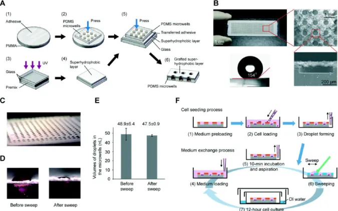
Figure 1.10. Superhydrophobic microwell array chip (SMARchip) and operation procedure. (A) Micrografting procedure for fabricating the
SMARchip. (B) Photos and scanning electron micrographs of the SMARchip. The SMARchip consists of a microfabricated polydimethylsiloxane (PDMS) microwell array and a superhydrophobic polymer layer. (C) A droplet array of culture medium spontaneously forming in the microwells due to the repelling effect of the superhydrophobic layer on the aqueous solution. (D) Typical side-views of the droplets in microwells before and after a sweep with a cell
spreader. (E) Average volumes and standard deviations of droplets in
46
47
Figure 1.12. a) Diagram of the layout of the microfluidic device. b) A sequence of micrographs illustrating the splitting of one microdroplet into a seed droplet and waste droplets with pre-programmed volumes. c) A sequence of
48
49 6
Ta
ble
1
.6
hi
P
S
C
-de
riv
e
d ne
uron t
y
pe
50
51
1.8: BIBLIOGRAPHY
1. Shoulson I, Schwid S, Hyson C, Oakes D, Gorbold E, Rudolph A, Shinaman A, Kamp C, Kieburtz K, Lang A et al. 2007. Mixed lineage kinase inhibitor cep-1347 fails to delay disability in early parkinson disease. Neurology.
69(15):1480-1490.
2. Pammolli F, Magazzini L, Riccaboni M. 2011. The productivity crisis in pharmaceutical r&d. Nature Reviews Drug Discovery. 10(6):428-438.
3. Gray M, Shirasaki DI, Cepeda C, Andre VM, Wilburn B, Lu XH, Tao JF, Yamazaki I, Li SH, Sun YE et al. 2008. Full-length human mutant huntingtin with a stable polyglutamine repeat can elicit progressive and selective
neuropathogenesis in bachd mice. Journal of Neuroscience. 28(24):6182-6195.
4. Miller J, Arrasate M, Shaby BA, Mitra S, Masliah E, Finkbeiner S. 2010.
Quantitative relationships between huntingtin levels, polyglutamine length, inclusion body formation, and neuronal death provide novel insight into huntington's disease molecular pathogenesis. Journal of Neuroscience. 30(31):10541-10550.
5. Lynch G, Kramar EA, Rex CS, Jia YS, Chappas D, Gall CM, Simmons DA. 2007. Brain-derived neurotrophic factor restores synaptic plasticity in a knock-in mouse model of huntington's disease. Journal of Neuroscience. 27(16):4424-4434.
6. Yero T, Rey JA. 2008. Tetrabenazine (xenazine), an fda-approved treatment option for huntington’s disease–related chorea. Pharmacy and Therapeutics. 33(12):690-694.
7. Ghosh A, Michalon A, Lindemann L, Fontoura P, Santarelli L. 2013. Drug
discovery for autism spectrum disorder: Challenges and opportunities. Nature Reviews Drug Discovery. 12(10):777-790.
8. Bear MF, Huber KM, Warren ST. 2004. The mglur theory of fragile x mental retardation. Trends Neurosci. 27(7):370-377.
9. Hoffman EP, Bronson A, Levin AA, Takeda S, Yokota T, Baudy AR, Connor EM. 2011. Restoring dystrophin expression in duchenne muscular dystrophy muscle progress in exon skipping and stop codon read through. American Journal of Pathology. 179(1):12-22.
52
11. Huard J, Mu X, Lu A. 2016. Evolving paradigms in clinical pharmacology and therapeutics for the treatment of duchenne muscular dystrophy. Clinical Pharmacology & Therapeutics. 100(2):142-146.
12. Hoffman EP, Brown RH, Kunkel LM. 1987. Dystrophin - the protein product of the duchenne muscular-dystrophy locus. Cell. 51(6):919-928.
13. Cirak S, Feng L, Anthony K, Arechavala-Gomeza V, Torelli S, Sewry C, Morgan JE, Muntoni F. 2012. Restoration of the dystrophin-associated glycoprotein complex after exon skipping therapy in duchenne muscular dystrophy. Molecular Therapy. 20(2):462-467.
14. Aartsma-Rus A, Janson AAM, Kaman WE, Bremmer-Bout M, den Dunnen JT, Baas F, van Ommen GJB, van Deutekom JCT. 2003. Therapeutic antisense-induced exon skipping in cultured muscle cells from six different dmd patients. Human Molecular Genetics. 12(8):907-914.
15. Asai A, Sahani N, Kaneki M, Ouchi Y, Martyn JAJ, Yasuhara SE. 2007. Primary role of functional ischemia, quantitative evidence for the two-hit mechanism, and phosphodiesterase-5 inhibitor therapy in mouse muscular dystrophy. Plos One. 2(8):16.
16. Carlson TJ, Pellerin A, Djuretic IM, Trivigno C, Koralov SB, Rao A, Sundrud MS. 2014. Halofuginone-induced amino acid starvation regulates stat3-dependent th17 effector function and reduces established autoimmune inflammation. Journal of Immunology. 192(5):2167-2176.
17. McGivern JV, Patitucci TN, Nord JA, Barabas MEA, Stucky CL, Ebert AD. 2013. Spinal muscular atrophy astrocytes exhibit abnormal calcium regulation and reduced growth factor production. Glia. 61(9):1418-1428.
18. Mehta NM, Newman H, Tarrant S, Graham RJ. 2016. Nutritional status and nutrient intake challenges in children with spinal muscular atrophy. Pediatric Neurology. 57:80-83.
19. Tisdale S, Pellizzoni L. 2015. Disease mechanisms and therapeutic approaches in spinal muscular atrophy. Journal of Neuroscience. 35(23):8691-8700. 20. 2015 sma researcher meeting summary: drug discovery. 2015. Cure SMA; [accessed]. http://www.curesma.org/news/2015-researcher-meeting-drug-discovery.html?referrer=https://www.google.com/).
21. Palacino J, Swalley SE, Song C, Cheung AK, Shu L, Zhang XL, Van Hoosear M, Shin Y, Chin DN, Keller CG et al. 2015. Smn2 splice modulators enhance u1-pre-mrna association and rescue sma mice. Nature Chemical Biology.
53
22. Naryshkin NA, Weetall M, Dakka A, Narasimhan J, Zhao X, Feng ZH, Ling KKY, Karp GM, Qi HY, Woll MG et al. 2014. Smn2 splicing modifiers improve motor function and longevity in mice with spinal muscular atrophy. Science.
345(6197):688-693.
23. Strawser CJ, Schadt KA, Lynch DR. 2014. Therapeutic approaches for the treatment of friedreich's ataxia. Expert Review of Neurotherapeutics. 14(8):949-957.
24. Martineau L, Noreau A, Dupre N. 2014. Therapies for ataxias. Current Treatment Options in Neurology. 16(7):13.
25. What is fa? Friedreich's Ataxia Research Alliance - CureFA.org; [accessed 2017]. http://www.curefa.org/whatis.
26. Diagnosis of ataxia. National Ataxia Foundation; [accessed 2017]. http://ataxia.org/learn/ataxia-diagnosis.aspx.
27. DiMasi JA, Grabowski HG, Hansen RW. 2016. Innovation in the pharmaceutical industry: New estimates of r&d costs. Journal of Health Economics. 47:20-33. 28. LaMattina JL. 2011. The impact of mergers on pharmaceutical r&d. Nature
Reviews Drug Discovery. 10(8):559-560.
29. Schnee JE. 1979. R and d strategy in the united-states pharmaceutical-industry. Research Policy. 8(4):364-382.
30. Scannell JW, Blanckley A, Boldon H, Warrington B. 2012. Diagnosing the decline in pharmaceutical r&d efficiency. Nature Reviews Drug Discovery. 11(3):191-200.
31. Scannell JW, Bosley J. 2016. When quality beats quantity: Decision theory, drug discovery, and the reproducibility crisis. Plos One. 11(2):21.
32. Russo E. 2003. Special report: The birth of biotechnology. Nature. 421(6921):456-457.
33. Dworkin RH, Kirkpatrick P. 2005. Pregabalin. Nature Reviews Drug Discovery. 4(6):455-456.
34. Richman DD. 2001. Antiretroviral activity of emtricitabine, a potent nucleoside reverse transcriptase inhibitor. Antiviral Therapy. 6(2):83-88.
54
36. Wlodkowic D, Faley S, Zagnoni M, Wikswo JP, Cooper JM. 2009. Microfluidic single-cell array cytometry for the analysis of tumor apoptosis. Analytical Chemistry. 81(13):5517-5523.
37. Dimov IK, Kijanka G, Park Y, Ducree J, Kang T, Lee LP. 2011. Integrated microfluidic array plate (imap) for cellular and molecular analysis. Lab on a Chip. 11(16):2701-2710.
38. Wang HY, Bao N, Lu C. 2008. A microfluidic cell array with individually
addressable culture chambers. Biosensors & Bioelectronics. 24(4):613-617. 39. Gao Y, Li P, Pappas D. 2013. A microfluidic localized, multiple cell culture array
using vacuum actuated cell seeding: Integrated anticancer drug testing. Biomedical Microdevices. 15(6):907-915.
40. Kim J, Taylor D, Agrawal N, Wang H, Kim H, Han A, Rege K, Jayaraman A. 2012. A programmable microfluidic cell array for combinatorial drug screening. Lab on a Chip. 12(10):1813-1822.
41. Zhou Y, Pang YH, Huang YY. 2012. Openly accessible microfluidic liquid handlers for automated high-throughput nanoliter cell culture. Analytical Chemistry. 84(5):2576-2584.
42. Du GS, Pan JZ, Zhao SP, Zhu Y, den Toonder JMJ, Fang Q. 2013. Cell-based drug combination screening with a microfluidic droplet array system.
Analytical Chemistry. 85(14):6740-6747.
43. Zhang PF, Zhang JX, Bian ST, Chen ZY, Hu YW, Hu RW, Li JQ, Cheng YC, Zhang XC, Zhou YM et al. 2016. High-throughput superhydrophobic microwell arrays for investigating multifactorial stem cell niches. Lab on a Chip.
16(16):2996-3006.
44. Popova AA, Demir K, Hartanto TG, Schmitt E, Levkin PA. 2016. Droplet-microarray on superhydrophobic-superhydrophilic patterns for high-throughput live cell screenings. Rsc Advances. 6(44):38263-38276. 45. Jakiela S, Kaminski TS, Cybulski O, Weibel DB, Garstecki P. 2013. Bacterial
growth and adaptation in microdroplet chemostats. Angewandte Chemie-International Edition. 52(34):8908-8911.
46. Bogojevic D, Chamberlain MD, Barbulovic-Nad I, Wheeler AR. 2012. A digital microfluidic method for multiplexed cell-based apoptosis assays. Lab on a Chip. 12(3):627-634.
55
48. Allman JM, Tetreault NA, Hakeem AY, Park S. 2011. The von economo neurons in apes and humans. American Journal of Human Biology. 23(1):5-21.
49. Steffenhagen C, Kraus S, Dechant FX, Kandasamy M, Lehner B, Poehler AM, Furtner T, Siebzehnrubl FA, Couillard-Despres S, Strauss O et al. 2011. Identity, fate and potential of cells grown as neurospheres: Species matters. Stem Cell Reviews and Reports. 7(4):815-835.
50. Grskovic M, Javaherian A, Strulovici B, Daley GQ. 2011. Induced pluripotent stem cells - opportunities for disease modelling and drug discovery. Nature Reviews Drug Discovery. 10(12):915-929.
51. Takahashi K, Tanabe K, Ohnuki M, Narita M, Ichisaka T, Tomoda K, Yamanaka S. 2007. Induction of pluripotent stem cells from adult human fibroblasts by defined factors. Cell. 131(5):861-872.
52. McNeish J, Roach M, Hambor J, Mather RJ, Weibley L, Lazzaro J, Gazard J, Schwarz J, Volkmann R, Machacek D et al. 2010. High-throughput screening in embryonic stem cell-derived neurons identifies potentiators of alpha-amino-3-hydroxyl-5-methyl-4-isoxazolepropionate-type glutamate receptors. Journal of Biological Chemistry. 285(22):17209-17217.
53. Di Giorgio FP, Boulting GL, Bobrowicz S, Eggan KC. 2008. Human embryonic stem cell-derived motor neurons are sensitive to the toxic effect of glial cells carrying an als-causing mutation. Cell Stem Cell. 3(6):637-648.
54. Ebert AD, Yu JY, Rose FF, Mattis VB, Lorson CL, Thomson JA, Svendsen CN. 2009. Induced pluripotent stem cells from a spinal muscular atrophy patient. Nature. 457(7227):277-U271.
55. Li XJ, Zhang XQ, Johnson MA, Wang ZB, LaVaute T, Zhang SC. 2009. Coordination of sonic hedgehog and wnt signaling determines ventral and dorsal telencephalic neuron types from human embryonic stem cells. Development. 136(23):4055-4063.
56. Eiraku M, Watanabe K, Matsuo-Takasaki M, Kawada M, Yonemura S,
Matsumura M, Wataya T, Nishiyama A, Muguruma K, Sasail Y. 2008. Self-organized formation of polarized cortical tissues from escs and its active manipulation by extrinsic signals. Cell Stem Cell. 3(5):519-532.
57. Chambers SM, Fasano CA, Papapetrou EP, Tomishima M, Sadelain M, Studer L. 2009. Highly efficient neural conversion of human es and ips cells by dual inhibition of smad signaling. Nature Biotechnology. 27(3):275-280.