THE EFFECTS OF HIP STRENGTH ON GLUTEAL MUSCLE ACTIVATION AMPLITUDES AND HOW THESE FACTORS PREDICT LOWER EXTREMITY KINEMATICS
Katie J. Homan
A thesis submitted to the faculty of the University of North Carolina at Chapel Hill in partial fulfillment of the requirements for the degree of Master of Arts in the Department of Exercise & Sport Science in the College of Arts & Sciences (Athletic Training).
Chapel Hill 2011
ii Abstract
KATIE J. HOMAN: The effects of hip strength on gluteal muscle activation amplitudes and how these factors predict lower extremity kinematics.
(Under the direction of Dr. J Troy Blackburn, PhD, ATC)
Objective: To evaluate the influence of hip strength on kinematic ACL injury risk factors and gluteal activation. Design: Cross-sectional. Setting: Research laboratory. Participants: Eighty-two healthy volunteers. Outcome Measures: Hip extension, external rotation, and abduction strength; gluteus maximus and medius electromyography (EMG); knee valgus, hip adduction, and hip internal rotation angles. Results: Peak knee valgus (p=0.016) and hip external rotation (p=0.023) angles were greater in individuals with weaker hip external rotators. Gluteus maximus EMG was greater in individuals with weaker hip extensors (p=0.031) and external rotators (p=0.043). Hip external rotation strength and gluteus
iii
Table of Contents
Chapter 1 ... 1
Introduction ... 1
Dependent/Criterion Variables ... 6
Kinematic Variables ... 6
Independent/Predictor Variables ... 6
Strength Variables ... 6
EMG Variables ... 6
Research Questions ... 7
Hip Strength and Kinematics ... 7
Hip Strength and EMG Amplitudes ... 7
Regressions ... 8
Research Hypothesis ... 9
Hip Strength and Kinematics ... 9
Hip Strength and EMG Amplitudes ... 9
Regressions ... 9
Operational Definitions ... 10
Assumptions ... 11
Limitations ... 11
Chapter 2 ... 12
Literature Review ... 12
Introduction ... 12
ACL Injury Epidemiology and Etiology ... 13
Epidemiology ... 13
Cadaver Studies ... 14
Etiology ... 15
iv
Biomechanical Risk Factors ... 16
Neuromuscular Risk Factors ... 18
Jump Landing Task ... 24
Electromyography ... 25
Clinical Significance ... 26
Chapter 3 ... 28
Methodology ... 28
Experimental Design ... 28
Subjects ... 28
Measurement and Instrumentation ... 29
Procedures ... 30
Isometric Muscle Strength Testing ... 30
Hip abduction strength ... 30
Hip extension strength ... 30
Hip external rotation strength ... 31
Electromyography ... 31
Jump Landing Task ... 32
Data Sampling and Reduction ... 33
Statistical Analysis ... 34
Chapter 4 ... 42
Results ... 42
Hip Strength and Kinematics ... 42
Hip Strength and EMG Amplitude ... 43
Regressions ... 43
Chapter 5 ... 59
Discussion ... 59
Clinical Relevance ... 69
Limitations ... 70
Conclusions ... 70
Appendix: Manuscript ... 74
v
List of Figures
Figure 3.1: Subject and dynamometer positioning for hip abduction isometric
vi List of Tables
Table 3.1. Subject Demographics ...39
Table 3.2: Intraclass correlation coefficients (ICC) and standard errors of the mean (SEM) ...40
Table 3.3. Statistical Analyses...41
Table 4.1. Muscle strength normalized to body mass and length of the segment. ...45
Table 4.2. Demographics of strength tertiles ...46
Table 4.3. Differences in kinematics and peak EMG amplitudes between high vs. low hip abduction strength ...47
Table 4.4. Differences in kinematics and peak EMG amplitudes between high vs. low hip external rotation strength ...48
Table 4.5. Differences in kinematics and peak EMG amplitudes between high vs. low hip extension strength ...49
Table 4.6. Dependent and predictor variables for regression analyses ...50
Table 4.7. Predictability of peak hip internal rotation based on hip external rotation strength and gluteus maximus activation ...51
Table 4.8. Predictability of knee valgus based on hip external rotation strength and gluteus maximus activation ...52
Table 4.9. Predictability of peak hip adduction based on hip abduction strength and gluteus medius activation ...53
Table 4.10. Predictability of peak knee valgus based on hip abduction strength and gluteus medius activation ...54
Table 4.11. Predictability of peak hip internal rotation based on hip extension strength and gluteus maximus activation ...55
Table 4.12. Predictability of peak knee valgus based on hip extension strength and gluteus maximus activation ...56
Table 5.1. Secondary Analyses ...72
vii
List of Abbreviations
ACL Anterior Cruciate Ligament
EMG electromyography
ICC Intraclass Correlation Coefficients
NMRL Neuromuscular Research Laboratory
MVIC Maximum Voluntary Isometric Contraction
SEM Standard Errors of Measurements
1
Chapter 1
Introduction
Approximately 250,000 anterior cruciate ligament (ACL) tears are estimated to occur annually in the United States, resulting in multiple short and long term effects including financial, psychological, and physical stresses (Hewett, et al., 1998). Financially, conservative estimations calculate that Americans spend $1.7 billion annually repairing these injuries (Hewett, Myer, & Ford, 2006; Miyasaka, 1991). Psychologically, an ACL tear can cause an athlete to miss 6-12 months of participation, which can be emotionally
challenging for an athlete. Athletes who have an injury for which they miss a significant amount of time have poorer academic performance (Freedman, Glasgow, Glasgow, & Bernstein, 1998). An ACL injury also results in degenerative changes in the knee. O’Neill (2001) found 11.6% of 225 subjects demonstrated osteoarthritis 6 to 11 years post-surgery, and Maletius and Messner (1999) found that 84% of 56 patients who had ACL surgery demonstrated mild to moderate osteoarthritis twenty years post-surgery. Due to the number, cost, and secondary effects of ACL injuries, it is important to understand factors that may predispose individuals to an ACL injury so that an effective injury prevention protocol can be created.
2
extensive amount of research has been performed to identify factors that predispose individuals to an ACL injury in an effort to develop injury prevention guidelines. This research has shown that there are multiple intrinsic and extrinsic risk factors that may predispose athletes to ACL injuries. A great deal of research has been focused on females, as studies show that females are 2-8 times more likely to experience an ACL injury (Arendt & Dick, 1995; Bjordal, Arnly, Hannestad, & Strand, 1997). Categories of intrinsic risk factors associated with ACL injuries include anatomical, hormonal, biomechanical, and
neuromuscular; a previous history of an ACL injury is also a potential intrinsic risk factor. Shoe to surface interface and contact with another player or object are both considered extrinsic risk factors for an ACL injury (Boden, Griffin, & Garrett, 2000; Griffin, et al., 2000; Hewett, et al., 2006; Ireland, 1999). However, only biomechanical and neuromuscular factors are considered to be modifiable through intervention programs, thus they have received the most attention in the research literature.
One biomechanical risk factor that has been widely researched is knee valgus. Hewett et al. (2005) found a significant difference in the knee valgus angle at initial contact (p<0.01) and maximum displacement (p<0.01) between subjects that went on to tear their ACL and an uninjured group. Specifically the injured cohort had an 8.4º greater knee valgus angle at initial ground contact and 7.6º greater knee abduction displacement. Knee valgus results from a combination of femoral adduction, femoral internal rotation, tibial external rotation, and tibial abduction (Claiborne, Armstrong, Gandhi, & Pincivero, 2006; Hewett, et al., 2006). Weak musculature that contributes to these specific motions has been
3
and internal rotation as effectively, thereby contributing to greater knee valgus motion during movement and potentially greater ACL injury risk. Accordingly, it has been noted in some studies that individuals with greater hip strength display less knee valgus (Claiborne, et al., 2006; Jacobs & Mattcola, 2005; Jacobs, Uhl, Mattacola, Shapiro, & Rayens, 2007; Willson, Ireland, & Davis, 2006).
Two muscles that control hip movements associated with knee valgus are the gluteus maximus and medius. The gluteus maximus is primarily responsible for hip extension and external rotation, while the gluteus medius is primarily responsible for hip abduction (Kendall, McCreary, Provance, Rodgers, & Romani, 2005). Theoretically a weak gluteus medius muscle would result in weak hip abduction strength resulting in greater hip adduction and associated knee valgus during functional movement. A weak gluetus
maximus muscle would result in weak hip extension and external rotation strength resulting in greater hip internal rotation and associated knee valgus during functional movement.
The literature regarding the influence of gluteal muscle strength on knee valgus motion is inconclusive. Some studies have reported that greater knee valgus is associated with muscle weakness, while other studies have not found a significant association (Bell, Padua, & Clark, 2008; Beutler, de la Motte, Marshall, Padua, & Boden, 2009; Claiborne, et al., 2006; Jacobs & Mattcola, 2005; Jacobs, et al., 2007; Padua, et al., 2009; Sigward, Ota, & Powers, 2008; Willson, et al., 2006). Beutler et al. (2009) found that muscle strength did not strongly predict landing error scoring system (LESS) values; a qualitative tool for
4
flexion and extension strength accounted for 74.3% of the variance in knee movement in the frontal plane during a single-leg squat. This study also reported a significant negative weak-to-moderate correlation between concentric hip abduction, knee flexion, and knee extension strength and knee valgus. However, Bell et al. (2008) found that individuals with excessive medial knee displacement, which is associated with knee valgus, while performing an overhead squat had significantly greater hip extension and hip external rotation strength compared to the control group who displayed a neutral alignment. The discrepancies in the previous studies indicate the need for further researched to determine specifically if muscle strength can predict lower extremity kinematics.
There has been a considerable amount of focus on the biomechanics of the lower extremity relating to injury prevention, but neuromuscular components are starting to be researched as well. A current conjecture is that people may have the strength to
dynamically control their extremity, but lack the appropriate level of muscle activation. With this said, dynamic tasks, such as landing, do not require maximal muscular force production, thus peak strength measures may not accurately predict kinematics. This indicates that the level of muscle activation may be a more important determinant of lower extremity
kinematics than muscle strength (Bell, et al., 2008).
These theories may explain 1) why some athletes sustain injury while others do not and 2) the discrepancies in the literature regarding the influence of gluteal strength on knee valgus motion (Bell, et al., 2008; Claiborne, et al., 2006; Jacobs, et al., 2007; Lawrence, Kernozek, Miller, Torry, & Reuteman, 2008). Multiple factors determine muscle force
5
requires greater activation to control the lower extremity and vice versa. This suggests that both muscle strength and activation are critical contributors to lower extremity kinematics
The current literature investigating the relationship or effect of gluteal muscle activation on knee valgus or kinematic factors associated with knee valgus is inconclusive and limited. Hollman et al. (2009) found that greater gluteus maximus activity was
moderately correlated with lesser knee valgus angle during a single-leg step-down task in 20 female subjects, indicating that those with greater gluteus maximus activity demonstrated lesser knee valgus. The only other study found that specifically investigated gluteus
maximus electromyography (EMG) and kinematics contradicted the previous study. Zeller, Kibler, & Uhl (2003) found no significant difference in gluteus maximus EMG activation during a single-leg squat between females and males, even though females demonstrated greater hip external rotation.
There has been more research investigating gluteus medius activation, however a lot of this research didn’t investigate kinematics. These studies contradict each other in that some found a significant difference in gluteus medius activation, while another study did not (Hanson, Padua, Blackburn, Prentice, & Hirth, 2008; Hart, Garrison, Kerrigan, Palmieri-Smith, & Ingersoll, 2007). Hanson et al. (2008) found that females had a greater gluteus medius activation compared to their male counterparts; since females experience ACL injuries at a much higher rate, this potential difference can be significant and needs to be further researched. However, the studies that have investigated gluteus medius activation and kinematics found no difference in activation amplitudes between genders even though the kinematics significantly differed (Russell, et al., 2006). Nonetheless, the contradictions in the previous literature and the limited studies that have investigated activation amplitudes and kinematics indicate the need for further research.
There have been multiple studies that have investigated how quadriceps and
6
there has been little research investigating how gluteal muscle strength and activation amplitudes affect these same risk factors. It is important to study these two muscles, as they primarily control hip movements associated with knee valgus. Also, most studies have reported the influence of muscle strength or muscle activation on ACL risk factors in
isolation, but few studies have evaluated these variables concomitantly. With the
contradicting results regarding how strength and activation amplitudes affect knee valgus, it is possible that it is a combination of these two factors that affects lower extremity
kinematics, especially knee valgus. The primary purpose of this study was to investigate the influence of gluteal muscle strength on gluteal EMG amplitudes, peak knee valgus, peak hip adduction, and peak hip internal rotation during a jump landing task. A secondary purpose was to determine if the combination of gluteal strength and EMG activity predicts knee and hip landing kinematics; specifically knee valgus, hip adduction and hip internal rotation.
Dependent/Criterion Variables
Kinematic Variables
1. Peak knee valgus angle
2. Peak hip internal rotation angle 3. Peak hip adduction angle
Independent/Predictor Variables
Strength Variables
1. Hip abductor strength measured by a hand-held dynamometer 2. Hip extensor strength measured by a hand-held dynamometer 3. Hip external rotator strength measured by a hand-held dynamometer
EMG Variables
7 2. Gluteus medius EMG amplitude
Research Questions
Hip Strength and Kinematics
1. Do hip and knee kinematics during the loading phase of a double-leg jump landing task differ between groups displaying high vs. low isometric hip abduction strength?
a. Peak knee valgus b. Peak hip adduction
2. Do hip and knee kinematics during the loading phase of a double-leg jump landing task differ between groups displaying high vs. low isometric hip extension strength?
a. Peak knee valgus
b. Peak hip internal rotation
3. Do hip and knee kinematics during the loading phase of a double-leg jump landing task differ between groups displaying high vs. low isometric hip external rotation strength?
a. Peak knee valgus
b. Peak hip internal rotation
Hip Strength and EMG Amplitudes
1. Does gluteus medius EMG amplitude during the loading phase of a double-leg jump landing task differ between groups displaying high vs. low isometric hip abduction strength?
8
3. Does gluteus maximus EMG amplitude during the loading phase of a double-leg jump landing task differ between groups displaying high vs. low isometric hip external rotation strength?
Regressions
1. Does the linear combination of isometric hip abduction strength and gluteus medius EMG amplitude predict peak hip adduction during the loading phase of a double-leg jump landing task?
2. Does the linear combination of isometric hip external rotation strength and gluteus maximus EMG amplitude predict peak hip internal rotation during the loading phase of a double-leg jump landing task?
3. Does the linear combination of isometric hip extension strength and gluteus maximus EMG amplitude predict peak hip internal rotation during the loading phase of a double-leg jump landing task?
4. Does the linear combination of isometric hip abduction strength and gluteus medius EMG amplitude predict peak knee valgus during the loading phase of a double-leg jump landing task?
5. Does the linear combination of isometric hip external rotation strength and gluteus maximus EMG amplitude predict peak knee valgus during the loading phase of a double-leg jump landing task?
9
Research Hypothesis
Hip Strength and Kinematics
1. There will be no difference in peak knee valgus and hip adduction angles during the loading phase of a jump landing task between those demonstrating high vs. low isometric hip abduction strength.
2. There will be no difference in peak knee valgus and hip internal rotation angles during the loading phase of a jump landing task between those demonstrating high vs. low isometric hip extension strength.
3. There will be no difference in peak knee valgus and hip internal rotation angles during the loading phase of a jump landing task between those demonstrating high vs. low isometric hip external rotation strength.
Hip Strength and EMG Amplitudes
1. Individuals with greater isometric hip abduction strength will display lower peak gluteus medius EMG amplitudes during the loading phase of a double-leg jump landing task.
2. Individuals with greater isometric hip extension strength will display lower peak gluteus maximus EMG amplitudes during the loading phase of a double-leg jump landing task.
3. Individuals with greater isometric hip external rotation strength will display lower peak gluteus maximus EMG amplitudes during the loading phase of a double-leg jump landing task.
Regressions
10
2. The linear combination of isometric hip external rotation strength and peak gluteus maximus EMG amplitude will predict a significant amount of variance in peak hip internal rotation.
3. The linear combination of isometric hip extension strength and peak gluteus maximus EMG amplitude will predict a significant amount of variance in peak hip internal rotation.
4. The linear combination of isometric hip abduction strength and peak gluteus medius EMG amplitude will predict a significant amount of variance in peak knee valgus. 5. The linear combination of isometric hip external rotation strength and peak gluteus
maximus EMG amplitude will predict a significant amount of variance in peak knee valgus.
6. The linear combination of isometric hip extension strength and peak gluteus maximus EMG amplitude will predict a significant amount of variance in peak knee valgus.
Operational Definitions
1. Jump landing task: Subjects performed a double-leg jump landing from a 30 centimeter box placed at a distance equal to half of their height from a force plate. Subjects were required to land with their dominant foot on the force plate, their non-dominant foot completely off the force plate, and to immediately jump for max height. 2. Active subjects: Individuals who were physically actively for at least 30 minutes, 3
times per week.
11
4. Knee valgus: the angle formed between the tibial and femoral shafts in the frontal plane
5. Muscle activation amplitude: Intensity of muscle activity as measured via EMG. 6. Loading phase: The time interval from initial ground contact to maximum knee flexion
(Blackburn & Padua, 2009; Shultz, Nguyen, Leonard, & Schmitz, 2009).
7. Initial ground contact: The instant during landing at which the vertical ground reaction force measured from the force plate exceeded 10 N.
8. Dominant leg: The leg that an individual would use to kick a soccer ball for a maximum distance.
Assumptions
1. Hip extension, abduction, and external rotation strength were measured accurately. 2. Subjects carried out the task to the best of their ability.
3. Subjects were truthful about the lack of surgery, injury, or current pain.
Limitations
1. Testing took place in a laboratory setting where the subjects may not have performed the task naturally due the testing parameters including multiple wires attached to them and being observed during the task.
2. The population chosen, healthy and active individuals who are between the ages of 18 and 30 and are affiliated with the University of North Carolina, Chapel Hill, do not represent all populations, particularly those in whom ACL injury risk is greatest. 3. Only the gluteus maximus and medius muscles were tested; muscles that were not
12
Chapter 2
Literature Review
Introduction
Conservative estimates indicate that 250,000 ACL injuries occur annually and that Americans spend $1.7 billion each year repairing these injuries (T.E. Hewett, et al., 2006; Miyasaka, 1991). Research has also shown that there has not been a significant amount of change in the occurrence of ACL injuries despite the increase knowledge about these injuries (Agel, Arendt, & Bershadsky, 2005; E. A. Arendt, Agel, & Dick, 1999; Mihata, Beutler, & Boden, 2006). Over the course of 13 years (1990-2002) there was not a significant change in the occurrence of ACL injuries in National Collegiate Athlete Association male and female soccer players (Agel, et al., 2005). The only significant difference demonstrated was the rate at which noncontact injuries in male soccer players occurred decreased. These findings suggest that while a considerable amount of research has been conducted in efforts to reduce ACL injury risk, only limited success has been achieved, thus emphasizing a continued need for research in this area.
13
to 11 years post-surgery. Maletius and Messner (1999) found that 84% of 56 patients who had ACL surgery had mild to moderate osteoarthritis 20 years post-surgery.
There has been a significant amount of research conducted in the past 20 years in hopes of understanding why ACL injuries occur at such an alarming rate, especially in females. Studies have been conducted to determine the epidemiology, etiology, risk factors, and potential prevention programs for this injury. Significant gains have been made in all of these topic areas, but gaps in the literature still exist. The objective of this literature review is to justify investigating the influence of gluteal muscle strength and activity on lower extremity kinematic factors. This literature review will focus on ACL injury etiology and epidemiology; biomechanical and neurological injury risk factors with a focus on knee valgus, muscle strength, and activation amplitude respectively; and the methods of a jump-landing task and EMG.
ACL Injury Epidemiology and Etiology
Epidemiology
14
Mihata et al. (2006) analyzed the rates of ACL tears from 1989-2004 in collegiate soccer, lacrosse, and basketball. The data was obtained from the NCAA Injury Surveillance System (ISS) in five-year increments. The injury rates for males and females in basketball and soccer were for the most part identical for each respective gender over the timeframe. For example, in women’s basketball the injury rates from 1989-1994 and 1999-2004 were 0.29 and 0.28, respectively, compared to 0.07 and 0.08 in men’s basketball during the same time periods (Mihata, et al., 2006). This pattern was similar across men’s and women’s soccer, thus supporting the data from Agel et al. (2005) and indicating that injury rates have not significantly changed over the years in basketball and soccer. The tertile values for lacrosse were not given, but over the course of 15 years, the male ACL injury rate was 0.17 and for females it was 0.18. This data indicates that females tear their ACLs at a much higher rate than males, men’s lacrosse is a much more high risk sport compared to men’s basketball and soccer, and ACL rates have not significantly changed over the course of the past fifteen years (Mihata, et al., 2006).
Cadaver Studies
15
factors predispose individuals to this kinematic motion, and what can be done to minimize this motion.
Etiology
Studies involving human subjects indicate that activities requiring cutting, sudden deceleration, pivoting, and awkward landings have been linked to ACL tears (Griffin, et al., 2000; Griffin, et al., 2006). Soccer, basketball, and lacrosse are examples of sports that meet this description. Arendt et al. (1999) demonstrated the following mechanisms of injuries in 49 ACL tears: landing from a jump (6), planting/pivoting (28), deceleration (6), going up for a jump (2), hyperextension (6), and one athlete was unsure of the mechanism. These findings demonstrate that most ACL injuries in sport occur due to a noncontact mechanism.
Risk Factors
Risk factors that can predispose athletes to ACL injuries can be classified as
extrinsic or intrinsic risk factors. Extrinsic factors are those that are external or outside of the body, such as shoe to surface interface and contact with another player or object; these risk factors are usually hard to moditfy or to control. Intrinsic risk factors are those that deal with the human body; some of which are modifiable. Anatomical, hormonal, neuromuscular, biomechanical, and a previous history of an ACL injury are all subgroups of intrinsic risk factors for ACL injuries (Boden, et al., 2000; Griffin, et al., 2000; T.E. Hewett, et al., 2006; Ireland, 1999).
Anatomical risk factors include smaller intercondylar notch, knee
16
correlate with ACL injuries in female athletes. However, the literature is inconclusive; therefore definitive conclusions cannot be made at this time (Boden, et al., 2000; Wojtys, Huston, Lindenfeld, Hewett, & Greenfield, 1998). A prior history of an ACL injury is another risk factor that cannot be controlled.
It is important to know what causes ACL injuries, but if the risk factors are not easily modifiable, the data is of little clinical significance. For this reason, a significant amount research focus has been on neuromuscular and biomechanical risk factors as these factors have been shown to be modifiable (J.D. Chappell & Limpisvasti, 2008; Chimera, Swanik, Swanik, & Straub, 2004). Some small and limited studies have even demonstrated some biomechanical and neuromuscular programs have even reduced injury rates (T. E. Hewett, Lindenfeld, Riccobene, & Noyes, 1999; Mandelbaum, et al., 2005). However, there are other studies that have shown that intervention programs have not reduced injury rates, indicating a continuing need in research to determine what specific components make an effective intervention program (Pfeiffer, Shea, Roberts, Grandstrand, & Bond, 2006; Soderman K, 2000). Biomechanical and neuromuscular risk factors are challenging to differentiate and usually are discussed together as it is believed that one set of risk factors influences the other.
Biomechanical Risk Factors
Biomechanical factors suggested as ACL injury risk factors include a lesser hip flexion angle, lesser knee flexion angle, greater knee valgus angle, and greater vertical ground reaction forces when landing from a jump or cutting. In general, since females have a higher rate of ACL tears compared to males, sex differences in biomechanical and
17
Lipfert, & van den Bogert, 2004). Landry et al. (2007) found that during a jump-stop task, males and females had similar hip flexion angles when taking off, but females landed with 48⁰ of flexion and males landed with 56⁰ of flexion. This same study found that females
demonstrated a lesser hip flexion compared to males during a side-cut.
Females have also demonstrated lesser knee flexion angles in multiple studies; therefore it is believe that decreased knee flexion is a predisposing factor for ACL injuries (Decker, Torry, Noonan, Riviere, & Sterett, 2002; Lephart, Ferris, Riemann, Myers, & Fu, 2002; Malinzak, Colby, Kirkendall, Yu, & Garrett, 2001; Sigward, et al., 2008). A lesser knee flexion angle is associated with a significantly greater force placed on the ACL (Li, et al., 2004). Li et al. (2004) specifically found the force placed on the ACL was greatest during full extension and 30 degrees of knee flexion, but the force significantly decreased at 60 degrees of flexion and higher angles. A lesser knee flexion angle also increases the patella tendon-tibia shaft angle thus increasing the anterior shear force at the proximal end of the tibia produced by quadriceps contraction (Yu & Garrett, 2007). Theoretically, these extra forces placed on the ACL during lesser knee flexion angles could potentially predispose the ACL to injury.
18
demonstrated greater knee valgus in multiple studies (Malinzak, et al., 2001; McLean, et al., 2004; Russell, et al., 2006). All of these studies indicate why it is significant to determine what components contribute to knee valgus and in turn how to better prevent this motion during functional activity.
Ground reaction forces have also been considered to influence the risk of ACL injury due to an increase in ACL loading. Yu & Garrett (2007) demonstrated that greater posterior ground reaction forces increased ACL loading. Hewett et al. (2005) prospectively
demonstrated that females who went on to tear their ACLs had significantly greater peak vertical ground reaction forces compared to those who did not.
In conclusion many biomechanical factors including lesser hip and knee flexion angles, along with greater knee valgus angles and ground reaction forces, have been considered as risk factors for ACL injuries. Hypothetically, correcting these biomechanical factors may potentially decrease the injury rate, but in order correct lower extremity
kinematics neuromuscular factors will need to be considered as well.
Neuromuscular Risk Factors
Neuromuscular risk factors associated with ACL injuries incorporate muscle strength, contraction amplitudes, activation patterns, proprioception, and fatigue. Theoretically all of these components can contribute to poor biomechanics, including knee valgus. A
considerable amount of research has been conducted to determine the effects of muscle strength and activation on knee biomechanics and how they, in turn, affect the ACL injuries.
Muscle Strength
19
Palmieri-Smith et al. (2009) found that due to these moment arms, the quadriceps and hamstrings can provide dynamic frontal-plane knee stability. This indicates that a weakness or inadequate activation patterns or amplitudes of the quadriceps and hamstrings could lead to excessive knee valgus.
A greater quadriceps to hamstring strength ratio has been suggested as an ACL injury risk factor. Quadriceps dominance, which is defined by Hewett et al. (2001) as “an imbalance between quadriceps and hamstring recruitment patterns in which the quadriceps are activated over the hamstrings in an attempt to stabilize the knee” is theorized to increase ATSF, leading to greater ACL loading. For this reason a lot of research has been done investigating the quadriceps and hamstrings. Quadriceps dominance can either be due to a difference in quadriceps and hamstring strength or a difference in activation amplitudes and patterns. In a study conducted by Ahmad et al. (2006), 123 recreational soccer players were split into four groups consisting of premenarchal girls, girls 2 or more years post-menarche, boys under the age of 13, and boys older than 14. It was found that mature females had a signifcantly greater quadriceps to hamstring ratio (2.06) compared to the other three groups. Ahmad et al. (2006) also found that mature girls had 44% greater quadriceps and a 27% greater hamstring strength compared to the premenarchal girls, while mature boys had a 148% greater quadriceps strength increase and a 179% hamstring strength compared to the boys under the age of thirteen. These findings indicate that through maturity, females do not develop the same strength gains as their male
20
et al. (2008) performed a study investigating the correlation of quadriceps and hamstring strength to ATSF, as well as the correlation of the quadriceps/hamstring ratio and anterior tibial shear force. None of the variables were found to predict anterior tibial shear force (Bennett, et al., 2008).
The gluteal muscles have also been studied in the literature, but not as frequently as the quadriceps and hamstring muscle groups. These muscle groups need to be examined more extensively due to the potential effect they have on knee movement in the frontal plane. The gluteus medius’ function is to abduct and rotate the hip. The anterior fibers of the gluteus medius medially rotate the hip and may assist with flexion, while the posterior fibers laterally rotate the hip and may assist with extension (Kendall, et al., 2005). The gluteus medius also supports the pelvis during single-leg stance and prevents contralateral hip drop and ipsilateral genu valgus (Russell, et al., 2006). If muscle strength does indeed affect kinematic factors of the lower extremity, then a weak gluteus medius would directly affect knee valgus potentially by permitting more hip adduction during movement, thus contributing to greater knee valgus. A weak gluteus medius could also affect knee valgus due to the rotational components of the muscle. The gluteus maximus’ function is to extend and laterally rotate the hip (Kendall, et al., 2005). Theoretically, a weak gluteus maximus would cause a decrease in hip external rotation strength, thus allowing the hip to internally rotate, which would contribute to knee valgus.
There have been a couple of studies that have examined the gluteal muscles and kinematics. Weakness of the hip abductors has been detected in those demonstrating greater knee valgus and has been associated with other knee injuries including
21
and knee extension strength, accounted for 74.3% of the variance in knee movement in the frontal plane during a single leg squat, and reported a significant negative weak-to-moderate correlation between concentric hip abduction, knee flexion, and knee extension strength and knee valgus. However, Bell et al. (2008) found that those with medial knee displacement, which can be associated with knee valgus, had significantly greater hip extension and hip external rotation strength compared to the control group, and Lawrence et al. (2008) found that hip external rotator strength did not significantly affect knee movement in the frontal plane. These contradictions indicate a continued need for research in this area. However, Bell et al. (2008) concluded that hip muscle strength does not influence lower extremity kinematics and instead suggested that muscle activation amplitudes and patterns could explain the differences in lower extremity kinematics.
Muscle Activation Amplitudes
A current conjecture is that people may have the strength to dynamically control their extremity, but lack the appropriate level of muscle activation. With this said, dynamic tasks, such as landing, do not require maximal muscular force production, so peak strength measures may not predict kinematics as much as once thought. Instead, the level of
muscle activation may be a more important determinant of lower extremity kinematics (D. R. Bell, et al., 2008). Improper activation patterns and amplitudes such as an imbalance between the lateral and medial musculature, pre-activation of protective muscle groups, and fatigue could affect lower extremity biomechanical factors that predispose individuals to ACL injuries.
22
contraction can apply an anterior shear force on the tibia through the patellar tendon, so if there is an imbalance in quadriceps and hamstring activation, with the quadriceps activating a high level, the ACL can be placed under greater stress compared to when there is equal activation among the muscle groups (Renstrom P., 1986). Shultz et al. (2009) found that even though a greater quadriceps activation amplitude was a significant predictor of greater anterior shear forces, quadriceps and hamstring activation patterns were not significant predictors of knee or hip movement in the sagittal plane. Palmieri-Smith et al. (2009) found that greater preparatory vastus medialis activity was associated with lesser knee valgus, while greater knee valgus was associated with greater preparatory activation of the vastus lateralis and lateral hamstring, indicating that activation patterns can affect knee kinematics. Malinzak et al. (2001) demonstrated that females had greater quadriceps activation and lesser hamstring activation compared to their male counterparts during athletic tasks such as running, side-cutting, and cross-cutting. Hanson et al. (2008) specifically found that females had a greater vastus lateralis and quadriceps to hamstring ratio activation amplitudes during the preparatory and loading phases of side-step cutting. Zebis et al. (2009) performed a study that used EMG to test 55 elite female soccer or handball player’s pre-activity levels of the vastus lateralis, vastus medialis, rectus femoris, semitendinosus, and biceps femoris during a side-cut. The five athletes that went on to tear their ACLs demonstrated reduced pre-activity levels of the semitendinosus and increased pre-activity of vastus lateralis compared to the uninjured cohort. All of these studies indicate that
activation patterns and amplitude could affect lower extremity kinematics.
23
task, but females displayed greater knee valgus. The authors concluded that it is possible the timing of the activation is more important than the level of activation. Contradicting the previous studies, Hart et al. (2007) and Hanson et al. (2008) found a significant difference in the gluteus medius activity between genders. Unfortunately both studies did not examine knee kinematics as the previous studies did, thus it is unclear how this difference in gluteal activity influenced knee valgus.
The gluteus maximus has also been investigated in the literature. Hollman et al. (2009) found that gluteus maximus activation was negatively correlated with knee valgus and that it accounted for 20 percent of the variance in knee valgus during a step-down task. Another study found that females activated their gluteus maximus more than their male counterparts post-contact during a single-leg landing task (Zazulak, et al., 2005).
Unfortunately kinematics were not measured in this study, again preventing the authors from identifying the influence of gluteal activity on knee valgus. The overall lack of research and the contradiction of the available research signifies the importance of continued research in this area. Inclusion of the gluteus maximus and medius research may aid in further
understanding of the neuromuscular patterns that relate to the biomechanical risk factors associated with ACL injuries, including knee valgus.
Most studies focus their inquiry solely on strength or activation patterns and
24
strength explains a portion of the variance in quadriceps and hamstring activation amplitudes (Shultz, et al., 2009).
It is important to understand the relationship between muscle strength and
activation and how they affect lower extremity kinematics. By knowing these relationships, better injury prevention programs can be created. With this said, gaps in the current literature include the effect of hip musculature strength on gluteal muscle activation amplitudes. How these factors affect or predict ACL risk factors, including knee valgus, is also not clearly understand. In conclusion there is contradicting literature that suggests gluteal strength and activation influence ACL injury risk factors. This contradicting literature may suggest that it is a combination of these two factors that influence ACL injury risk factors instead of the factors in isolation. It is this theory that will be tested in this study.
Jump Landing Task
25
stimulates game-like conditions. For these reasons, this specific jump landing task will be used for this study.
Electromyography
EMG is often used in the literature to assess muscle activation patterns and amplitudes of different hip and knee musculature (Ayotte, Stetts, Keenan, & Greenswa, 2007; Blackburn & Padua, 2009; Boudreau, et al., 2009; Landry, et al., 2007; Palmieri-Smith, et al., 2009; Shields, et al., 2005; Shultz, et al., 2009). EMG provides an indication of the neural drive sent from the central nervous system to the muscle. An amplifier magnifies the muscle action potentials and smoothes out ambient noise (Pease, Lew, & Johnson, 2007). It is imperative to understand that EMG is not a measure of muscle strength, but a measure of the electricity activity in a muscle.
When performing EMG studies it is important to place the electrodes parallel to the muscle fibers and in the middle of the muscle belly so that they are not close to the
neuromuscular junction (Ayotte, et al., 2007). It is also critical to clean and abrade the skin in order to remove any residue of oils, lotions, perfumes, and dead skin. Lotions and perfumes are poor conductors of electricity and dead skin offers high impedance (Pease, et al., 2007).
26
Leads or cables attach the electrodes to an amplifier. The amplifier is probably the most important component of the EMG system as it magnifies the potential difference between the active and reference inputs. An analog to digital converter is then used to measure the EMG signal at regular time intervals which are then plotted and connected by a line (Pease, et al., 2007).
The Nyquist theorem states that EMG signals should be sampled at a rate that is at least twice the frequency of its highest harmonic order (Merletti & Parker, 2004). Filters are then used to attenuate noise. A high-pass filter is a lower frequency filter that accepts data above a certain frequency, while a low-pass filter is a higher frequency filter that accepts data below a certain frequency. A high-pass filter is usually around 10-20 Hz because harmonics of unwanted artifacts in surface electrodes occur around the 0-20 Hz range. A low-pass filter is usually around 400-450 Hz (Merletti & Parker, 2004). A bandpass filter has a low and high setting to smooth out “noise”. Decreasing the high-frequency and increasing the low-frequency, thus reducing the bandwidth, affects the EMG signal indicating the importance of understanding the EMG waveform being collected. A notch filter is used to smooth out specific amplitudes and is typically used around 50-60 Hz. At this specific amplitude power-lines operate which is why it is important filter out this signal, as it could compromise the sample.
Clinical Significance
Injury prevention is a very important role of a clinician, as it not only saves time lost by the athlete, but it also saves money. As stated previously, a significant amount of money is spent each year in repairing and rehabilitating ACL injuries. ACL research has come a long way in offering a better understanding of ACL injuries and associated risk factors, but there are areas that still need to be investigated. The effect of hip strength on
27
inconclusive. There is also little research incorporating how muscle strength relates to muscle activation amplitudes. This study will add to the literature on how hip strength affects lower extremity kinematics, specifically knee valgus and factors associated with knee valgus. More insight on how hip strength affects gluteus maximus and gluteus medius activation amplitudes will be also obtained, along with how these two factors predict knee valgus and the associated movements. The outcomes of this study could also contribute to current injury prevention programs; specifically if gluteus maximus and medius
28
Chapter 3
Methodology
Experimental Design
This cross sectional investigation utilized a casual-comparative design to evaluate the influence of gluteal strength and EMG amplitudes on peak knee valgus, hip adduction, and hip internal rotation angles. Hip extension, abduction, and external rotation strength were always measured first via a hand-held dynamometer, with the order of muscle testing being randomized. Lower extremity kinematics were then measured using an
electromagnetic motion capture system as the subjects performed a double-leg jump landing task. Gluteus medius and maximus EMG data were also sampled during each of these tasks. All data was sampled from the subject’s dominant limb, which was defined as the leg that would be used to kick a ball for maximal distance.
Subjects
29
chronic or neurological disorders. Upon reporting to the Neuromuscular Research Laboratory (NMRL), subjects read and sign an informed consent document approved by the Institutional Review Board at the University of North Carolina at Chapel Hill. Anthropometric measurements consisting of the subject’s height (cm) and mass (kg) were then recorded.
Measurement and Instrumentation
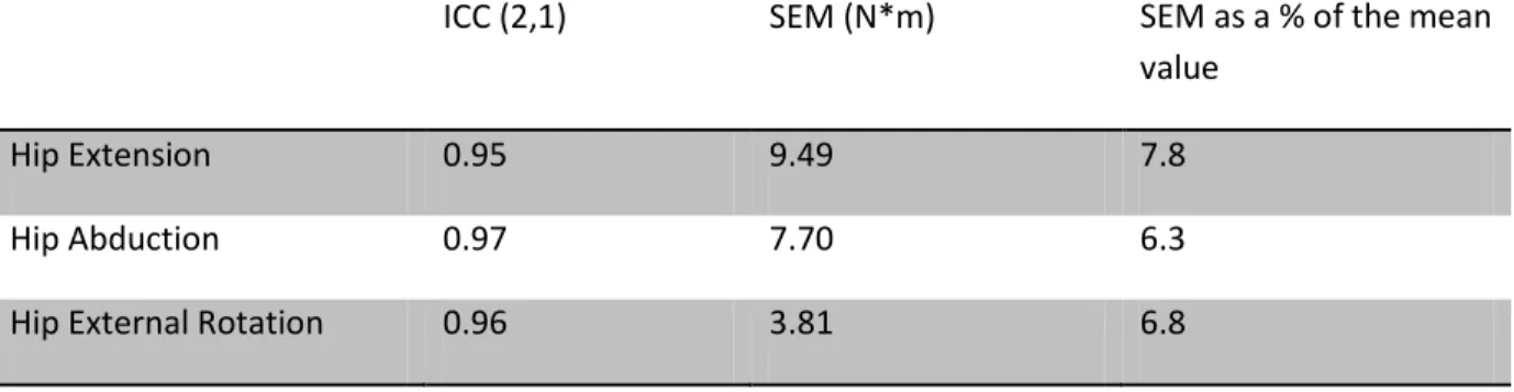
A hand-held dynamometer (Chatillon CSD 300, Amteck, Inc., Largo, FL) was used to measure peak muscle force during maximum voluntary isometric contractions (MVICs) of the hip abductors, extensors, and external rotators. All strength data was collected by the same researcher (MFN) to enhance reliability. Table 3.2 provides the intraclass correlation coefficients (ICC) and standard errors of measurements (SEMs) for these strength
measurements.
An electromagnetic motion capture system (MotionStar, Ascension Technology Corp., Burlington, VT) was used to assess kinematic data including peak knee valgus, hip adduction, and hip internal rotation angles. All kinematic data was collected at 120Hz. A non-conductive force plate (Model 4060-NC; Bertec Corporation, Columbus, OH) was used to capture ground reaction forces simultaneously at 1,200Hz.
Neural activity of the gluteus maximus and gluteus medius was measured during the MVIC and kinematic data collection using pre-amplified/active surface EMG electrodes, which have an interelectrode distance of 10mm. The signals were amplified by a factor of 10,000 (DelSys Bagnoli-8, DelSys Inc., Boston, MA). The signal amplifier features a
30
Procedures
Isometric Muscle Strength Testing
The MVICs for each subject’s dominant hip abductor, extensor, and external rotator were measured in a randomized order via a hand-held dynamometer. Three trials were conducted for each muscle group and measured by the same investigator (MFN) to improve reliability. Subjects were instructed to maximally contract for 5 seconds as the investigator resisted the motion, thus maintaining an isometric contraction. All subjects received verbal encouragement. Peak force measured using the dynamometer was multiplied by segment length to calculate peak torque. Results were then normalized to the product of body weight and height (Nm).
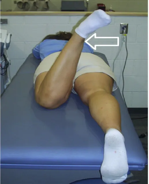
Hip abduction strength
Hip abduction strength was measured with the subject side-lying on the
non-dominant side. The non-non-dominant leg was slightly flexed at the knee and hip in order to help the subject remained balanced. The subject was asked to abduct the dominant hip without flexing, extending, or rotating the hip and to keep the ipsilateral knee fully extended. The dynamometer was placed on the lateral aspect of the subject’s thigh just proximal to the lateral joint line of the knee. The subject was asked to push into the dynamometer with maximal effort while the investigator resisted motion (Hislop, Mongomery, Connelly, & Daniels, 1995). Figure 3.1 demonstrates the subject and dynamometer positioning used for this measurement.
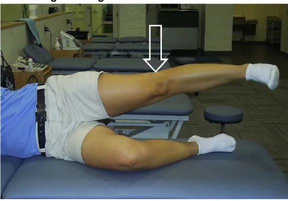
Hip extension strength
31
position was selected in order to isolate the gluteus maximus muscle and minimize
contributions from the hamstrings. The investigator placed the dynamometer immediately superior to the knee, over the posterior thigh. The subject was asked to push into the dynamometer with maximal effort while the investigator resisted this motion (Hislop, et al., 1995). Figure 3.2 demonstrates the subject and dynamometer positioning used for this measurement.
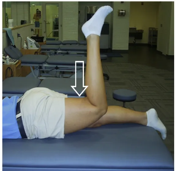
Hip external rotation strength
Hip external rotation strength was measured with the subject prone, the knee flexed to 90 degrees, and the hip in a neutral position. The subject was instructed to maximally externally rotate the dominate hip against the dynamometer positioned over the medial aspect of the shank, just proximal to the medial malleolus. The investigator applied a force over the medial aspect of the ankle in the lateral direction (Hislop, et al., 1995). Figure 3.3 demonstrates the subject and dynamometer positioning used for this measurement.
Electromyography
EMG electrodes were placed over the gluteus maximus and gluteus medius muscle bellies parallel with the muscle fibers (Ayotte, et al., 2007). All sites for electrode placement were prepared by shaving, abrading, and cleaning the skin with alcohol pads in order to reduce impedance.
32
have the same shape. The reference electrode was placed on the tibial tuberosity.
Intraclass correlation coefficients using this technique for EMG normalization for the gluteus medius and maximus have been reported to be 0.98 and 0.95 respectively (Norcross, Blackburn, & Goerger, 2009).
Jump Landing Task
Subjects performed a double-leg jump landing from a 30 cm box positioned a
distance equal to 50% of their height away from the force plate. Subjects took three practice jumps, but were permitted to take as many practice trials needed in order to become
comfortable with the task. Subjects then performed 5 successful trials with 30 seconds between each trial in order to reduce the likelihood of fatigue. Successful trials required the subjects to land with the dominant foot completely on the force plate and the non-dominant foot completely off the force plate, and to immediately jump vertically for maximum height (Padua, et al., 2009) Unsuccessful trials were repeated until 5 successful trials were obtained.
33
Data Sampling and Reduction
Kinematic data was sampled at 120 Hz and low pass filtered at 12 Hz (4th order
zero-phase-lag Butterworth), while kinetic and EMG data were sampled at 1200 Hz. The kinematic data was time-synchronized to the EMG and kinetic data and re-sampled to 1,200 Hz. Knee and hip kinematic angles were calculated using Euler angles in a Y X’ Z’’ rotation sequence. Euler angles were defined as flexion and extension occurring about the Y-axis, adduction and abduction occurring about the X-axis, and internal and external rotation occurring about the Z-axis. Motion about the knee was defined as the shank relative to the thigh and about the hip as the thigh relative to the sacrum. EMG data was corrected for DC bias and band-pass (20-350 Hz) and notch (59.5-60.5 Hz) filtered (4th order
zero-phase-lag Butterworth) using custom software (LabVIEW, National Instruments, Austin, TX). Peak hip adduction, hip internal rotation, and knee valgus angles, along with the peak EMG amplitudes for the gluteus medius and maximus were calculated during the loading phase of the landing task. If a subject did not reach hip adduction, hip internal rotation, or knee valgus during the task, the value closest to these positions was recorded. The loading phase was defined as the time interval from initial ground contact to maximum knee flexion (Blackburn & Padua, 2009). Initial ground contact was defined as the instant at which the vertical ground reaction force exceeded 10N. The peak amplitudes during the MVIC trials were also analyzed. The averages across the 3 trials for each respective muscle were used to normalize the peak EMG amplitudes of the respective muscles during the jump landing task. The normalized peak EMG amplitudes were reported as a
34
we normalized all gluteus maximus EMG amplitudes to the hip extension MVIC EMG amplitudes.
Statistical Analysis
The 82 subjects were arranged into tertiles based on hip strength (n = 27 for the high and low tertiles; n = 28 for the middle tertile). Gluteus medius and maximus EMG
amplitudes, peak knee valgus, peak hip adduction, and peak hip internal rotation angles were compared between the highest and lowest tertiles for each strength test via
independent-samples t-tests. The middle subjects were excluded from these analyses in an effort to create two groups with disparate strength values.
Multiple linear regression analyses were used to evaluate the relationships between peak kinematic values during the jump landing task and the linear combinations of muscle strength and EMG amplitude. Specifically, separate multiple linear regression models were used to evaluate
1) the relationship between peak hip adduction and the linear combination of hip abduction strength and gluteus medius EMG amplitude
2) the relationship between peak hip internal rotation and the linear combination of hip external rotation strength and gluteus maximus EMG amplitude
3) the relationship between peak hip internal rotation and the linear combination of hip extension strength and gluteus maximus EMG amplitude
35
5) the relationship between peak knee valgus and the linear combination of hip external rotation strength and gluteus maximus EMG amplitude
6) the relationship between peak knee valgus and the linear combination of hip extension strength and gluteus maximus EMG amplitude.
36
37
38
39
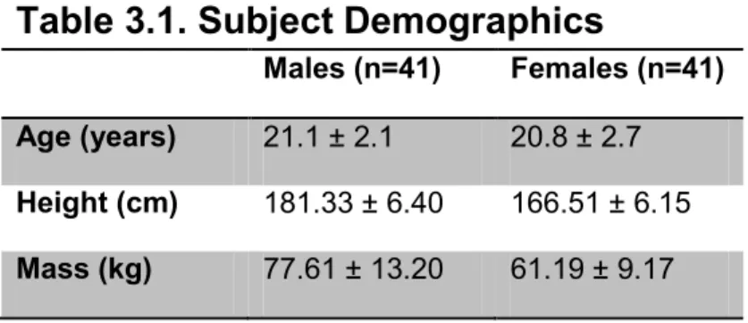
Table 3.1. Subject Demographics
Males (n=41) Females (n=41)
Age (years) 21.1 ± 2.1 20.8 ± 2.7
40
Table 3.2: Intraclass correlation coefficients (ICC) and standard
errors of the mean (SEM)
ICC (2,1) SEM (N*m) SEM as a % of the mean value
Hip Extension 0.95 9.49 7.8
Hip Abduction 0.97 7.70 6.3
41
Table 3.3. Statistical Analyses
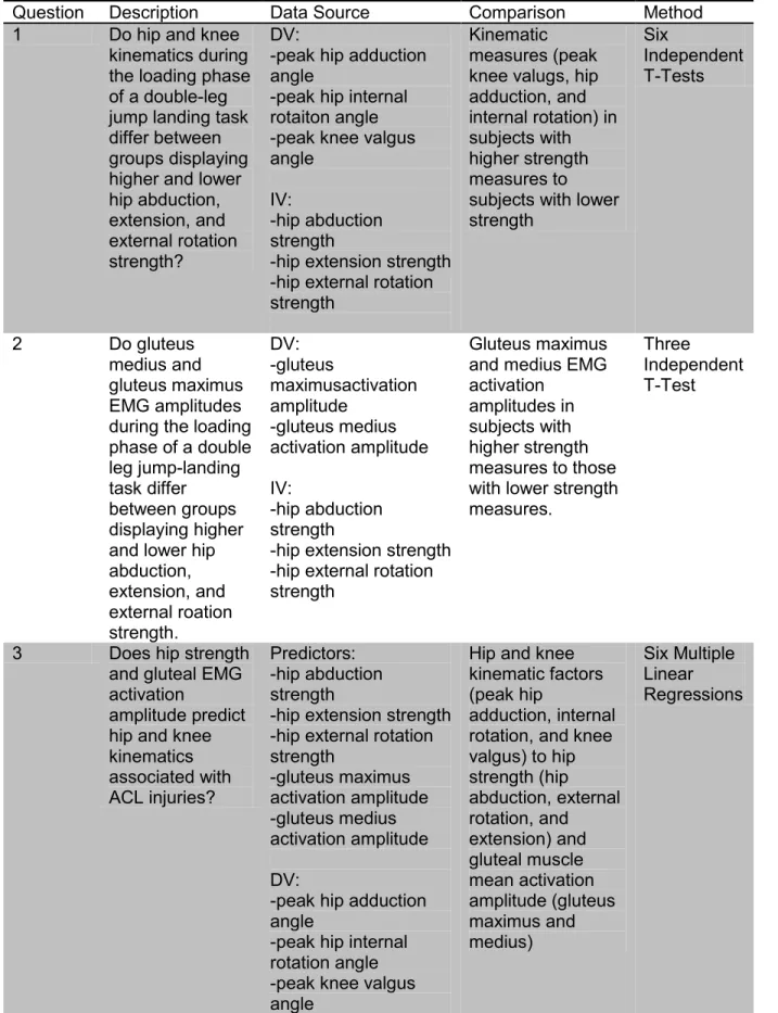
Question Description Data Source Comparison Method
1 Do hip and knee
kinematics during the loading phase of a double-leg jump landing task differ between groups displaying higher and lower hip abduction, extension, and external rotation strength?
DV:
-peak hip adduction angle
-peak hip internal rotaiton angle -peak knee valgus angle
IV:
-hip abduction strength
-hip extension strength -hip external rotation strength
Kinematic measures (peak knee valugs, hip adduction, and internal rotation) in subjects with higher strength measures to subjects with lower strength
Six
Independent T-Tests
2 Do gluteus
medius and gluteus maximus EMG amplitudes during the loading phase of a double leg jump-landing task differ between groups displaying higher and lower hip abduction, extension, and external roation strength. DV: -gluteus maximusactivation amplitude -gluteus medius activation amplitude IV: -hip abduction strength
-hip extension strength -hip external rotation strength
Gluteus maximus and medius EMG activation
amplitudes in subjects with higher strength measures to those with lower strength measures.
Three Independent T-Test
3 Does hip strength
and gluteal EMG activation
amplitude predict hip and knee kinematics associated with ACL injuries? Predictors: -hip abduction strength
-hip extension strength -hip external rotation strength -gluteus maximus activation amplitude -gluteus medius activation amplitude DV:
-peak hip adduction angle
-peak hip internal rotation angle -peak knee valgus angle
Hip and knee kinematic factors (peak hip
42
Chapter 4
Results
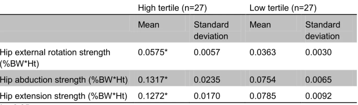
Independent t-tests signify that the high and low strength tertile groups were significantly different for hip external rotation (t52=16.916, p<0.001), hip abduction
(t52=12.013, p<0.001), and hip extension (t52=13.064, p<0.001), indicating that there was a
significant difference in the “strong” group and “weak” groups. Means and standard
deviations for each strength comparison are detailed in Table 4.1. It is important to note that the strong groups were primarily composed of males and the weak groups were primarily composed of females. The demographics for each tertile are detailed in Table 4.2. The results for the independent t-tests used to evaluate differences in lower extremity kinematics and peak EMG amplitudes between high and low hip strength groups are describe below; as well as the results for the regression analyses investigating if strength and peak EMG
amplitudes of the gluteal muscles predict peak knee valgus angle and other kinematic factors associated with knee valgus including peak hip adduction and internal rotation.
Hip Strength and Kinematics
43
at 0.05 thus we conducted secondary analyses to evaluate the observed power and
determine the number of subjects that would have been necessary to achieve a priori power of 0.80. These analyses indicated generally a low observed power and that a much higher number of subjects (80-90 subjects per group) would have been necessary to provide adequate power. There were a significant differences in peak hip external rotation and knee valgus angles between the high and low hip external rotation strength groups. Those with greater peak isometric hip external rotation strength demonstrated lesser peak hip external rotation (t52=2.033, p=0.0236) and knee valgus (t52=2.209, p=0.0158) compared to those
with lesser peak isometric hip external rotation strength. All of these results can be viewed in Tables 4.3, 4.4, and 4.5
Hip Strength and EMG Amplitude
There was no significant difference in the gluteus medius EMG amplitudes during the loading phase of a double-leg jump landing between those demonstrating high vs. low isometric hip abduction strength. There were, however, significant differences in gluteus maximus EMG amplitude between the hip extension strength groups (t52=-1.902, p=0.031)
and hip external rotation strength groups (t52=-1.749, p=0.043), with stronger individuals
demonstrating less gluteus maximus EMG activity. The results for the means and standard deviations for all the independent t-tests run can be seen in Tables 4.3, 4.4, 4.5.
Regressions
44
transverse plane compared to gluteal maximus activation amplitude (standardized Beta-weight = 0.024). Similarly, isometric hip rotation strength (standardized Beta-Beta-weight = 0.348) was a stronger predictor of peak knee valgus angle compared to gluteal maximus activation amplitude (standardized Beta-weight = 0.124). The results from these significant findings can be seen in Figures 4.1 and 4.2. No other regressions were found to be
45
Table 4.1. Muscle strength normalized to body mass and length of
the segment.
High tertile (n=27) Low tertile (n=27)
Mean Standard
deviation
Mean Standard
deviation Hip external rotation strength
(%BW*Ht)
0.0575* 0.0057 0.0363 0.0030
Hip abduction strength (%BW*Ht) 0.1317* 0.0235 0.0754 0.0065
Hip extension strength (%BW*Ht) 0.1272* 0.0170 0.0785 0.0092
46
Table 4.2. Demographics of strength tertiles
High tertile (n=27) Low tertile (n=27)
Males Females Males Females
Hip external rotation strength 22 5 4 23
Hip extension strength 19 8 7 20
47
Table 4.3. Differences in kinematics and peak EMG amplitudes
between high vs. low hip abduction strength
Dependent Variable High Strength
Tertile (n=27)
Low Strength Tertile (n=27)
Observed Power
Subjects/group necessary for 0.80 priori power Peak Hip Adduction
Angle (º) † 1.98 ± 8.31 3.19 ± 6.77 0.161
481
Peak Knee Valgus
Angle (º) ‡ -15.34 ± 9.00 -18.92 ± 9.34 0.372 90
Peak Gluteus Medius EMG (%MVIC)
101.08 ± 117.15
155.10 ±
177.36 0.367 93
† Negative value indicates hip abduction and a positive value indicates hip adduction ‡Negative value indicates knee valgus and a positive value indicates knee varus
48
Table 4.4. Differences in kinematics and peak EMG amplitudes
between high vs. low hip external rotation strength
Dependent Variable High Strength
Tertile (n=27)
Low Strength Tertile (n=27)
Observed Power
Subjects/group necessary for 0.80 priori power Peak Hip Internal
Rotation Angle (º) †
-3.21 ± 9.92* -8.39 ± 8.75 _ _
Peak Knee Valgus Angle (º) ‡
-14.65 ± 11.28*
-21.17 ± 10.39 _ _
Peak Gluteus Maximus EMG Amplitude (%MVIC)
117.39 ± 63.20*
307.00 ± 559.75
_ _
† Negative value indicates hip external rotation and a positive value indicates hip internal
rotation
‡ Negative value indicates knee valgus and a positive value indicates knee varus
49
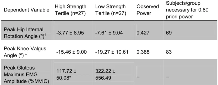
Table 4.5. Differences in kinematics and peak EMG amplitudes
between high vs. low hip extension strength
Dependent Variable High Strength
Tertile (n=27)
Low Strength Tertile (n=27)
Observed Power
Subjects/group necessary for 0.80 priori power
Peak Hip Internal
Rotation Angle (º)† -3.77 ± 8.95 -7.61 ± 9.04 0.427 69
Peak Knee Valgus
Angle (º) ‡ -15.46 ± 9.00 -19.27 ± 10.61 0.388 83
Peak Gluteus Maximus EMG Amplitude (%MVIC)
117.72 ± 50.08*
322.22 ±
556.49 _ _
† Negative value indicates hip external rotation and a positive value indicates hip internal
rotation
‡ Negative value indicates knee valgus and a positive value indicates knee varus
50
Table 4.6. Dependent and predictor variables for regression
analyses
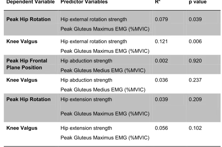
Dependent Variable Predictor Variables R2 p value
Peak Hip Rotation Hip external rotation strength 0.079 0.039 Peak Gluteus Maximus EMG (%MVIC)
Knee Valgus Hip external rotation strength 0.121 0.006
Peak Gluteus Maximus EMG (%MVIC) Peak Hip Frontal
Plane Position
Hip abduction strength 0.002 0.920
Peak Gluteus Medius EMG (%MVIC)
Knee Valgus Hip abduction strength 0.036 0.237
Peak Gluteus Medius EMG (%MVIC)
Peak Hip Rotation Hip extension strength 0.039 0.209
Peak Gluteus Maximus EMG (%MVIC)
Knee Valgus Hip extension strength 0.056 0.102
51
Table 4.7. Predictability of peak hip internal rotation based on hip
external rotation strength and gluteus maximus activation
Variable Parameter
Estimate
SE Standardized
Coefficient
t Value p Value
Hip Rotation
Intercept -18.248 5.262 -3.468 0.001
Hip external rotation strength
280.880 108.367 0.284 2.592 0.011
Peak Gluteus Maximus EMG (%MVIC)
52
Table 4.8. Predictability of knee valgus based on hip external
rotation strength and gluteus maximus activation
Variable Parameter
Estimate
SE Standardized
Coefficient
t Value p Value
Knee Valgus
Intercept -35.703 5.750 -6.210 < 0.001
Hip external rotation strength
383.977 118.408 0.348 3.243 0.002
Peak Gluteus Maximus EMG (%MVIC)
53
Table 4.9. Predictability of peak hip adduction based on hip
abduction strength and gluteus medius activation
Variable Parameter
Estimate
SE Standardized
Coefficient
t Value p Value
Frontal Plane Hip Position
Intercept 3.931 3.310 1.187 0.239
Hip abduction strength
-10.357 30.551 -0.038 -0.339 0.736
Peak Gluteus Medius EMG (%MVIC)
54
Table 4.10. Predictability of peak knee valgus based on hip
abduction strength and gluteus medius activation
Variable Parameter
Estimate
SE Standardized
Coefficient
t Value p Value
Knee Valgus
Intercept -24.707 4.626 -5.341 <0.001
Hip abduction strength
72.899 42.689 0.189 1.708 0.092
Peak Gluteus Medius EMG (%MVIC)
55
Table 4.11. Predictability of peak hip internal rotation based on hip
extension strength and gluteus maximus activation
Variable Parameter
Estimate
SE Standardized
Coefficient
t Value P Value
Hip Rotation
Intercept -13.626 5.041 -2.703 0.008
Hip extension strength
82.725 46.705 0.202 1.771 0.080
Peak Gluteus Maximus EMG (%MVIC)
56
Table 4.12. Predictability of peak knee valgus based on hip
extension strength and gluteus maximus activation
Variable Parameter
Estimate
SE Standardized
Coefficient
t Value P Value
Knee Valgus
Intercept -28.871 5.587 -5.167 <0.001
Hip extension strength
108.185 51.765 0.236 2.090 0.040
Peak Gluteus Maximus EMG (%MVIC)
57
Figure 4.1. Correlation between hip strength and hip rotation
during a jump landing task
*Positive values indicate internal rotation and negative values indicate external rotation p=0.011
58
Figure 4.2. Correlation between hip strength and knee valgus
during a jump landing task
*Positive values indicate knee varus and negative values indicate knee valgus p=0.003
59
Chapter 5
Discussion
The primary findings of this study include the following: individuals with greater peak isometric hip external rotation strength demonstrated lesser peak hip external rotation and knee valgus angles, and those with stronger hip extensors and external rotators
demonstrated a lesser peak gluteus maximus amplitudes than those with weaker hip extensors and external rotators respectively. Additionally, isometric hip external rotation strength and gluteus maximus EMG amplitude explained a significant, though limited, amount of variance in peak hip rotation and knee valgus angles during the loading phase of a double-leg jump landing task. Furthermore, hip abduction and extension strength, and gluteus medius EMG activity did not influence peak hip or knee kinematics.
Some of these findings agree with previous literature, while others differ from the already controversial literature on the influences of gluteal muscle strength and activation on hip and knee biomechanics. While a number of previous investigations evaluated