IRON DEFICIENCY AND IRON SUPPLEMENTATION CONSPIRE TO MEDIATE SUSCEPTIBILITY TO ERYTHROCYTIC STAGE PLASMODIUM FALCIPARUM INFECTION
Martha Ann Clark
A dissertation submitted to the faculty at the University of North Carolina at Chapel Hill in partial fulfillment of the requirements for the degree of Doctor of Philosophy in the Department
of Microbiology and Immunology in the School of Medicine.
Chapel Hill 2014
Approved by:
Anthony Richardson
Steven Meshnick
ABSTRACT
MARTHA ANN CLARK: Iron Deficiency and Iron Supplementation Conspire to Mediate Susceptibility to Erythrocytic Stage Plasmodium falciparum Infection
(Under the direction of Steven Meshnick)
Malaria and iron deficiency are interconnected public health concerns, which
disproportionally affect children and pregnant women. Malariacauses an estimated 250 million
infections and 1 million deaths per year. Plasmodium falciparum is the most virulent species of the malaria parasite that infects humans. Anemia, predominantly iron deficiency anemia, is the most common nutritional deficiency worldwide, and affects up to 50% of populations in the
developing world. The World Health Organization recommends universal iron supplementation
in regions where malnutrition is common. This recommendation has been complicated by
clinical evidence that iron deficiency protects against malaria infection, and that iron
supplementation increases susceptibility to malaria infection. The mechanisms underlying the
interaction between malaria, host iron, and iron supplementation remain unclear. Here, I’ve
employed the in vitro system for cultivating erythrocytic stage P. falciparum to assess first, the impact of extracellular iron on parasite growth as well as the bioavailable iron content of
parasitized erythrocytes. I have found that extracellular iron is incorporated into parasitized
erythrocytes but does not have affect parasite growth. Second, I assessed the capacity of
iv
merozoites within iron deficient erythrocytes. I additionally observe that P. falciparum propagation is recovered in erythrocytes donated by iron supplemented iron deficient donors.
Furthermore, I attribute the recovery of P. falciparum erythrocyte propagation to the replacement of iron deficient erythrocytes with young iron-replete erythrocytes that are produced in response
to iron supplementation. These results are consistent with clinical observations that iron
deficiency is protective against malaria infection and iron supplementation increases the risk of
malaria infection. Moreover, my results suggest that iron mediated alterations to erythrocyte
physiology and intra-host erythrocyte population dynamics as well as potentially altered serum
iron levels contribute to the underlying mechanisms governing the relationship between the
ACKNOWLEDGMENTS
“These are just physiological dispositions and yet again nature taunts our convention, our intuition, our fear. You have to be brave to study her brilliance.”
– Janna Levin, theoretical cosmologist
Above all I would like to thank Carla Cerami for teaching me to be fearless in science as
well as life. For teaching me that revelation cannot occur unless you are willing to let go of your
assumptions and your biases. Thank you for being an incredible mentor, welcoming me into your
family when the distance from my own family was especially painful, and being an invaluable
friend and confidante. I will never forget our spirited discussions of a newly discovered paper, a
recent result or a new idea. With both sadness and excitement I anticipate leaving the embrace of
the scientific family you’ve created here at UNC with Steve and joining my new scientific family
in Boston. I take solace in the fact that I know this is not an end to our brilliant association and
friendship, but rather a new chapter. I look forward to sharing my scientific journey with you for
many years to come.
I would like to thank Steve Meshnick for sharing his vast expertise and experience in the
malaria field. The small little pocket of malaria research you’ve created at UNC very special and
exceptionally unique. You’ve assembled an excited and talented multidisciplinary team that is
fearless bridging the chasm that currently exists between basic science and epidemiology. Thank
vi
most importantly mentoring. It is people like you who are responsible for inspiring and preparing
the next generation of explorers, innovators, and creators; in a word scientists.
I would like to thank Steve Taylor and Jon Juliano for instilling in me an appreciation for
the beauty of a clearly communicated scientific story, and helping me find my own scientific
voice. The countless lab meetings spent pulling apart presentations and specific aims pages seem
like they have been seamlessly incorporated into the very rhythm of my own scientific voice. I
guess I’ve picked up y’alls scientific twang.
I would like to thank my committee for challenging me to develop scientifically, and
discover the fine balance between confidently communicating my scientific story and
acknowledging the perpetual unknowns that exists in scientific research. Because of you I was
able to discover an inner confidence I never knew existed. I would not be where I am today as a
scientist without you.
I would like to thank Morgan Goheen, my scientific sister, as well as each of the beautiful
smart women I have been blessed to train with and be mentored by here at UNC. Morgan,
Cheryl, Blaire, Megan, Caitlin, Maria, Liz, and Eliza thank you for playing and working so hard
side by side with me each and every step of the way. Because of you I have been able to discover
the sisterhood that exists between each and every woman. I love y’all and look forward to many
more long conversations and equally long bike rides, runs, and walks.
I would like to thank my amazing family. I know that more often than not I have
stubbornly pushed forward doggedly brandishing my independence. Doubt not though your
importance in every aspect and every minute of these past six years. It was learning to live life
joyfully and ever rolling with the punches with Mary, Paul, Charlotte, and Daniel that got me to
means to have true comradeship. It was mom day after day demonstrating that rewards that can
be found in one’s career and the virtue of perseverance. Because of your example mom, I’ve
found a career I love and I learned that to reap the wards one must dig in and push through even
in the hardest of times. It was dad repeatedly challenging me to ask questions, and me obviously
not doing so (most of the time). Who would have guessed I’d come to the point where I can’t
stop asking questions. Because of your example dad, I’ve discovered the transformative gift of
wonder. Life has never looked more beautiful.
Finally, I would like to thank Nicholas Spidale, my partner and my anchor. Because of
you I was able to keep my sanity in the insanity that is graduate school. It is so easy to forget
every other aspect of our lives (food, bills, family and friends) amid the excitement of a new idea
or a surprising result. Your daily presence beside me has kept me grounded; ever reminding me
nothing is as important as the people (and pups) we love. I cannot express how excited and
happy I am to embark with you on the next big adventure of both our professional and personal
viii PREFACE
This work would not have been possible without the contribution of many talented
scientists, skilled nurses, and patient study participants. The experiments outlined in Chapters 2,
3, and 4 were made possible through our collaboration with Dr. Raj Kasthuri. With Raj’s
assistance we were able to identify and enroll participants into our study.
Chapter 2 represents a publication for which I was the first author. I conducted all the
experimentation contained in Chapter 2. Dr. Carla Cerami and Dr. Nancy Fisher provided
assistance with experimental design and Dr. Carla Cerami helped write the resulting paper. The
paper was published prior to writing this thesis with the following citation.
Clark, M., Fisher, N. C., Kasthuri, R., and Cerami Hand, C. (2013). Parasite maturation
and host serum iron influence the labile iron pool of erythrocyte stage Plasmodium
falciparum. Br. J. Haematol. 161, 262–269.
Permission to include the article in its entirety in a PhD dissertation was retained from John
Wiley & Sons Ltd. (publisher of The British Journal of Haematology) as explained at
https://s100.copyright.com/CustomerAdmin/PLF.jsp?ref=17c65dfb-78bf-4cfc-87f0-f86c38ab9926.
As with Chapter 2, I conducted all the experimentation contained in Chapter 3. Dr. Carla
Cerami, Dr. Tony Fulford, and Nick Spidale provided assistance with experimental design. Dr.
Carla Cerami and Nick Spidale helped write the resulting paper, which has been reviewed by
The experimentation contained within Chapter 4 is primarily my own work. Morgan
Goheen provided assistance with experimentation. As mentioned previously Dr. Raj Kasthuri
assisted in identifying and enrolling study participants. Michael Nicholoson of Precision
Biosciences (Durham, NC) provided access to the Beckman Quanta Flow Cytometer used to
analyze RBC volume experiments (Figure 4.7A). Dr. Tony Fulford performed statistical analysis
of growth data (Figure 4.3C and D). Dr. Carla Cerami, Dr. Tony Fulford, and Dr. Steve Taylor
assisted with experimental design, data analysis, and helped write the resulting paper. The
x
TABLE OF CONTENTS
LIST OF TABLES ... xv
LIST OF FIGURES ... xvi
LIST OF ABBREVIATIONS AND SYMBOLS ... xviii
CHAPTER ONE ... 1
Introduction ... 1
1.1 A relationship revealed – host iron status and malaria ... 1
1.2 Iron deficiency and iron deficiency anemia ... 2
1.3 Malaria ... 4
1.4 Clinical studies linking iron and malaria infection ... 4
1.5 Biological importance of iron ... 6
1.6 Iron metabolism in the malaria parasite ... 7
1.7 Iron chelators and their contribution to the elucidation of malaria iron biology ... 9
1.8 Host iron reservoirs available to erythrocytic stage malaria ... 10
1.9 The relationship between serum iron and erythrocytic stage malaria ... 11
1.10 The relationship between intra-erythrocytic iron and erythrocytic stage malaria ... 12
1.11 Microcytic iron-deficient RBCs and malaria ... 14
1.12 Perturbations in erythropoiesis and malaria ... 15
1.13 Objectives of this dissertation ... 17
CHAPTER TWO ... 38
Parasite maturation and host serum iron influence the labile iron pool of erythrocyte stage P. falciparum ... 38
2.1 Overview ... 38
2.2 Introduction ... 39
2.3 Materials and Methods ... 41
P. falciparum culture and growth assays... 41
P. falciparum LIP assay ... 42
Flow Cytometry Analysis ... 43
Microscopy ... 43
2.4 Results ... 44
Assessing the effect of extracellular iron on erythrocytic stage P. falciparum growth ... 44
Detection of LIP in P. falciparum infected erythrocytes by flow cytometry ... 44
Characterization of LIP during maturation of P. falciparum within host RBCs ... 46
Investigation of the impact of host serum iron sources on LIP in P. falciparum infected erythrocytes ... 47
2.5 Discussion ... 48
REFERENCES ... 60
CHAPTER THREE ... 64
A three-color invasion assay allows for the study of erythrocyte population dynamics and P. falciparum merozoite invasion ... 64
xii
Three-color invasion assay ... 67
Enzyme treatment of RBCs ... 68
Microscopy ... 68
Flow Cytometry Analysis ... 69
Statistical Methods ... 69
3.4 Results ... 70
Labeling RBCs with CellTrace dyes DDAO-SE and Violet allows for the direct comparison of P. falciparum invasion into different RBC populations. ... 70
The three color P. falciparum invasion assay allows for the direct comparison of P. falciparum invasion into physiologically different RBC populations. ... 71
The relationship between different RBC populations and P. falciparum invasion is revealed by the three-color invasion assay. ... 72
3.5 Discussion ... 74
REFERENCES ... 87
CHAPTER FOUR ... 89
Host iron status and iron supplementation conspire to mediate host susceptibility to the erythrocytic stage of Plasmodium falciparum ... 89
4.1 Overview ... 89
4.2 Introduction ... 90
4.3 Materials and Methods ... 91
Clinical ... 91
Parasite Culture ... 92
Growth Assays ... 92
Invasion assay ... 93
Density Separation ... 94
Flow cytometry ... 94
Statistical Methods ... 95
4.4 Results ... 96
Iron deficient RBCs are protective against P. falciparum infection ... 96
Erythrocytic stage P. falciparum growth is increased in RBCs from iron supplemented donors. ... 97
Changing RBC population dynamics impacts susceptibility to erythrocytic stage P. falciparum infection... 99
4.5 Discussion ... 104
REFERENCES ... 121
CHAPTER FIVE ... 125
Discussion... 125
5.1 Where we stand in the grand scheme of things ... 125
5.2 Determining the impact of host iron status on virulence of erythrocytic stage P. falciparum ... 125
Transferrin and ferric citrate, which are found in human serum, contribute to the bioavailable iron of erythrocytic stage P. falciparum. ... 125
Unraveling erythrocytic stage P. falciparum iron biology and the parasites dependence on host iron... 127
5.3 Defining the influence of changing host RBC population dynamics on erythrocytic stage P. falciparum infection, specifically in the context of host iron deficiency and iron supplementation. ... 130
xiv
Pursuing mechanisms by which microcytic iron deficient RBCs are protective
against P. falciparum infection. ... 131
Pursuing the effect of changing RBC population dynamics in the human host on
susceptibility to malaria infection in vivo ... 133
5.4 Beyond iron as a growth factor and mediator of RBC population
dynamics – exploration of alternate mechanisms governing the relationship
between iron deficiency, iron supplementation and malaria. ... 134
Probing the effect of iron deficiency and iron supplementation on important mediators of malaria pathogenesis – phagocytosis of pRBCs and pRBC
endothelial cytoadhesion ... 135
5.5 Looking forward ... 136
LIST OF TABLES
Table 1.1 – Summary of clinical studies on iron deficiency,
iron supplementation, and malaria ... 20
Table 1.2 – Relationship between host serum iron and P. falciparum ... 24 Table 1.3 – Relationship between RBC iron and P. falciparum ... 26 Table 4.1 – Iron status of donors enrolled in IDA, IDA-Fe, IR,
and IR-Fe study groups ... 120
Table 5.1 – Questions for future investigation of erythrocytic stage
P. falciparum utilization of host iron ... 138 Table 5.2 – Questions for future investigation of the protection
provided by microcytic iron deficient and old RBCs against
erythrocytic stage P. falciparum infection ... 139 Table 5.3 – Questions for future investigation of the relationship
between host RBC population dynamics and erythrocytic
xvi
LIST OF FIGURES
Figure 1.1 – Host iron available to erythrocytic stage P. falciparum. ... 27 Figure 1.2 – Impact of iron deficiency anemia and iron supplementation
on erythropoietic rate and erythrocyte physiology and hypothesized
effect on P. falciparum erythrocytic infection. ... 28 Figure 2.1 – The effect of extracellular iron on erythrocytic stage
P. falciparum infection is unclear. ... 52 Figure 2.2 – Calcein and SYTO 61 fluorescence does not exhibit spectral
overlap, and SYTO 61 does not interfere with Calcein iron binding ... 53
Figure 2.3 – Measurement of the basal levels of CA-AM fluorescence ... 55
Figure 2.4 – Measurement of LIP in P. falciparum infected RBCs using
Flow Cytometry ... 56
Figure 2.5 – LIP increases as parasite matures inside host red blood cell ... 57
Figure 2.6 – Addition of human transferrin or ferric citrate increases LIP in parasitized RBCs, and transferrin co-localizes with late stage
parasitized RBCs ... 58
Figure 3.1 – RBCs stained with CellTrace DDAO-SE and Violet can be combined to directly compare P. falciparum invasion in a three-color
invasion assay ... 80
Figure 3.2 – Labeling RBCs with CellTrace Violet does not impact
P. falciparum invasion ... 81 Figure 3.3 – Direct comparison of P. falciparum strains 3D7, Dd2 and
FCR3-FMG invasion into RBCs labeled with DDAO-SE and
CellTrace Violet ... 82
Figure 3.4 – Comparison of two and three color P. falciparum invasion
assay ... 83
Figure 3.5 – Comparison of invasion of P. falciparum into untreated
and neuraminidase, treated RBCs ... 84
Figure 3.6 – Comparison of invasion of P. falciparum into untreated
and trypsin treated RBCs ... 85
Figure 3.7 – Comparison of invasion of P. falciparum into untreated
Figure 4.1 – Serial in vitro growth assay design for P. falciparum... 108 Figure 4.2 – P. falciparum erythrocytic stage propagation is attenuated
in microcytic IDA RBCs as compared to normocytic iron-replete
RBCs in vitro ... 109 Figure 4.3 – Effect of iron supplementation of IR and IDA donors on
erythrocytic stage P. falciparum growth in vitro ... 111 Figure 4.4 – The potential effect of altered host RBC population dynamics
that result from iron deficiency and iron supplementation, on
susceptibility to erythrocytic stage malaria infection ... 113
Figure 4.5 – Replacement of RBCIDA with RBCIR increases the invasion
and growth rate of erythrocytic stage P. falciparum ... 114 Figure 4.6 – P. falciparum prevalence is higher in CD71+ reticulocytes
from iron supplemented iron deficient donors ... 115
Figure 4.7 – Young RBCs sustain greater P. falciparum growth due to increased risk of young RBCs to invasion and greater capacity of P. falciparum produce infectious daughter merozoites within young
RBCs ... 117
Figure 4.8 – Elevated frequency of RBCY in a RBC population sustains an elevated P. falciparum invasion rate, and replacement of RBCY
xviii
LIST OF ABBREVIATIONS AND SYMBOLS
ACM – Albumax II Complete Media
ATP – Adenosine triphosphate
C – Chymotrypsin treatment
CA-AM – Calcein acetoxymethyl ester
CI – Confidence interval
DFO – Deferoxamine
DNA – Deoxyribonucleic acid
DV – Digestive vacuole
ER – Endoplasmic reticulum
EC – Endothelial cell
Fe2+ – Ferrous iron
Fe3+ – Ferric iron
G6PD – Glucose-6-phosphate dehydrogenase
Hgb – Hemoglobin
IDA – Iron deficiency anemia
IDA-Fe – Iron deficiency anemia iron supplemented
IR – Iron-replete
IR-Fe – Iron-replete iron supplemented
IRB – Internal review board
IRE – Iron responsive element
IRP – Iron responsive protein
MA – Mature adult
MACS – Magnetic activated cell sorting
MCHC – Mean corpuscular hemoglobin content
MCV – Mean corpuscular volume
N – Neuraminidase treatment
NTBI – Non-transferrin bound iron
O – Old
O+ RBC – O positive RBC
Pf – Plasmodium falciparum pRBC – Parasitized RBC
RBC – Red blood cell
RDW – RBC Distribution Width
ROS – Reactive oxygen species
RPMI – Roswell park medical institute
SD – Standard deviation
SI – Susceptibility index
T – Trypsin treatment
VY – Very young
WHO – World Health Organization
Y – Young
CHAPTER ONE
Introduction 1.1 A relationship revealed – host iron status and malaria
Iron deficiency and malaria are significant co-morbidities in large portions of the
developing world, and both maladies disproportionately affect pregnant women and children.
Malaria causes an estimated 250 million infections and 500,000 deaths annually. Iron deficiency
is estimated to affect one quarter of the world’s populations causing substantial morbidity.
Fortunately, iron deficiency is easily treated with iron supplementation (Okebe et al., 2011).
Accordingly the World Health Organization (WHO) recommends routine iron supplementation
for children and adults in areas with high prevalence of iron deficiency (Haider et al., 2013; Low
et al., 2013). However, the wisdom of universal iron supplementation campaigns in malaria
endemic regions has recently been questioned due to clinical evidence that suggests iron
deficiency protects against malaria, and that iron supplementation of women and children may
increase the incidence of malaria when given without malaria prophylaxis or access to adequate
health care (Sazawal et al., 2006; Tielsch et al., 2006; Veenemans et al., 2011; Zlotkin S et al.,
2013; Esan et al., 2013; Nyakeriga et al., 2004; Gwamaka et al., 2012; Jonker et al., 2012;
Kabyemela et al., 2008; Senga et al., 2011). This situation has created a dilemma for health
2
Despite these clinical and epidemiological studies, the extent to which the human host’s
iron status affects risk to and severity of malaria infection is unknown. Differences in study
design and confounding factors (such as acquired immunity to malaria and hemoglobinopathies)
have made the clinical and epidemiological studies difficult to interpret (Prentice et al., 2007).
Furthermore, though iron and malaria have been and continue to be studied the exact biological
relationship between host iron and malaria virulence remains largely unclear.
1.2 Iron deficiency and iron deficiency anemia
Iron deficiency is a condition in which there is insufficient iron in the body to maintain
normal physiologic functions. Iron deficiency can be categorized into three stages: iron
deficiency without anemia, iron deficiency with mild anemia, and iron deficiency with severe
anemia. Iron deficiency anemia occurs when iron stores are exhausted and the supply of iron to
tissue is compromised; this condition is defined as anemia with biochemical evidence of iron
deficiency. Iron deficiency is most prevalent and severe in young children and women of
reproductive age, but can also occur in older children, adolescents, adult men, and the elderly.
The WHO estimates that 50% of pregnant women and 40% of preschool children in the
developing world are iron-deficient (WHO | Assessing the iron status of populations; Kassebaum
et al., 2013) Often, iron deficiency develops slowly and is not clinically diagnosed until severe
anemia is apparent (Stoltzfus, 2003).
Studies suggest that iron deficiency impairs the growth, cognition, and neurological
development of children from infancy through adolescence, impairs immune function, and is
associated with increased morbidity rates (Wang et al., 2013; De-Regil et al., 2011, 2013). Iron
deficiency during pregnancy is associated with multiple adverse outcomes for both mother and
and low birth weight (Peña-Rosas et al., 2012a, 2012b). Iron deficiency anemia can be a direct
cause of death or contribute indirectly. For example, during childbirth an anemic mother cannot
afford to lose more than 150 mL of blood, compared with a healthy mother who can lose up to 1
liter of blood and still survive. Thus, the WHO recommends iron supplementation for all men,
women and children in areas where malnutrition is prevalent (WHO | Guidelines on food
fortification with micronutrients).
Host iron metabolism is intimately linked to the host response to infection and
inflammation. With the high incidence of infection in the developing world, the study and
treatment of iron deficiency anemia becomes challenging. In the face of infection and
inflammation, the human host protein hepcidin becomes elevated and initiates signaling which
results in reduced iron absorption into the body along with the redistribution of body iron stores.
As a consequence of the effect of infection and inflammation on human iron metabolism, many
of the biomarkers utilized to assess host iron status are sensitive to both iron as well as infection.
For example, low serum ferritin (serum ferritin reflects total body iron reservoirs) is indicative of
iron deficiency. However, ferritin is also an acute phase protein, which is elevated in the context
of infection, and as a result is not a reliable marker of human iron status in the presence of
infection or inflammation. Like serum ferritin, transferrin saturation and transferrin receptor
levels are biochemical markers of human iron status that are also sensitive to infection and
inflammation. As a result evaluating an individual’s iron status in the context of infection has
proven difficult (Aguilar et al., 2012), and the scientific community has struggled to establish
4 1.3 Malaria
In 2012 malaria caused an estimated 207 million infections and over 600,000 deaths;
90% of these deaths occurred in sub-Saharan Africa, and 77% occurred in children under five
(WHO | World Malaria Report 2013). At least five species of the eukaryotic Apicomplexan parasite from the genus Plasmodium cause malaria in humans with Plasmodiumfalciparum being the most common and deadly. Malaria parasites are transmitted to the human host by
mosquitoes. Following the bite of a malaria parasite infected mosquito, the sporozoite stage of
the parasite enters the bloodstream and travels to the liver, where it subsequently infects liver
hepatocytes. Malaria replication in the liver is asymptomatic. Next, the merozoite form of the
parasite leaves the liver and enters into circulation to infect host red blood cells (RBCs). During
the erythrocytic stage of infection, the parasite repeatedly invades, replicates within, and egresses
from host RBCs. This erythrocytic stage of infection is responsible for all symptoms of disease
(Miller et al., 2013), and the severity of disease is directly associated with parasite burden
(Chotivanich et al., 2000; Dondorp et al., 2005).
A wide range of symptoms can be observed in malaria patients. Clinically however,
malaria is categorized as either uncomplicated or complicated. Complicated malaria is further
divided into three overlapping syndromes: cerebral malaria, severe anemia, and metabolic
acidosis. The clinical syndrome observed in each individual patient is influenced by multiple
variables: parasite species, host immune status and genetic background, as well as the use and
timing of antimalarial drugs (Taylor et al., 2010).
1.4 Clinical studies linking iron and malaria infection
Host iron has received significant attention at the clinical level as a major factor that may
examined the relationship between host iron status and malaria risk are reviewed in three
meta-analyses (Oppenheimer, 2001; Shankar, 2000; Gera and Sachdev, 2002). In the interim, two
large iron supplementation trials as well as several smaller clinical studies have shed further light
on the relationship between host iron status and malaria infection (Table 1). Clinical trials that have examined the relationship between host iron and malaria fall into two basic categories:
those that observe the rate of malaria in individuals with iron deficiency anemia, and those that
look at the rate of malaria infection in individuals given iron supplementation. Differences in
study design exist within both study types, and include: the definition of study participant iron
status, the administration of iron alone or with folate, and access to health care. Despite
differences in study design, assessment of the outcome of the clinical studies has led to the
general consensus that iron deficiency is protective against malaria, and iron supplementation
increases malaria risk in the absence of access to adequate health care (Spottiswoode et al., 2012;
Prentice and Cox, 2012; Stoltzfus, 2012).
While these clinical studies and meta-analyses have been indispensable for determining
the relationship between host iron status and malaria risk, it is not clear how iron deficiency
protects and why iron supplementation increases risk. Immunity to malaria and high prevalence
of genetic traits such as Glucose-6-phosphate dehydrogenase (G6PD) deficiency and
hemoglobinopathies in the study populations limit the capacity of clinical studies to parse out
causation. Furthermore, relatively little is known with regards to the role host iron plays in
malaria pathogenesis. Iron impacts a broad range of biological processes that have the potential
6
iron deficiency against malaria and (ii) the increased risk of malaria associated with iron
supplementation is critical for managing iron supplementation campaigns in malaria endemic
regions.
1.5 Biological importance of iron
Iron is an essential nutrient for nearly every living organism including humans and the
malaria parasite. Iron impacts a broad range of biological processes; including host and parasite
cellular function, erythropoiesis and immune function. The capacity of iron to fluctuate between
two oxidation states, ferrous (Fe2+) and ferric (Fe3+), makes it indispensable for many critical
biological processes, including DNA replication, cellular respiration, and oxygen transport.
However, the same useful biphasic properties of iron, which make it indispensable, also
contribute to its high cytotoxicity. As a result the human host tightly regulates iron availability
and usage.
Access to iron is particularly important in the context of host-pathogen interactions.
When confronted with infection and inflammation the human host reallocates its iron reservoirs
in an effort to deprive invading pathogens of iron. As described, the human protein hepcidin – a
rheostat of systemic iron homeostasis – signals the body to decrease absorption of iron in the
proximal duodenum and orchestrates the movement of iron from serum into storage within the
liver and macrophages (Roy, 2013). As a result of reduced serum iron, erythropoiesis—a process
exquisitely sensitive to iron levels—slows in the face of infection as well as inflammation. The
human host’s active reduction in bioavailable iron protects against a wide range of pathogens
(Armitage et al., 2011). Not surprisingly, as many pathogens require access to host iron sources
acquisition systems of many bacterial and fungal species have been well described (Skaar, 2010).
By comparison how the malaria parasite acquires, regulates, and utilizes iron remains a mystery.
1.6 Iron metabolism in the malaria parasite
Iron is essential for the survival of the malaria parasite. The parasite multiplies 8-32 times
in the course of a single intra-erythrocytic lifecycle. Iron is an essential cofactor for the DNA
replication enzyme ribonucleotide reductase, and as a result iron is required to fuel this rapid
intra-erythrocytic proliferation (Rubin et al., 1993). Iron is also utilized by the parasite for
pyrimidine (Krungkrai et al., 1990; van Dooren et al., 2006) and heme biosynthesis (Sato et al.,
2004; Nagaraj et al., 2010, 2013, 2009; Sato and Wilson, 2002; Dhanasekaran et al., 2004;
Nagaraj et al., 2008). As with the human host, the malaria parasite must also balance its need for
iron against the cytotoxicity of iron.
The malaria parasite metabolizes host hemoglobin in its acidic digestive vacuole in order
to acquire necessary amino acids; however, as discussed below (The relationship between intra-erythrocytic iron and erythrocytic stage malaria) the parasite does not utilize the iron in host heme. Plasmodium aspartic and cysteine proteases degrade host hemoglobin and release large quantities of toxic iron-laden heme (Goldberg et al., 1990; Subramanian et al., 2009).
Apicoblast parasites neutralize the cytotoxic heme produced during hemoglobin metabolism by
sequestering the heme in an inert crystal, hemozoin (Rudzinska et al., 1965; Chugh et al., 2013).
Despite neutralizing a substantial portion of host heme into hemozoin, some residual heme
remains free and becomes oxidized, generating free oxygen radicals (Francis et al., 1997). The
8
replicates within host erythrocytes (Fu et al., 2010). In fact, many antimalarials, including
artemisinin, appear to target the parasite’s ability to detoxify reactive oxygen species (ROS)
(Rosenthal and Meshnick, 1996; Klonis et al., 2013; Ariey et al., 2014). For example, it was
recently found that mutations in PF3D7_1343700 (Kelch) can confer resistance to artemisinin.
The authors speculate that these mutations cause a disruption of the parasite’s ability to detoxify
reactive oxygen species because the efficacy of artemisinin depends on its ability to generate
oxygen radicals and some kelch-containing proteins in other organisms have been shown to be
involved in the regulation of cytoprotection (Ariey et al., 2014).
Given the relationship between iron, heme, and ROS, it is possible that perturbations in
host iron regulation might also affect the malaria parasite’s redox equilibrium. Iron responsive
proteins and their accompanying iron responsive elements are critical for maintaining cellular
iron homeostasis in the human host. Iron responsive proteins and iron responsive elements are
responsible for mobilizing iron when demands are high and moving iron into storage when
excess iron may promote ROS formation (Hentze et al., 2010). Loyevsky et al. identified and
characterized a P. falciparum iron responsive protein (IRP), the expression of which was
affected by iron starvation as well as iron supplementation (Hodges et al., 2005; Loyevsky et al.,
2001, 2003). However, a search of gene databases failed to identify Plasmodium homologues of ferritin, ferroportin, metallothione, a ferrioxamine based transport system or ferredoxin or
siderophore biosynthesis pathways – all proteins and processes utilized by other organisms to
acquire, regulate, and store iron (Scholl et al., 2005a). Clearly, much remains unknown regarding
1.7 Iron chelators and their contribution to the elucidation of malaria iron biology Realizing the importance of iron for the malaria parasite, researchers have invested
extensive time and effort into the investigation of the anti-malarial activity of iron chelating
agents. These studies have also provided insight into malaria parasite iron biology. In contrast to
mammalian cells, which are sensitive to millimolar concentrations of iron chelators, erythrocytic
stage malaria parasites are sensitive to micromolar concentrations of iron chelators in vitro and in animal models (Cabantchik et al., 1996). The cytotoxicity of iron chelators is dependent upon the
stage of intra-erythrocytic maturation of the malaria parasite and the hydrophobicity of the iron
chelator (Lytton et al., 1994). For example, the hydrophilic chelator hydroxamate-based
deferoxamine (DFO) has cytostatic activity against the ring stage and cytotoxic activity against
the late trophozoite and schizont erythrocytic stages of the parasite (Whitehead and Peto, 1990;
Lytton et al., 1994; Cabantchik et al., 1999).
The cytotoxicity of iron chelators against the malaria parasite suggests that the
mechanism of action of iron chelators is more complex than simple iron deprivation. Alternative
mechanisms have been suggested for some chelators, including the direct inhibition of parasite
ribonucleotide reductase activity (Lederman et al., 1984; Lytton et al., 1994). Furthermore, as
iron chelators can modulate host immune function, iron chelator anti-malarial activity may be a
result of modification of the host immune response (Li et al., 2012; Golenser et al., 2006).
Caution must be taken when considering the use of iron chelators to inform our
understanding of the biological relationship between iron deficiency and malaria infection. The
10
transferrin. Because the iron saturation of each of these host iron reservoirs are reduced in iron
deficiency, iron chelators are not suitable for studying the effect of host iron reduction on the
malaria parasite.
That said, evidence that chelation of chelatable extracellular and intra-erythrocyte iron
does not impact erythrocytic stage P. falciparum growth, suggests that chelatable host iron is not necessary for the erythrocyte stage of infection (Scott et al. 1990). Furthermore, work by
Moormann et al. shows that parasite nuclear and mitochondrial transcripts decrease in the
presence of the iron chelator DFO (Moormann et al., 1999). These results are consistent with a
normal cellular response to iron deprivation. In conclusion, iron chelators are obviously
indispensable in the study of iron biology. However, in the case of malaria caution must be
taken.
1.8 Host iron reservoirs available to erythrocytic stage malaria
It is inarguable that iron is essential to erythrocytic stage malaria and therefore possible
that alterations in host iron levels may tip the balance between inhibiting or promoting parasite
growth and virulence. Consequently, the question of how the parasite acquires host iron becomes
central. A healthy iron-replete human has 3 – 4 total grams of iron, which is distributed in
hemoglobin contained within circulating RBCs (2.5 g), in iron containing proteins (400 mg), in
serum bound to transferrin (3 – 7 mg), and in storage proteins such as ferritin (1 g). Host iron
reservoirs available to erythrocytic stage malaria parasite include: (1) transferrin and
non-transferrin bound iron (NTBI) in the serum and (2) intra-erythrocytic iron contained within
hemoglobin, ferritin, as well as trace amounts freely bioavailable iron in the RBC cytosol
Iron deficiency affects these host iron reservoirs by significantly reducing the availability
of both serum iron and intra-erythrocytic iron. Iron supplementation results in brief spikes in
serum iron levels (Schümann et al., 2012, 2013), but has little immediate effect on
intra-erythrocyte iron. However, approximately two weeks following iron supplementation, average
intra-erythrocyte iron levels slowly begin improving as new iron-replete RBCs enter into
circulation. It is well documented that virulence of many bacteria is directly associated with the
availability of host iron, and as a result iron supplementation can exacerbate infections (Doherty,
2007). Whether described changes in serum and intra-erythrocyte iron stores affect erythrocytic
stage malaria infection remains unknown.
1.9 The relationship between serum iron and erythrocytic stage malaria
The relationship between host serum iron and parasitized RBCs (pRBCs) is especially
intriguing (Table 2). Because transferrin has an extremely high affinity for iron (1023M-1 at pH 7.4), NTBI is scarce in healthy individuals. There is strong evidence that transferrin associates
with pRBCs but not uninfected RBCs. Work by Pollack et al. shows that pRBCs take up Fe59
bound to human transferrin, and a recent publication by our own group demonstrates that
incubation of pRBCs with transferrin and ferric citrate increases the bioavailable iron in pRBCs
(Pollack and Fleming, 1984; Clark et al., 2013). The idea that the parasite is able to acquire
transferrin bound iron is further supported Surolia et al. who demonstrated that gelonin toxicity
towards P. falciparum is 25 times greater when the gelonin is bound to transferrin (Surolia and Misquith, 1996). Moreover, Fry et al. report transferrin reductase activity associated with pRBCs
12
binding of pRBCs is non-specific (Pollack and Schnelle, 1988), and additional studies were
unable to detect any acquisition of transferrin bound iron by pRBCs (Peto and Thompson, 1986;
Sanchez-Lopez and Haldar, 1992).
Despite strong evidence that transferrin associates with pRBCs, neither iron depletion nor
iron supplementation of malaria culture media has any observable effect on parasite growth (Peto
and Thompson, 1986; Sanchez-Lopez and Haldar, 1992; Scott et al., 1990, unpublished data
Clark et al.). These results challenge the idea that serum iron, specifically transferrin bound iron,
contributes to the protection of iron deficiency from malaria and the increased risk of malaria
associated with iron supplementation. Yet, it should be noted that malaria culture media contains
tenfold less iron than human sera and all existing studies have utilized culture adapted P. falciparum laboratory lines. It is possible laboratory lines have adapted to an iron-starved extracellular environment. Furthermore, because hemoglobin is an essential nutrient for
erythrocytic stage malaria, it is impossible to “starve” the parasite of iron in vitro and this may in turn limit the ability to study the effect of serum iron on P. falciparum.
1.10 The relationship between intra-erythrocytic iron and erythrocytic stage malaria Much less is known about the ability of the malaria parasite to access intra-erythrocytic
iron (Table 3). An individual RBC contains 100 fg (20mM) of iron, the majority of which is contained within hemoglobin. It is estimated that if the parasite were able to access only 1% of
this hemoglobin iron all of its iron demands would be fulfilled (Gabay and Ginsburg, 1993;
Hershko and Peto, 1988). However, as discussed above, the parasite incorporates the majority of
heme released as a result of hemoglobin digestion into hemozoin (Chugh et al., 2013). Despite
activity nor possess a canonical heme oxygenase pathway (Sigala et al., 2012). Even without
inherent heme oxygenase activity, it remains possible that non-enzymatic mechanisms release
enough iron from trace heme to meet the iron requirements of the parasite. Possible mechanisms
include heme breakdown by glutathione or hydrogen peroxide, the conditions for which are
predicted to exist within erythrocytic stage parasites (Loria et al., 1999; Ginsburg et al., 1998).
However, as the parasite synthesizes heme de novo, it does not seem likely that the parasite
draws iron from host heme (Nagaraj et al., 2013).
In addition to hemoglobin, RBCs contain residual amounts of bioavailable iron (1-10
µM) as well as iron stored within ferritin (0.7 nM), and it is possible that the parasite is capable
of utilizing one or both of these erythrocyte iron reservoirs. Currently, however, there is no
reported evidence to either support or refute these possibilities (Scholl et al., 2005). However,
despite a lack of evidence that the parasite accesses host intra-erythrocytic iron, recent work by
our own group has shown that pRBC bioavailable iron content increases as the parasite matures
from ring stage to schizont. This observation suggests that iron is released from some form of
storage as the parasite develops within host RBCs (Clark et al., 2013). Whether the iron is
released from parasite or host storage remains an open question.
Although the precise host iron source(s) the malaria parasite acquires remains unclear, all
the potential host iron reservoirs (serum and intra-erythrocyte) available to erythrocytic stage
malaria are affected by iron deficiency as well as iron supplementation. Therefore, it is
reasonable to hypothesize that iron deprivation and excess iron contribute to the relationship
14
environment in the human body. As such it is alternatively possible that neither iron deficiency
nor iron supplementation perturb iron reservoirs enough to significantly impact the parasite.
1.11 Microcytic iron-deficient RBCs and malaria
In addition to affecting host iron reservoirs, iron deficiency also induces changes in RBC
physiology. One such difference between iron-replete and iron-deficient RBCs is the substitution
of zinc for iron in hemoglobin when iron is limiting. This results in zinc protoporphoryin IX
levels ten times higher in iron-deficient as compared to iron-replete RBCs (Wong et al., 1996).
As zinc protoporphoryin IX inhibits hemozoin extension in vitro; it is reasonable to hypothesize that that elevated zinc protoporphoryin IX in iron-deficient erythrocytes provides protection
against malaria infection by impeding parasite growth (Iyer et al., 2003).
Additional changes to RBC physiology caused by iron deficiency include: microcytosis,
greater susceptibility to oxidative stress, reduced ATP content, and decreased deformability
(Nagababu et al., 2008; Acharya et al., 1991; Yip et al., 1983; Brandão et al., 2009).
Furthermore, iron-deficient RBCs experience enhanced eryptotic cell death (Kempe et al., 2006).
The altered physiology of microcytic iron-deficient RBCs may therefore protect against
erythrocytic stage malaria infection. Research by Koka et al. indicates that propagation of the
erythrocytic stage of P. falciparum strain BinH is reduced in iron-deficient RBCs (Koka et al., 2007). However, earlier work by Luzzie et al. observed abnormal parasite morphology but no
difference in the growth of P. falciparum strain UPO in deficient as compared to iron-replete RBCs (Luzzi et al., 1990). The differences between these studies may be explained by the
Accelerated host clearance of iron-deficient pRBCs is an additional explanation for the
protection afforded by iron deficiency against malaria. Results from two studies that examined
malaria infection in deficient mice both observed a higher clearance rate of pRBCs in
iron-deficient as compared to iron-replete mice (Koka et al., 2007; Matsuzaki-Moriya et al., 2011).
Specifically, Matsuzaki et al. observed elevated phagocytosis of pRBCs in iron-deficient as
compared to iron-replete mice, and proposed that the increased phagocytosis rate may be
attributable to greater phosphatidylserine levels on deficient pRBCs as compared to
iron-replete pRBCs. Koka et al. similarly observed greater phosphatidylserine levels on P. falciparum human iron-deficient pRBCs. Ultimately, these limited data suggests that iron deficiency may provide protection against malaria infection by both impeding erythrocytic stage
malaria growth and increasing phagocytosis of iron-deficient pRBCs. However, only further
investigation will reveal the true relationship between iron-deficient RBCs and P. falciparum. 1.12 Perturbations in erythropoiesis and malaria
In the absence of sufficient iron for heme synthesis, the human host’s erythropoietic rate
decreases. Conversely, iron supplementation of individuals with iron deficiency anemia results in
a strong erythropoietic response; because the body attempts to recover RBC numbers and replace
less viable iron-deficient RBCs (Figure 2). It is well known that P. vivax exclusively infects the very youngest RBCs (reticulocytes). However, P. vivax is not the only Plasmodium species that prefers young RBCs. In fact many species of Plasmodium, including P. falciparum,
preferentially infect young RBCs, and furthermore young RBC support greater parasite
16
context of pregnant women, who are at greater risk of malaria infection than their non-pregnant
counterparts and experience increased erythropoietic rates to meet the oxygen demands of the
growing fetus. The authors report that P. falciparum growth is significantly greater in the on average younger RBCs taken from pregnant women as compared to the on average older RBCs
taken from non-pregnant women (Tian et al., 1998).
Murine models have additionally been used to shed light on the relationship between
erythropoiesis and malaria infection. Interestingly, when Chang et al. manipulated the timing of
erythropoiesis during the course of a malaria infection it was observed that reticulocytosis early
in infection significantly increased infection and morbidity, while reticulocytosis late in infection
decreased mortality (Chang et al., 2004). These observations are consistent with recent work by
Zhao et al. showing that lipocalin 2, which is elevated during malaria infection, provides
protection from malaria infection in mice by limiting reticulocytosis (Zhao et al., 2012).
Furthermore, mathematical modeling by Cromer et al. makes several key predictions that
support a role for erythropoiesis in driving the protection from malaria associated with iron
deficiency anemia and increased risk associated with iron supplementation. First, their model
predicts that low reticulocyte production rate – as would be observed in iron deficiency – in
combination with a parasite that prefers reticulocytes, could result in a less severe infection.
Second, high reticulocyte production – as would be observed in iron-deficient individuals
responding to iron supplementation – could increase severity of malaria infection (Cromer et al.,
2009). These results indicate that limiting reticulocytosis early in infection is important for
limiting erythrocytic stage malaria infection and further support the hypothesis that iron
Together, these observations provide insight into potential cellular mechanisms
contributing to the protection of iron deficiency against malaria, and the increased risk of malaria
associated with iron supplementation. With regard to iron deficiency, altered RBC physiology
may limit P. falciparum propagation within deficient RBCs and increase clearance of iron-deficient pRBCs. Furthermore, the reduced erythropoietic rate and subsequent reduction in an
iron-deficient individual’s hematocrit may additionally contribute to protection. Conversely, the
increased erythropoietic rate triggered by iron supplementation paired with the preference of P. falciparum for young RBCs may be partially responsible for the increased risk of malaria infection that is associated with iron supplementation.
1.13 Objectives of this dissertation
The relationship between host iron status and malaria revealed by clinical and
epidemiological studies has halted much needed nutritional iron supplementation campaigns in
malaria endemic regions. As discussed previously, the biological mechanisms governing the
relationship between host iron status and susceptibility to malaria remain largely unknown. As
the erythrocytic stage of malaria infections is responsible for all symptoms of disease, I
hypothesized that iron deficiency and iron supplementation directly impact the erythrocytic stage
of malaria infection. Specifically that iron deficiency limits propagation of erythrocytic stage P. falciparum infection while iron supplementation would exacerbate the erythrocytic stage of P. falciparum infection.
I have focused on two distinct avenues by which host iron status might impact
18
providing or limiting the availability of essential nutritional iron to the parasite, focused
specifically on serum iron bound to transferrin or loosely chelated to citrate. To study the effect
iron has on erythrocyte physiology and erythropoiesis, I isolated exclusively RBCs from blood
donated by (i) iron-deficient (ii) iron supplemented iron-deficient, and (iii) iron supplemented
iron-replete individuals and compared erythrocytic stage P. falciparum propagation in the RBCs of each group to propagation in RBCs from iron-replete non-supplemented donors.
The first aim of this thesis was designed to determine the capacity of serum iron to serve
as a growth factor for erythrocytic stage P. falciparum and affect the bioavailable iron content of pRBCs. The second two aims were constructed to study specifically the effect of exclusively
RBCs from iron-deficient and iron supplemented donors on erythrocytic stage P. falciparum. Defining the biological parameters of the (i) protection associated with iron deficiency and (ii)
the increased risk conferred by iron supplementation on malaria susceptibility is essential for
informing public health policy on iron supplementation in malaria endemic areas.
Hypothesis: Iron deficiency and iron supplementation affect erythrocytic stage P. falciparum
infection by either or both (i) altering the availability of host iron, which the parasite requires for growth (ii) altering the physiological parameters of the host RBC population and as a result changing the susceptibility of host RBCs to parasite infection.
Aim 1: To determine the effect of serum iron on erythrocytic stage P. falciparum, I assessed the effect of physiologic levels of transferrin and non-transferrin bound iron on erythrocytic stage P. falciparum growth and bioavailable iron.
Aim 3: To determine the susceptibility of erythrocytes from iron-deficient and iron supplemented individuals to erythrocytic stage P. falciparum infection, I assessed the
propagation of P. falciparum in RBCs from iron-deficient donors as well as iron supplemented iron-replete and iron-deficient donors as compared to propagation of the parasite in RBCs
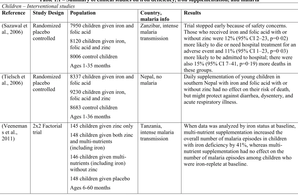
Table 1.1 – Summary of clinical studies on iron deficiency, iron supplementation, and malaria Children – Interventional studies
Reference Study Design Population Country,
malaria info Results (Sazawal et al., 2006) Randomized placebo controlled
7950 children given iron and folic acid
8120 children given iron, folic acid and zinc 8006 control children Ages 1-35 months
Zanzibar, intense malaria
transmission
Trial stopped early because of safety concerns. Those who received iron and folic acid with or without zinc were 12% (95% CI 2–23, p=0·02) more likely to die or need hospital treatment for an adverse event and 11% (95% CI 1–23, p=0·03) more likely to be admitted to hospital; there were also 15% (95% CI 7–41, p=0·19) more deaths in these groups. (Tielsch et al., 2006) Randomized placebo controlled
8337 children given iron and folic acid
9230 children given iron, folic acid and zinc 8683 control children Ages 1-36 months
Nepal, no malaria
Daily supplementation of young children in southern Nepal with iron and folic acid with or without zinc had no effect on their risk of death, but might protect against diarrhea, dysentery, and acute respiratory illness.
(Veeneman s et al., 2011)
2x2 Factorial trial
145 children given zinc only 148 children given both zinc and multi-nutrients
(including iron)
146 children given multi-nutrients (including iron) without zinc
148 children given placebo Ages 6-60 months
Tanzania, intense malaria transmission
When data was analyzed by iron status at baseline, multi-nutrient supplementation increased the overall number of malaria episodes in children with iron deficiency by 41%, whereas multi-nutrient supplementation had no effect on the number of malaria episodes among children who were iron-replete at baseline.
(Zlotkin et
al., 2013) Cluster randomized, double blind
967 children given
micronutrient powder with iron
991 children given micronutrient powder without iron
Ages 6-35 months
Ghana, intense malaria
transmission Insecticide treated bed nets provided at enrollment
Malaria incidence was significantly lower in the iron group compared with the no iron group during the intervention period (risk ratio [RR], 0.87; 95% CI, 0.78-0.96). In secondary analyses, these differences were no longer statistically significant after adjusting for baseline iron deficiency and anemia status overall (RR, 0.87; 95% CI, 0.75-1.01)
Subgroup analysis of 704 children who had anemia at baseline and for whom additional blood samples were obtained at the end of the
intervention period found only a small mean increase in hemoglobin in the iron group (mean change of 0.08 g/dL measured), indicating that the micronutrient powder had limited efficacy in this trial.
(Esan et al.,
2013) 2-arm, double-blind, randomized
100 children received multivitamins plus iron 96 children received multivitamins alone HIV infected children aged 6-59 months with moderate anemia (Hgb=7.0-9.9 g/dL); 3 months of treatment, 6 months follow up
Malawi, intense malaria
transmission
Children who received iron had a better CD4 percentage response at 3 months, but an increased incidence of malaria at 6 months (incidence rate, 120.2 vs. 71.7; adjusted incidence rate ratio [aIRR], 1.81 [95% CI, 1.04-3.16]; p = .04), especially during the first 3 months (incidence rate, 78.1 vs. 36.0; aIRR, 2.68 [95% CI, 1.08-6.63]; p = .03).
22 Children – Observational Studies
Reference Study Design Population Country,
malaria info Results (Nyakeriga et al., 2004) 2 Cross sectional studies Study 1: Iron-replete (n=95) Iron-deficient (n=78) Study 2: Iron-replete (n=104) Iron-deficient (n=91) Ages 8 months-8 years
Kenya, intense malaria
transmission
Incidence of clinical malaria was significantly lower among children with iron deficiency anemia (incidence-rate ratio [IRR], 0.70; 95% confidence interval [CI], 0.51-0.99; P<.05).
(Gwamaka et al., 2012)
Longitudinal 785 children monitored for 3
years Tanzania, intense malaria transmission
Iron deficiency anemia at routine, well-child visits significantly decreased the odds of subsequent parasitemia (23% decrease, p < .001) and subsequent severe malaria (38% decrease, p = .04). Iron deficiency anemia was also associated with 60% lower all-cause mortality (p = .04) and 66% lower malaria-associated mortality (p = .11). (Jonker et
al., 2012)
Longitudinal 727 children monitored for 1 year
Malawi, intense malaria
transmission
Children with iron deficiency anemia at baseline had a lower incidence of malaria parasitemia and clinical malaria during a year of follow-up; adjusted hazard ratios 0.55 (95% CI: 0.41-0.74) and 0.49 (95% CI: 0.33-0.73), respectively.
Pregnant women - observational
Reference Study Design Population Country,
malaria info
Results (Kabyemel
a et al., 2008)
Cross sectional
445 pregnant women (120 primigravidae, 112
secundigravidae, and 213 multigravidae)
Tanzania, intense malaria transmission
Iron deficiency decreased the risk of placental malaria.
(Senga et al., 2011)
Case-Control Pregnant women
(112 cases with placental malaria, 110 controls with no evidence of placental
infection)
Malawi, intense malaria
transmission
Iron deficiency decreased risk of acute, chronic and past placental malaria. The association was greater in the multigravidae group.
24
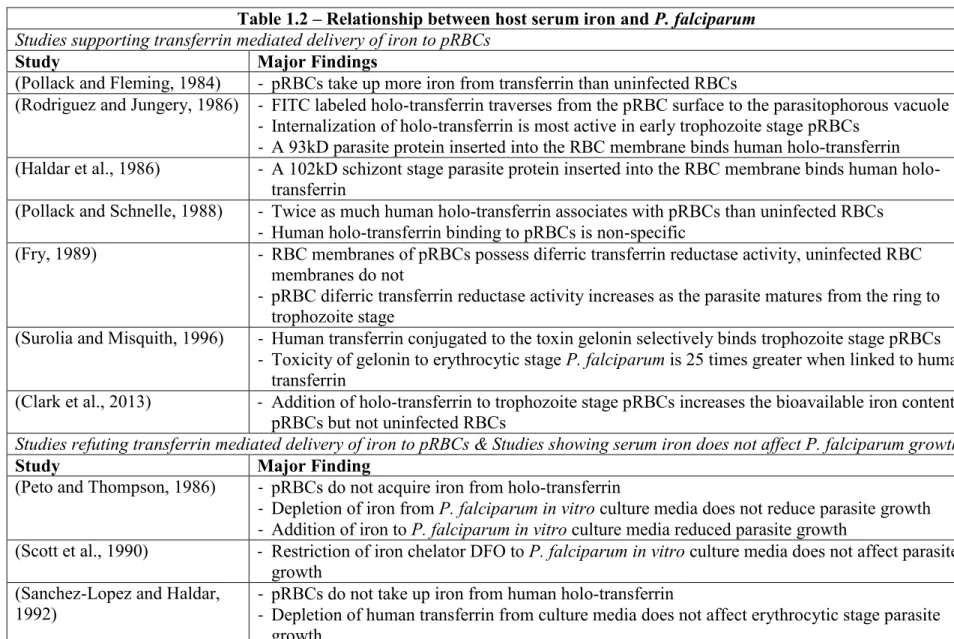
Table 1.2 – Relationship between host serum iron and P. falciparum
Studies supporting transferrin mediated delivery of iron to pRBCs
Study Major Findings
(Pollack and Fleming, 1984) - pRBCs take up more iron from transferrin than uninfected RBCs
(Rodriguez and Jungery, 1986) - FITC labeled holo-transferrin traverses from the pRBC surface to the parasitophorous vacuole - Internalization of holo-transferrin is most active in early trophozoite stage pRBCs
- A 93kD parasite protein inserted into the RBC membrane binds human holo-transferrin
(Haldar et al., 1986) - A 102kD schizont stage parasite protein inserted into the RBC membrane binds human holo-transferrin
(Pollack and Schnelle, 1988) - Twice as much human holo-transferrin associates with pRBCs than uninfected RBCs - Human holo-transferrin binding to pRBCs is non-specific
(Fry, 1989) - RBC membranes of pRBCs possess diferric transferrin reductase activity, uninfected RBC
membranes do not
- pRBC diferric transferrin reductase activity increases as the parasite matures from the ring to
trophozoite stage
(Surolia and Misquith, 1996) - Human transferrin conjugated to the toxin gelonin selectively binds trophozoite stage pRBCs
- Toxicity of gelonin to erythrocytic stage P. falciparum is 25 times greater when linked to human transferrin
(Clark et al., 2013) - Addition of holo-transferrin to trophozoite stage pRBCs increases the bioavailable iron content of
pRBCs but not uninfected RBCs
Studies refuting transferrin mediated delivery of iron to pRBCs & Studies showing serum iron does not affect P. falciparum growth
Study Major Finding
(Peto and Thompson, 1986) - pRBCs do not acquire iron from holo-transferrin
- Depletion of iron from P. falciparum in vitro culture media does not reduce parasite growth - Addition of iron to P. falciparum in vitro culture media reduced parasite growth
(Scott et al., 1990) - Restriction of iron chelator DFO to P. falciparum in vitro culture media does not affect parasite
growth (Sanchez-Lopez and Haldar,
1992) -- pRBCs do not take up iron from human holo-transferrin Depletion of human transferrin from culture media does not affect erythrocytic stage parasite growth
Studies supporting acquisition of NTBI by pRBCs
Study Major Finding
(Peto and Thompson, 1986) - pRBCs take up NTBI (Sanchez-Lopez and Haldar,
1992) - pRBCs take up of free, non-transferrin bound iron (NTBI), but not any more than uninfected RBCs
- pRBC NTBI acquisition is time, concentration, and temperature but not energy dependent
(Clark et al., 2013) - Addition of ferric citrate (NTBI) to trophozoite stage pRBCs increases the bioavailable iron
content of pRBCs but not uninfected RBCs
26
Table 1.3 – Relationship between RBC iron and P. falciparum
RBC hemoglobin
Study Major Findings
(Rudzinska et al., 1965) - P. falciparum metabolizes host RBC hemoglobin - P. falciparum inserts host heme into hemozoin
(Okada, 2009) - P. falciparum has a heme oxygenase homolog
(Sigala et al., 2012) - P. falciparum lacks both heme oxygenase activity and a canonical heme oxygenase pathway
(Loria et al., 1999) - Hydrogen peroxide degrades host heme under conditions that are analogous to the
microenvironment of the parasite food vacuole RBC ferritin – unknown
RBC bioavailable iron – unknown
Figure 1.1 – Host iron available to erythrocytic stage P. falciparum. During the erythrocytic stage of infection P. falciparum the parasite travels through the hosts vascular system within RBCs. Host iron immediately available to the parasite include serum and RBC iron. Serum iron ranges from 10-27 µM. Iron deficiency anemia is characterized by a significant decline in serum iron.
Transferrin bound iron is the predominant form of iron in the serum, though trace amounts of non-transferrin bound iron (NTBI) are present. In some pathologic conditions such as hemochromatosis, NTBI may be significantly greater. In the RBC iron is found within hemoglobin (20mM), ferritin (0.7nM), and as bioavailable iron (1-10µM). Iron deficiency anemia significantly reduces RBC iron, specifically hemoglobin iron. Shown in the figure are: P. falciparum (Pf), digestive vacuole (DV), parasite nucleus (N), and endothelial cell (EC).
28
Figure 1.2 – Impact of iron deficiency anemia and iron supplementation on erythropoietic rate and erythrocyte physiology and hypothesized effect on P. falciparum erythrocytic infection.
REFERENCES
Acharya, J., Punchard, N. A., Taylor, J. A., Thompson, R. P., and Pearson, T. C. (1991). Red cell lipid peroxidation and antioxidant enzymes in iron deficiency. Eur. J. Haematol. 47, 287–291.
Aguilar, R., Moraleda, C., Quintó, L., Renom, M., Mussacate, L., Macete, E., Aguilar, J. L., Alonso, P. L., and Menéndez, C. (2012). Challenges in the diagnosis of iron deficiency in children exposed to high prevalence of infections. PloS One 7, e50584.
Ariey, F., Witkowski, B., Amaratunga, C., Beghain, J., Langlois, A.-C., Khim, N., Kim, S., Duru, V., Bouchier, C., Ma, L., et al. (2014). A molecular marker of artemisinin-resistant Plasmodium falciparum malaria. Nature 505, 50–55.
Armitage, A. E., Eddowes, L. A., Gileadi, U., Cole, S., Spottiswoode, N., Selvakumar, T. A., Ho, L.-P., Townsend, A. R. M., and Drakesmith, H. (2011). Hepcidin regulation by innate immune and infectious stimuli. Blood 118, 4129–4139.
Brandão, M. M., Castro, M. de L. R. B., Fontes, A., Cesar, C. L., Costa, F. F., and Saad, S. T. O. (2009). Impaired red cell deformability in iron deficient subjects. Clin. Hemorheol. Microcirc. 43, 217–221.
Cabantchik, Z. I., Glickstein, H., Golenser, J., Loyevsky, M., and Tsafack, A. (1996). Iron chelators: mode of action as antimalarials. Acta Haematol. 95, 70–77.
Cabantchik, Z. I., Moody-Haupt, S., and Gordeuk, V. R. (1999). Iron chelators as anti-infectives; malaria as a paradigm. FEMS Immunol. Med. Microbiol. 26, 289–298.
Chang, K.-H., Tam, M., and Stevenson, M. M. (2004). Modulation of the course and outcome of blood-stage malaria by erythropoietin-induced reticulocytosis. J. Infect. Dis. 189, 735– 743.
Chotivanich, K., Udomsangpetch, R., Simpson, J. A., Newton, P., Pukrittayakamee, S., Looareesuwan, S., and White, N. J. (2000). Parasite multiplication potential and the severity of Falciparum malaria. J. Infect. Dis. 181, 1206–1209.
Chugh, M., Sundararaman, V., Kumar, S., Reddy, V. S., Siddiqui, W. A., Stuart, K. D., and Malhotra, P. (2013). Protein complex directs hemoglobin-to-hemozoin formation in Plasmodium falciparum. Proc. Natl. Acad. Sci. U. S. A. 110, 5392–5397.
30
Dhanasekaran, S., Chandra, N. R., Sagar, B. K. C., Rangarajan, P. N., and Padmanaban, G. (2004). δ-Aminolevulinic Acid Dehydratase from Plasmodium falciparum; Indigenous versus imported. J. Biol. Chem. 279, 6934–6942.
Doherty, C. P. (2007). Host-Pathogen Interactions: The Role of Iron. J. Nutr. 137, 1341–1344. Dondorp, A. M., Desakorn, V., Pongtavornpinyo, W., Sahassananda, D., Silamut, K.,
Chotivanich, K., Newton, P. N., Pitisuttithum, P., Smithyman, A. M., White, N. J., et al. (2005). Estimation of the total parasite biomass in acute falciparum malaria from plasma PfHRP2. PLoS Med. 2, e204.
Van Dooren, G. G., Stimmler, L. M., and McFadden, G. I. (2006). Metabolic maps and functions of the Plasmodium mitochondrion. FEMS Microbiol. Rev. 30, 596–630.
Esan, M. O., van Hensbroek, M. B., Nkhoma, E., Musicha, C., White, S. A., Ter Kuile, F. O., and Phiri, K. S. (2013). Iron supplementation in HIV-infected Malawian children with anemia: a double-blind, randomized controlled trial. Clin. Infect. Dis. Off. Publ. Infect. Dis. Soc. Am.
Fontecave, M., Nordlund, P., Eklund, H., and Reichard, P. (1992). The redox centers of ribonucleotide reductase of Escherichia coli. Adv. Enzymol. Relat. Areas Mol. Biol. 65, 147–183.
Francis, S. E., Sullivan, D. J., Jr, and Goldberg, D. E. (1997). Hemoglobin metabolism in the malaria parasite Plasmodium falciparum. Annu. Rev. Microbiol. 51, 97–123.
Fry, M. (1989). Diferric transferrin reductase in Plasmodium falciparum-infected erythrocytes. Biochem. Biophys. Res. Commun. 158, 469–473.
Fu, Y., Tilley, L., Kenny, S., and Klonis, N. (2010). Dual labeling with a far red probe permits analysis of growth and oxidative stress in P. falciparum-infected erythrocytes. Cytom. Part J. Int. Soc. Anal. Cytol. 77, 253–263.
Gabay, T., and Ginsburg, H. (1993). Hemoglobin denaturation and iron release in acidified red blood cell lysate--a possible source of iron for intraerythrocytic malaria parasites. Exp. Parasitol. 77, 261–272.
Gera, T., and Sachdev, H. P. S. (2002). Effect of iron supplementation on incidence of infectious illness in children: systematic review. BMJ 325, 1142.
Ginsburg, H., Famin, O., Zhang, J., and Krugliak, M. (1998). Inhibition of glutathione-dependent degradation of heme by chloroquine and amodiaquine as a possible basis for their
antimalarial mode of action. Biochem. Pharmacol. 56, 1305–1313.