Chemoenzymatic Synthesis of Heparan Sulfate
Renpeng Liu
A dissertation submitted to the faculty of the University of North Carolina at Chapel Hill in partial fulfillment of the requirements for the degree of Doctor of Philosophy in the Eshelman School of Pharmacy (Medicinal Chemistry and Natural Product)
Chapel Hill 2010
Approved by
Jian Liu, Ph.D.
Qisheng Zhang, Ph.D.
Harold Kohn, Ph.D.
Michael Jarstfer, Ph.D.
ABSTRACT
Renpeng Liu: Chemoenzymatic synthesis of heparan sulfate
(Under the direction of Jian Liu, Ph.D.)
Heparan sulfate (HS) participates in a variety of biological functions and has been
exploited for its ability to be utilized as a HS-based drug. Chemical synthesis of HS remains
extremely challenging. Previous research has proven the feasibility of using a HS
enzyme-based approach to synthesize HS structures with unique biological activities. Our central
hypothesis is that all subsequent modifications following N-sulfation during HS biosynthesis
are governed by the number and position of the GlcNS residue. In this dissertation, a
fluorous affinity tag-assisted chemoenzymatic synthesis technique has been developed to
build a HS octasaccharide library with defined N-sulfo glucosamine (GlcNS) positions. The
HS backbone was synthesized by heparosan biosynthetic enzymes. N-acetyl glucosaminyl
transferase from E.coli K5 (KfiA) was used to transfer either GlcNAc or GlcNTFA (N
-trifluoroacetylglucosamine) residues to the growing chain. Heparosan synthase from
pasteurella (PmHS2) was used to transfer the GlcUA residues. A selective
de-trifluoroacetylation was performed because under these conditions, the GlcNTFA is labile
and will be converted to glucosamine (GlcNH2) while the GlcNAc residue remains intact.
The resultant GlcNH2 is then converted to a GlcNS residue by N-sulfotransferase (NST). N
also prepared. Furthermore, we prepared oligosaccharide capable of binding to antithrombin
(AT), which correlates to HS anticoagulant activity. In this study, an AT-binding
dodecasaccharide was prepared and its structure was proven. The continuation of this
dissertation will allow us to not only investigate enzymatic approaches to synthesize
HS-based anticoagulant drugs, but also develop a general method for synthesizing structurally
defined HS oligosaccharides that could aid in the discovery of novel HS-based therapeutic
ACKNOWLEDGEMENTS
First and foremost, I would like to thank my advisor, Dr. Jian Liu, for his support,
enthusiasm, motivation, and guidance. He has been instrumental in my success as a Ph.D.
student and gives me confidence that I will carry with me throughout the rest of my academic
career. I would also like to thank the other members of my doctoral committee, Drs. Harold
Kohn, Qisheng Zhang, Marcey Waters, and Michael Jarstfer for their constructive advice.
My labmates, especially Dr. Yongmei Xu and Dr. Miao Chen, also deserve my
gratitude for always giving me guidance when called upon no matter how trivial the
problems were. I wish to extend my thanks to all my other labmates, Dr. Michael Duncan,
Dr. Ronald Copeland, Dr. Jinghua Chen, Dr. Ding Xu, Dr. Tanya Burch, Dr. Kai Li, Dr.
Juzhen Sheng, Dr. Xianxuan Zhou, Courtney Jones, Sherket Peterson, Elizabeth Pempe,
Heather Bethea, Ryan Bullis, Justin Roberts, Lan Yu, Xinan Lu and Truong Quang Pham.
They provided hands-on assistance and friendly discussion on my project which made me
feel like we are a big family.
Furthermore, I would like to say thank you to Dr. Arlene Bridges for her assistance in
MS and LC-MS, Dr. Robert Linhardt for providing me UDP-GlcNTFA in large quantities,
Dr. Qian Shi for her assistance in my preparations of fluorous-tagged disaccharides, as well
as Dr. Dan Cline and Dr. Ole Hindsgaul for their assistance in solid phase synthesis.
throughout my Ph.D. career. I also thank my parents, Chifu Liu and Tianyu Chang, and my
sister, Zhimin Liu for their support in my life, especially during difficult times. My parents-
in-law, Yuanjun Li and Meiying Ding also deserve my gratitude for their support during my
TABLE OF CONTENTS
Page
ABSTRACT...ii
ACKNOWLEDGEMENTS...iv
TABLE OF CONTENTS...vi
LIST OF TABLES...xii
LIST OF FIGURES ... xiii
ABBREVIATIONS ...xvi
Chapter I INTRODUCTION ...1
Structure of heparan sulfate ...1
Biological function of heparan sulfate...4
Anticoagulant activity...4
Assisting virus infection ...6
Stimulating cell proliferation...8
Inflammation...8
Tumor growth, metastasis and heparanase ...9
PF4 and thrombocytopenia ...12
Structure and activity relationship of HS...13
AT-binding domain ...13
HSV-1 gD binding domain...14
Biosynthesis of HS...16
Chain initiation ...17
Chain polymerization...18
NDST...19
C5-epimerase...21
2OST...22
6OST...22
3OST...23
Substrate recognition of 2OST and 3OSTs ...26
Biosynthesis of heparosan...27
Heparosan synthases from E.coli...28
Heparosan synthases from P. multocida...28
Chemical and enzymatic synthesis of HS...29
Chemical synthesis of AT-binding oligosaccharides ...29
Chemical synthesis of peptide/HS oligosaccharide conjugate mCD4-HS12 ...32
Enzymatic synthesis of biological active polysaccharides ...32
Enzymatic synthesis of biological active oligosaccharides ...37
Unnatural UDP donors ...41
Structural characterization of HS...42
Disaccharide analysis...42
Mass spectrometry...44
Statement of problems ...46
Chapter II MATERIALS AND METHODS ...49
Preparation of substrate acceptors and UDP-donors ...49
Preparation of heparosan from E. coli K5 ...49
Preparation of disaccharide acceptors ...50
Synthesis of [3H]-labeled UDP-N-acetylglucosamine...51
Preparation of UDP-N-trifluoroacetylglucosamine ...52
Preparation of biosynthetic enzymes ...53
Expression of HS biosynthetic enzymes in E. coli...53
Expression of KfiA ...54
Expression of PmHS2...55
Expression of NST...55
Expression of C5-epi ...57
Expression of 2OST...57
Expression of 6OST1 and 6OST3 ...58
Expression of 3OST1...59
Expression of GlmU ...60
Preparation of N-sulfo tagged octasaccharide library...60
Preparation of tagged disaccharide...60
Preparation of fluorous tagged octasaccharide backbones ...62
Selective de-N-trifluoroacetylation of GlcNTFA units ...62
Preparation of N-sulfo tagged octasaccharides 3-6...62
Preparation of HS untagged oligosaccharide backbones...63
Preparation of N-sulfo decasaccharide 11, undecasaccharide 12 and dodecasaccharide 13...64
Preparation of N-sulfo-6-O-sulfo decasaccharide 14 and dodecasaccharide 15...65
Preparation of N-sulfo-3, 6-O-sulfo decasaccharide 16 and dodecasaccharide 17...65
Structural analysis...66
Nitrous acid degradation of untagged oligosaccharides ...66
HPLC analysis ...66
Microdialysis of oligosaccharides ...67
Liquid chromatography linked mass spectrometry analysis...67
Mass spectrometry analysis ...68
Activity analysis...69
Antithrombin binding assay...69
Determination of the binding affinity of oligosaccharides to AT...69
Chapter III SYNTHESIS OF N-SULFO OCTASACCHARIDE LIBRARY...70
Introduction...70
Optimization of fluorous tagged saccharides...72
Preparation of tagged glucose...72
Removal of fluorous tag by catalytic hydrogenolysis ...73
Optimization of fluorous tag...74
Preparation of tagged disaccharide...75
Susceptibilities of tagged disaccharide to KfiA and pmHS2...76
Susceptibilities of UDP-GlcNTFA as the donor to KfiA ...80
Preparation of N-sulfo hexasaccharide ...82
Selective de-N-trifluoroacetylation of tagged hexasaccharide ...82
Preparation of N-sulfo tagged hexasaccharide ...84
Optimization of mass spectrometry...84
Preparation of N-sulfo octasaccharide library...86
Preparation of HS octasaccharide library ...86
Characterization of N-sulfo octasaccharide library ...88
Conclusion ...92
Chapter IV SYNTHESIS OF 6-O-SULFO OLIGOSACCHARIDES...94
Introduction...94
N-sulfo-6-O-sulfo oligosaccharides...94
Preparation of N-sulfo-6-O-sulfo hexasaccharides...94
Characterization of N-sulfo-6-O-sulfo hexasaccharides...96
Preparation of N-sulfo-6-O-sulfo octasaccharide ...99
Fluorous tag does not interact with AT ...101
Conclusion ...102
Chapter V SYNTHESIS OF AT-BINDING OLIGOSACCHARIDES...103
Introduction...103
Preparation of AT-binding oligosaccharides ...104
Preparation of N-sulfo oligosaccharides...105
Preparation of N-sulfo-6-O-sulfo deca and dodecasaccharide ...107
Preparation of oligosaccharides binding to AT ...110
The AT-binding affinities ...116
Conclusion ...117
Chapter VI SOLID PHASE SYNTHESIS OF HS ...119
Introduction...119
Solid phase synthesis ...121
Preparation of Resins 1 and 2...121
Preparation of cleavable linker 33 and spacer 34...123
Preparation of cleavable resin 3...125
Installation of spacer...125
Immobilization of disaccharide to the solid support...127
Solid phase KfiA assay...128
Conclusion ...128
Chapter VII CONCLUSION...130
APPENDIX...133
LIST OF TABLES
Table
1. List of HS biosynthetic enzymes expressed in E. coli ………56
2. Binding affinity of fluorous tag………75
3. AT binding assay for 30, 32 and [35S] PAPS………101
4. List of the synthesized oligosaccharide backbones………105
5. Summary of Disaccharide analysis of decasaccharide 14 and dodecasaccharide 15 ………108
LIST OF FIGURES
Figure
1. Disaccharide repeating units of heparan sulfate and heparin………2
2. Coagulation cascade………5
3. Substrate specificity and inhibitor for heparanase………11
4. The structure of AT-binding pentasaccharide………14
5. Chemical structures of gD, FGF-2 and FGF-1 binding sites………15
6. HS biosynthetic pathway………19
7. The HS chain initiation and elongation………20
8. Proposed mechanism of C5-epimerase………21
9. Substrate specificity of 6OSTs………23
10. Substrate specificity of 3OSTs………25
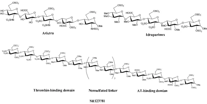
11. The chemical structure of Arixtra, Idraparinux and SR123781………30
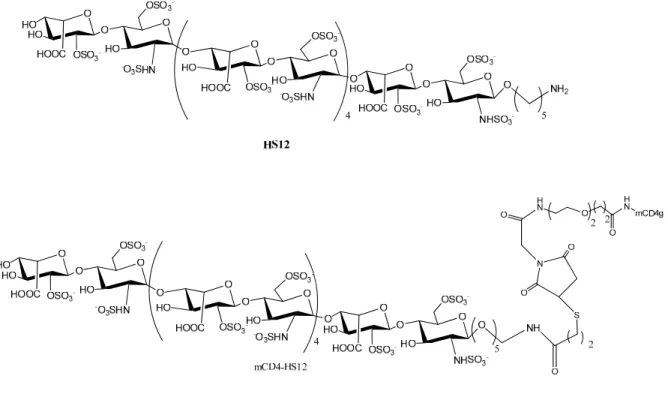
12. The chemical structures of HS12, CD4-HS12 and mCD4g………32
13. PAPS regeneration system………34
14. Enzymatic synthesis of anticoagulant CDSNS heparin………35
15. Enzymatic synthesis of Recomparin……….37
16. Enzymatic synthesis of AT-binding pentasaccharide………39
17.Preparation of 3-O-sulfated octasaccharide inhibiting the entry of HSV-1…………40
18. Heparin lyases degradation reaction………43
19. High and low pH degradation of HS………44
21. Enzymatic synthesis of UDP-GlcN[3H]Ac………52
22. Chemoenzymatic synthesis of UDP-GlcNTFA………53
23. Synthesis and characterization of tagged glucose 18………73
24: Catalytic hydrogenolysis to remove the fluorous tag………74
25. Synthesis and characterization of tagged disaccharide 7………76
26. Tagged oligosaccharides are substrates of KfiA and pmHS2………78
27. Structural characterization of hexasaccharide 23………79
28. Structural characterization of pentasaccharide 24………81
29. Structural characterization of hexasaccharide 25 and 26………83
30. Structural characterization of N-sulfated hexasaccharide 27………85
31. Scheme for the synthesis of octasaccharide library 3-6………87
32. Structural characterization of octasaccharide 3………89
33. Structural characterization of octasaccharide 4………90
34. Structural characterization of octasaccharide 5………91
35. Structural characterization of octasaccharide 6………92
36. Preparation of N-sulfo, 6-O-sulfo hexasaccharide 29 and 30………95
37. HPLC analysis of 6-O-sulfo hexasaccharide 29 and 30………96
38. Determination of the structure for 6-O-sulfo hexasaccharide 29……….98
39. Structural characterization of hexasaccharide 30………99
40. Structural characterization of octasaccharide 31………100
41. Structure of nonasaccharide 32……….……….101
42. Scheme for the synthesis of oligosaccharides………104
44. Structural characterization of decasaccharide 14 and dodecasaccharide 15………109
45. Preparation of 3-O-sulfo decasaccharide 16………111
46. Determination of the structures of peak I for decasaccharide 16………112
47. Determination of the structures of peak II for decasaccharide 16………113
48. Preparation of 3-O-sulfo dodecasaccharide 17………114
49. Determination of the structures of peak I for dodecasaccharide 17………115
50. Determination of the structures of peak II for dodecasaccharide 17………116
51. Solid phase oligosaccharide synthesis………120
52. Preparation of hydroxylamine resin 1………122
53. Preparation of hydrazide resin 2………123
54. Total synthesis of cleavable Linker 33 and Spacer 34………124
55. Preparation of cleavable hydroxylamine resin 3………126
56. Installation of spacer 34………127
ABBREVIATIONS
2OST uronosyl 2-O-sulfotransferase
3OST glycosaminyl 3-O-sulfotransferase
6OST glycosaminyl 6-O-sulfotranferase
ADP adenosine diphosphate
AMP adenosine monophosphate
AnMan 2, 5-anhydromannitol
AST-IV arylsulfotransferase-IV
AT antithrombin
ATP adenosine triphosphate
C5-epi glucoronyl C5-epimerase
calcd calculated
CS chondroitin sulfate
DS dermatan sulfate
ECM extracellular matrix
equiv. equivalent
ESI electrospray mass spectroscopy
FGF(R) fibroblast growth factor(receptor)
GAG glycosaminoglycan
Gal galactose
GalNAc N-acetylated galactosamine
GalTI/II galactosyltransferase I/II
gC herpes simplex virus -1 glycoprotein C
gD herpes simplex virus -1 glycoprotein D
Glc glucose
GlcNS N-sulfated glucosamine
GlcNH2 N-unsubstituted glucosamine
GlcNAc N-acetylatedglucosamine
GlcNAcTI/II HS GlcNAc transferase I/II
GlcUA glucuronic acid
GlcUATI/II HS glucuronosyltransferase
HA hyaluronic acid
HIT heparin induced thrombocytopenia
HCV hepatitis virus C
HIV human immunodeficiency virus
HS heparan sulfate
HSPG heparan sulfate proteoglycan
HSV herpes simplex virus
IdoUA iduronic acid
IdoUA2S 2-O-sulfated iduronic acid
KS keratan sulfate
LB luria-bertani
MALDI matrix-assisted laser desorption/ionization
MS mass spectrometry
PAP 3’-phosphoadenosine 5’-phosphate
PAPS 3’-phosphoadenosine 5’-phosphosulfate
PNP p-nitrophenol
PNPS p-nitrophenyl sulfate
PF4 platelet factor 4
ST sulfotransferase
UDP uridine diphosphate
UTP uridine triphosphate
VEGF(R) vascular endothelial growth factor (receptor)
XT xylosyltransferase
Xyl xylose
ΔUA deoxy-α-L-threo-hex-enopyranosyluronic acid
k kilo
L liter(s)
M moles per liter
m/z mass to charge ratio (MS)
Chapter I. INTRODUCTION
Section 1. Structure of heparan sulfate
Heparan sulfate (HS) is a highly sulfated polysaccharide that represents a unique class
of natural products. Heparin is a special type of HS synthesized within mast cells. It was
discovered in 1918 (1) and has been widely used as an anticoagulant drug for decades.
HS/heparin consists of 50 to 100 disaccharide units carrying sulfo groups. HS has an average
molecular weight of about 30 kDa ranging from 5 to 50 kDa while heparin has an average
molecular weight of 12 kDa ranging from 5-40 kDa (2-4). HS/heparin belongs to the
glycosaminoglycan family. Depending on the structures of the disaccharide repeating units,
glycosaminoglycans are classified as HS, heparin, chondroitin sulfate (CS), dermatan sulfate
(DS), keratan sulfate (KS) and hyaluronic acid (HA). HS and heparin consist of highly
sulfated repeating disaccharide units of 1→4 linked glucosamine (GlcN) and glucuronic
(GlcUA)/iduronic acid (IdoUA) (Figure 1). CS and DS consist of sulfated repeating
disaccharide units of 1→3 linked N-acetylgalactosamine (GalNAc) and GlcUA/IdoUA, and
KS consists of sulfated repeating disaccharide units of 1→3 linked galactose (Gal) and N
-acetylglucosamine (GlcNAc). HA consists of repeating disaccharide units of 1→3-linked
Fig 1. Disaccharide repeating units of HS and heparin. Sulfation (R= -SO3) at Carbon 6 (known as 6-O
-sulfated glucosamine, GlcN6S) of glucosamine is common. Sulfation (R’= -SO3) at Carbon 2 of iduronic
acid (known as 2-O-sulfated iduronic acid, IdoUA2S) is common. Sulfation at Carbon 3 of glucosamine
(known as 3-O-sulfated glucosamine, GlcN3S) is rare. Both N-acetylated (R’’=acetyl, GlcNAc) and N
-sulfated (R’’= -SO3, GlcNS) are common. N-unsubstituted glucosamine (R’’= -H, GlcNH2) is a low
abundant component. IdoUA (2S) is presented in both 1C4 and 2S0 conformation. Both conformations are
HS is widely expressed on the cell surface and within the extracellular matrix on HS
proteoglycans (HSPGs, which contain a core protein and polysaccharide side chains).
HSPGs are involved in numerous biological processes, including blood coagulation, wound
healing, embryonic development, and regulation of tumor growth, as well as assisting viral
and bacterial infections (5-13). HS polysaccharides play an essential role in HSPGs
functions. The wide ranges of biological functions of HS attract considerable interest in
exploiting HS-based anticoagulant, antiviral and anticancer drugs.
The majority of glucosamine residues are either N-acetylated (GlcNAc) or N-sulfated
(GlcNS) (14-16). However, up to 7% of the glucosamine residues in HS are present as N
-unsubstituted glucosamine (GlcNH2) which may play an important biological role (17). For
example, it is known that the GlcNH2 unit is involved in the binding of herpes simplex virus
1’s (HSV-1) glycoprotein D (gD) (11, 18). The 6-O-sulfo glucosamine (GlcN6S) and 2-O
-sulfo iduronic acid (IdoUA2S) units are common sulfated monosaccharides, and these units
play critical roles in binding to fibroblast growth factors (FGFs), fibroblast growth factor
receptors (FGFRs) (19) and platelet factor 4 (PF4) (20). 3-O-sulfo glucosamine is a rare
component of HS and plays an important role in binding to antithrombin (AT) (3) as well as
binding to HSV-1 gD (21). The distribution of different sulfo groups determines the
biological function of HS. Although the overall structures of heparin and HS are similar;
heparin has a higher content of IdoUA and more sulfo groups per disaccharide unit (3, 4).
HS contains 0.6 sulfo groups per disaccharide unit, and 40% uronic acid of HS is iduronic,
while heparin contains 2.6 sulfo groups per disaccharide unit and 90% uronic acid of heparin
Section 2. Biological function of heparan sulfate
Anticoagulant activity
Heparin has remained the main anticoagulant drug on the market since it was
introduced in the 1930s (2). The coagulation cascade consists of a series of proteases and
their precursors. The anticoagulant action of heparin is due to the activation of the serine
protease inhibitor AT. The AT/heparin complex inhibits the activity of factor Xa and
thrombin (or named as factor IIa), two key proteases in controlling the blood coagulation
cascade (Figure 2). The advantages of heparin as an anticoagulant drug include the
following: heparin is the only drug can inhibit both factor Xa and thrombin activities, heparin
has a fast anticoagulant response, and excessive anticoagulant activity can be reversed by
protamine (3).
Heparin is an exclusive product of mast cells, and is released during degranulation of
mast cells. Therefore, HS, rather than heparin, is considered to be the “natural anticoagulant”
in humans. Pharmaceutical grade heparin is derived from slaughtered domesticated animal
tissues such as the porcine intestines. The purity of heparin heavily depends on the quality of
the pig resource (13). It is estimated that 30-40 tons of heparin are prepared each year
worldwide from 400 to 700 million pigs (22). The majority is from China.
Heparin’s complex structure causes many of the unwanted side effects of heparin,
including hemorrhaging and heparin-induced thrombocytopenia (HIT) (23). Furthermore,
since heparin supply chain is long, it could be contaminated by human factors. Most recently,
a contaminated heparin drug made by Scientific Protein Labs (SPL) caused severe side
effects including life-threatening anaphylactic reactions resulting in abnormally low blood
worldwide have suffered the severe reactions linked to this contaminated heparin and this
accident has led to 81 deaths. This accident resulted in a major heparin recall in the USA,
countries of the European Union and Japan. Oversulfated chondroitin sulfate (OSCS) was
identified as the contaminant in heparin (25, 26), and it is believed to have been intensionally
added for illegal profits. This tragic event suggested that the heparin supply chain is
vulnerable. Thus, a synthetic heparin that can be manufactured in a confined facility remains
a high priority. The cofined facility can increase the purity and decrease the chances of
contamination.
Intrinsic pathway activation Extrinsic pathway activation
II IIa
Antithrombin Inhibit
Inhibit
(thrombin)
Fibrinogen Fibrin
Clot
XII XIIa
VII VIIa
XI XIa
IX IXa
X
Tissue factor Trauma
Trauma
X Xa
Heparin
Fig 2. Coagulation cascade. The anticoagulant action of heparin is due to the activation of the serine
Assisting virus infection
In order to establish an infection, a virus must first make close contact with the host
cell. Since HSPGs are widely expressed on the human cell surface, it is not surprising that
HS could function as docking and/or receptor sites for different viruses to attach to and/or
invade host cell. HSV-1, human immunodeficiency virus (HIV) and hepatitis virus C (HCV)
are known to utilize HS to establish the infection (11). Therefore, the mechanism of
HS-involvement in viral interaction with host cells has attracted considerable interest from both
scientific and pharmaceutical communities.
The involvements of HS in HSV-1 and HIV infections are the most thoroughly
studied. HSV-1 is a widespread virus which can cause facial mucocutanous lesions, keratitis,
and occasionally, life-threatening encephalitis. The involvement of cell surface HS in HSV-1
infection was discovered in 1989 (27). It is believed that HSV-1 first attaches to target cells
through an interaction between its viral envelope glycoprotein C (gC), or in some cases of
glycoprotein B (gB) and HS on the surface of host cells (11, 21). Structural analysis of HS
involvement in gC binding indicated that a saccharide sequence containing IdoUA2S and
GlcNS(or Ac)6S is required for this interaction. Once contact is established, the binding of
viral envelope protein gD and the cell surface entry receptors triggers fusion between viral
particles and their target cells, permitting the viral particles to penetrate the target cell
membrane (21). Studies indicated that the 3-O-sulfo glucosamine of HS plays an important
role in binding to HSV-1 gD. It is important to note that the 3OST1-modified HS does not
bind to gD while 3OST3 and 3OST5-modified HS binds to gD, suggesting that 3-O-sulfated
Several lines of research revealed that HS may play multiple roles in assisting HIV
infection (11). First, it has been known for a long time that HS interacts with HIV envelop’s
glycoprotein gp120, the key viral protein for cell entry, and that this interaction facilitates the
binding of HIV to host cells (31). HS binding to gp120 has been localized on the V3 loop. A
V3 loop is a major epitope of gp120 which carriess variable positive charges. Although the
V3 loop is not involved in the initial gp120-CD4 binding, it plays an essential role in
subsequent steps that lead to membrane fusion for viral entry (4, 32 and 33). Recent studies
showed other domains, including the cryptic and CD4-induced coreceptor binding surface,
contribute to gp120-HS binding (34). Structural selectivity studies of heparin involved in the
binding of gp120 and CD4 have been carried out. These studies showed O-sulfation,
especially 6-O-sulfation, and N-substitution (N-sulfation or acetylation) are both essential for
binding. Also, the minimum size of gp-120 binding heparin is a decasaccharide (4, 35). In
addition to the role of facilitating initial binding of HIV to the target cell, HS promotes the
spreading of HIV. A recent study shows HIV relies on HS to attach to the surface of sperm
and these HIV viruses are transmitted to physiological target cells of HIV, such as dendritic
cells, T cells and macrophages (36). Finally, HS facilitates the internalization of HIV
transactivator protein, Tat, an etiologic agent of AIDS. Tat is released from HIV infected
cells, and internalized Tat causes damage to the cells and tissues (11, 37-39). It is found that
Tat binds to heparin or HS, and minimum size of the Tat binding domain is a hexasaccharide.
The binding affinity increases with increasing oligosaccharide size, and about eighteen
saccharide residues are required to match the full affinity of heparin (4, 11 and 40).
Therefore, HS and heparin could be a potential multi-target agent in the therapy and
Stimulating cell proliferation
The FGFs are a family of growth factors involved in angiogenesis, wound healing, and
embryonic development (4, 19). A total of 22 different isoforms of FGF have been reported
(41). FGFs are key players in proliferation and differentiation of a wide variety of cells and
tissues. The FGF signaling pathway is initiated by the binding of FGF to its receptor on the
cell surface, triggering the dimerization of FGFR and downstream phosphorylation/activation
of enzymes (42). It is well known that FGFs are heparin-binding proteins and interactions
with cell-surface associated HS proteoglycans have been shown to be essential for FGF
signal transduction. The FGF, FGFR and HS interaction has been elucidated by crystal
structures. The crystal structure of a dimeric ternary complex of 2:2:2 FGF-2, a heparin
decasaccharide and FGFR-1 demonstrated that heparin makes contact with both FGF-2 and
FGFR-1 in each FGF-FGFR complex to stabilize FGF-FGFR binding. The FGFR-1 of the
adjacent FGF-FGFR complex also makes contact with heparin to promote FGFR
dimerization (4, 43). In contrast, the crystal structure of a complex of 2:2:1 FGF-1, FGFR-2
and a heparin decasacchride indicated a different dimerization pattern. In this complex, the
heparin molecule links to two FGF-1 ligands into a dimer. The 2:1 FGF-1 heparin complex
acts as a bridge between the two FGFR-2 ligands (4, 44).
Inflammation
Inflammation is a complex biological process in response of vascular tissues reaction
to harmful stimuli such as irritants, pathogen infection and tissue injury. Inflammation is a
healing process for the tissue. Inflammation is a multi-step process involving chemokine
generation at the infected tissue (45). Chemokines are a group of small proteins with a
variety of biological functions including selective recruitment and activation of cells during
inflammation. Chemokines migrate into the luminal side of the endothelial cells and bind to
leukocytes within the blood vessel. This leukocyte-chemokine interaction triggers leukocyte
extravasation and migration towards the infected tissue. The roles of heparin and HS
involved in regulating inflammatory responses have been noticed recently (46). It has been
shown that chemokines are heparin binding proteins (47); the HS and chemokine interactions
have been determined to be essential for the presence of chemokines on the luminal surface
of endothelial cells (47). HS also binds to cell adhesion molecules (such as L- and
P-selectins), as well as, growth factors and growth factor receptors to regulate leukocyte
migration through the blood vessel wall (48, 49). Although the mechanism is not fully
understood, it is generally accepted that the sulfation patterns of HS direct the interactions of
L-selectin and chemokines (50, 51). For example, the heparin carrying 6-O-sulfo groups
exhibit anti-inflammatory effects by blocking the binding of HS and L- and P-selectins (52).
Therefore, developing HS-based anti-inflammatory agents attracts a lot of interest.
Tumor growth, metastasis and heparanase
Clinical studies have clearly demonstrated the benefits of treating cancer patients with
heparin, especially LMW heparin. Tumor cells release procoagulant molecules to activate
factors X and VII. The interaction between immune and malignant cells activates platelets,
factor XII, and factor X, leading to thrombin production and thrombosis. Thus, the
studies also indicate that heparin’s antiangiogenic and antimetastatic properties may also
contribute to its anticancer activity. For example, studies have found that the administration
of LMW heparin has improved the survival rate of the patients with small cell lung cancer
from 29.5% to 51.3%, while another commonly used anticoagulant drug, warfarin, did not
exhibit the anticancer activity (22, 53 and 54). However, clinical evidence for heparin use as
an antimetastatic and antiangiogenesis agent is not conclusive and anticoagulant side effect
limits the anticancer application of HS (55).
Heparanase is an endo-β-D glucuronidase that cleaves HS into fragments ranging from
10 to 20 saccharide units (56-59) (Figure 3). Heparanase is highly conserved and so far only
a single form has been found. Heparanase is involved in several physiological and
pathological processes, such as wound healing, embryonic development and HS cleavage.
Heparanase is attributed to cancer by two aspects: firstly, heparanase degrades the HS in the
extracellular matrix, facilitating cell invasion and metastasis; secondly, by degrading HS,
heparanase is involved in stimulating angiogenesis through the release of HS-bound growth
factorssuch as vascular endothelial growth factor (VEGF) and FGFs. Therefore, the role of
heparanase in tumorigenesis makes it an attractive anti-cancer target (60). Progen
Pharmaceuticals has developed phosphomannopentose sulfate (PI-88), a mixture of highly
sulfated monophosphorylated mannose oligosaccharides to inhibit heparanase activity
(Figure 3). PI-88 also competes with HS for binding to growth factors such as FGF-1 and
FGF-2, consequently reducing their ability to stimulate tumor angiogenesis. Unfortunately,
although phase II studies of PI-88 in patients with numerous cancers showed promising
reasons such as modest results to treat malignancies, lack of a global distributor, and its
immune-mediated thrombocytopenia (22, 61).
Fig 3. Substrate specificity and inhibitor for heparanase. Panel A is the substrate specificity of heparanase.
Panel B is the Structure of PI-88, the inhibitor of heparanase.
Although the molecular mechanism for the linking cancer and HS is not fully
understood, it is suspected that the saccharide structures exhibiting anticancer activity are
Because the anticoagulant properties of HS/heparin could limit dosing for its anticancer
purposes, synthesis of non-anticoagulant HS with antiangiogenic and antimetastatic
properties attracts considerable interest in exploiting HS or HS-like molecules for the
development of anticancer drugs.
PF4 and thrombocytopenia
PF4, a polypeptide with 70 amino acids, belongs to the chemokine family (4). PF4 is
released from platelets and is believed to associate with inflammation and wound healing
which is a result of its ability to neutralize the activity of heparin and HS (4). However,
when heparin binds to PF4, it can induce heparin-induced thrombocytopenia (HIT), a serious
side effect of heparin. HIT occurs in approximately 3% of patients receiving unfractionated
heparin and about 0.2% in those patients receiving low molecular weight heparin (63). HIT
is an immunologically induced loss of platelets. In HIT, the immune system forms
antibodies against heparin when heparin is bound to PF4. The antibodies form a complex
with heparin and PF4 in the bloodstream. The tail of the antibody then binds to the FcγIIa
receptor, a protein on the surface of the platelet. This results in platelet activation and the
formation of platelet microparticles, which initiate the formation of blood clots; the platelet
count falls as a result of this clotting (64, 65). It is believed that PF4 recognizes a long
sulfated domain in HS and the IdoUA2S residue in HS has been identified to be important for
binding to PF4 (4, 66 and 67). The optimal size of oligosaccharide needed to form a
complex with PF4 is about a hexadecasaccharide, although the minimal size for PF4 binding
conformation on the backbone of heparin (4, 68). Based on this observation, it is viable to
design anticoagulant heparin derived oligosaccharides without binding to PF4. For example,
Petitou and colleagues have successfully synthesized anticoagulant oligosaccharide without
binding to PF4 (71).
Section 3. Structure and activity relationship of HS
AT-binding domain
Only a fraction of HS binds to AT and exhibits anticoagulant activity. About 1-10%
of HS binds to AT while about 30% of heparin binds to AT, suggesting that AT recognizes a
special sequence of HS (3). Studies have revealed that the anticoagulant drug heparin and
HS contain a structurally defined AT-binding pentasaccharide sequence with the structure of
–GlcNS (or Ac) 6S-GlcUA-GlcNS3S6S-IdoUA2S-GlcNS6S- (72-74) (Figure 4). The
AT-binding site is the essential motif for the anticoagulant activity of heparin and HS. Binding
to this pentasaccharide unit will trigger a conformational change of AT and accelerate the
inhibition of factor Xa. However, the pentasaccharide unit only inhibits the activity of factor
Xa. The structural requirements needed for HS binding to AT were proven via chemically
synthesized pentasaccharides with various combinations of sulfo groups and carboxyl groups.
The 3-O-sulfation of a glucosamine residue (3-O-sulfation is circled) is the critical
modification to generate the AT binding site. Removal of 3-O-sulfo group decreased the
binding affinity to AT by nearly 20, 000 fold (3).
A larger oligosaccharide is required to inhibit both anti-Xa and anti-thrombin activity.
thrombin-binding domain. Heparin/HS acts as a template to facilitate the AT-mediated inhibition of
thrombin. Petitou et al proved that the order of the three domains is essential for thrombin
inhibition, namely, the AT-binding domain must be positioned at the reducing end of the
linker region and the thrombin-binding domain must be located at the nonreducing end of the
linker region. The linker region forms a bridge that has no interaction with either of the
proteins. Compare to the AT-binding domain, the interaction of thrombin and the
thrombin-binding domain is less specific. The minimum size to inhibit both factor Xa and thrombin is
found to be a pentadeca-saccharide (71).
Fig 4. The structure of the AT-binding pentasaccharide. The 3-O-sulfation of the glucosamine residue
(3-O-sulfo group is circled) is the critical modification to generate the AT binding site. GlcNS(Ac)6S,
GlcUA, GlcNS3S6S, IdoUA2S and GlcNS6S represent the abbreviation of the individual monosaccharide residues.
HSV-1 gD binding domain
The minimum size of HS that will bind to gD is found to be an octasaccharide (18,
21). An octasaccharide binding to gD was isolated from a 3OST3-modified octasaccharide
octasaccharide was determined to be ΔUA-GlcNS-IdoUA2S-GlcNAc-GlcUA2S (or
IdoUA2S)-GlcNS-IdoUA2S-GlcN3S6S (∆UA is deoxy-α-L-threo-hex-enopyranosyluronic
acid) (Figure 5). It is very interesting to find that a rare GlcNH2 unit is involved in the
binding of HSV-1 (gD), and that the binding octasaccharide contains an unique GlcN3S6S
linked to a IdoUA2S at the non-reducing end (18).
FGF binding domain
HS had been shown to enhance the formation of FGF-FGFR complexes and stabilize
FGFR oligomers. The most well studied FGFs are FGF-1 and FGF-2. Studies revealed that
the minimal heparin structure binding to FGF-2 is a pentasaccharide of
–UA-GlcNS-UA-GlcNS-IdoUA2S-, while the HS sequence binding to FGF-1 is 5-7 monosaccharide units and
contains a critical trisulfated disaccharide unit with the sequence IdoUA2S-GlcNS6S. It is
interesting to find that 6-O-sulfation is only necessary for FGF-1 binding (4, 22) (Figure 5).
The studies for HS binding to FGFR indicate that FGFR contains a binding site interacting
with GlcNS6S. Based on the crystal structure of FGF, FGFR and the heparin decasaccharide
and biochemical studies, the active HS fragment must be at least a decasaccharide in order to
trigger the dimerization of FGFR and initiate downstream cell signaling (4, 41-44).
Section 4. Biosynthesis of HS
Heparin and HS share the same biosynthetic pathway. Understanding the
biosynthetic mechanism of HS provides a tool for altering the synthesis of HS in the cells,
and helps to delineate the contribution of HS in a specific biological process. Consequently,
the results can be employed to improve the pharmacological drug properties of anticoagulant
heparin and aid in the development of HS/heparin-based therapeutic agents with anticancer
and antiviral activities. It should be noted that unlike proteins and nucleic acids, the synthesis
of polysaccharides does not have a template; the specific saccharide sequences are governed
by the expression level of HS biosynthetic enzymes (8, 13). The biosynthesis of HS is
apparatus, although the core protein is synthesized in the endoplasmic reticulum (ER). The
biosynthesis of HS is initiated as a copolymer of GlcUA and GlcNAc, which is catalyzed by
copolymerases (EXT1 and EXT2) (7). The backbone is then modified by a C5-epimerase
(C5-epi) and different sulfotransferases. The first modification is N-deacetylation/N-sulfation
to form GlcNS by N-deacetylase/N-sulfotransferase (NDST). NDST is a dual function
enzyme that catalyzes the removal of the acetyl group from a GlcNAc residue and the
transfer of a sulfo group to form a GlcNS residue. NDST has four different isoforms (75).
After the N-sulfated backbone is generated, C5-epi converts the neighboring GlcUA unit on
the reducing side to an IdoUA unit. The chain modification proceeds with 2-O-sulfation of
IdoUA/GlcUA (with a preference to IdoUA), 6-O-sulfation of glucosamine, and 3-O
-sulfation of glucosamine by different O-sulfotransferases (OSTs). C5-epi and 2-O
-sulfotransferase (2OST) only have one isoform, while 6-O-sulfotransferase (6OST) has three
(76) and 3-O-sulfotransferase (3OST) has seven isoforms (3, 13).
Chain initiation
The biosynthesis of HS is initiated by the formation of a tetrasaccharide which links
to the core protein: GlcUAβ1-3Galβ1-3Galβ1-4Xylβ-O-Ser. This tetrasaccharide also serves
as the linkage for the biosynthesis of CS (14) (Figure 7). The formation of this
tetrasaccharide linkage unit is catalyzed by the sequential actions of four glycotransferases:
xylotransferase (XT), galactosyltransferase I (GalTI), galactosyltransferase II (GalTII) and
glucuronosyltransferase I (GlcUATI). XT is the first enzyme to intiate the synthesis of the
linkage tetrasaccharide at a specific serine residue of the core protein (77). Although the
still unclear, a Ser-Gly repeating dipeptide seems to be the minimum structural requirement
for the xylosylation to occur, and a Glu or Asp residue of the core protein is often present in
the vicinity of the Ser-Gly sequence (78). Once the xylose is transferred to the core protein,
two galactosyltransferases, GalTI and GalTII, transfer two galactoses onto xylose to form
Galβ1-3Galβ1-4Xylβ1-O-Ser (79, 80). The last enzyme GlcUATI transfers a GlcUA
molecule to the existing glycan chain to form GlcUAβ1-3Galβ1-3Galβ1-4Xylβ1-O-Ser (14).
Chain polymerization
The tetrasaccharide linkage to the core protein is the common precursor for the
biosynthesis of both HS and CS polysaccharide chains. The critical step that differentiates
polymerization of HS from that of CS is the addition of a GlcNAcα1-unit instead of the
GalNAcβ1-unit to the nonreducing end of the tetrasaccharide. The enzyme HS GlcNAc
transferase II (GlcNAcTII) adds GlcNAc to the growing HS chain (81). Once the GlcNAc
unit is coupled to the linkage tetrasaccharide, polymerization for HS synthesis is committed
by adding GlcUA and GlcNAc alternatively. The enzymes encoded by the members of the
Exostosin genes, EXT1 and EXT2; carry out the polymerization of the HS backbone (Figure
7). EXT1 and EXT2 function as a hetero-oligomeric complex, exhibiting both GlcNAC
transferase and GlcUA transferase activities (82, 83). The complete loss of both EXT genes
is embryonic lethal due to the shorter HS chain links to the core protein in EXT knockout
mice. Partial loss in either isoform leads to bone exostosis, a genetically heterogeneous
human disease characterized by bony outgrowths near the ends of the long bones (22, 84 and
Fig 6. HS biosynthetic pathway. Synthesis is initiated with a copolymer of GlcUA and GlcNAc by HS
copolymerases (EXT1 and EXT2). The first modification is to form the N-sulfo glucosamine unit (GlcNS)
by N-deacetylase/N-sulfotransferase (NDST). The C5-epimerase then converts the neighboring GlcUA on
the reducing side to an IdoUA unit. Chain modification proceeds with 2-O-sulfation at iduronic acid (or to
a lesser extent at a GlcUA), 6-O-sulfation at glucosamine, and 3-O-sulfation at glucosamine by different
O- sulfotransferases. The reactions involved in polymer elongation are not shown (13). PAPS is the
natural sulfo donor.
NDST
The synthesis of GlcNS by NDST is the very first step of a series of modifications of
the backbones. NDST has four different isoforms which determine the N-sulfation pattern of
O-sulfation and epimerization, which consequently dectates the biological function of HS
(86-88). Results from cell-based assays demonstrated that the cells that lack of NDST1 or
NDST4 synthesize low sulfated HS (89). Targeted gene knockouts of NDSTs also revealed
the physiological significance of GlcNS in the biosynthesis of HS. For example, mice
deficient in NDST1 displayed dramatically reduced amounts of sulfated HS, and died shortly
after birth (90). Studies of the conditional knockout of the NDST1 gene revealed that GlcNS
is essential for synthesizing the HS that regulates L-selectin- and chemokine-mediated
neutrophil trafficking (91). NDST2 null mice are viable but lack heparin synthesis in mast
cells (92). NDST3 null mice are fertile and exhibit only minor hematological and behavioral
phenotypes (93). The distinct phenotypes of different NDST knockout mice strongly suggest
that unique GlcNS distribution directs the synthesis of HS with specific physiological
functions.
C5-epimerase
The first modification of GlcUA residue is catalyzed by HS C5-epi, which converts
the configuration of the proton at the C5 position, thus, generating an IdoUA unit (94) (Figure
8). Another possible mechanism is the generation of an enol-intermediate rather than a
carbanion intermediate; however, there is no literature report to support this notion. C5-epi
has only one isoform in almost all species examined (14). A GlcNS unit is required at the
non-reducing end of the GlcUA for the action of C5-epi, suggesting the C5-epimerization
rigorously follows N-sulfation. IdoUA has been suggested to give HS a more flexible
structure. In addition, IdoUA is a much more favorable substrate for HS 2OST (95-97), and
the resultant IdoUA2S plays an essential tole for HS biological functions. HS isolated from
C5-epi null mice lacked IdoUA and the predominant sulfated disaccharide is
GlcUA-GlcNS6S. The lack of C5-epi is also lethal to mice due to an immature lung phenotype,
abundant skeletal abnormalities, and kidney agenesis (98).
2OST
2OST transfers a sulfo group to the 2-OH position of IdoUA or GlcUA (with a
preference to IdoUA) within HS and 2-O-sulfation is the only sulfation that occurs on the
uronic acid units. Just as C5-epi has only one isoform, so does 2OST in most species which
forms a complex with C5-epi in vivo (99). The sequence of 2OST is highly conserved across
species. Human 2OST shares 97% sequence identity with mouse and 92% identity with
chicken (100). It is interesting to note that the phenotypes displayed by 2OST knockout mice
are similar to those observed in C5-epi null mice. However, in contrast to C5-epi null mice,
the loss of 2-O-sulfation in mutants is compensated by a 13% increase in 6S and a 9%
increase in NS (101, 102). Moreover, unlike C5-epi-null mice, 2OST knockout mice survive
until birth, but die soon after due to kidney agenesis (103). Similar to C5-epi, GlcNS at the
non-reducing end of the acceptor residue significantly enhances the susceptibility of IdoUA
or GlcUA to 2OST modification.
6OST
HS 6OST catalyzes the transfer of a sulfo group to the C6 position of glucosamine
residue (GlcN) to form 6-O-sulfo glucosamine. 6-O-sulfation occurs predominantly at the
GlcNS residue, generating a GlcNS6S moiety. However, in some cases, it can also occur at
the GlcNAc residue, generating a GlcNAc6S moiety; 6-O-sulfation is the only type of
sulfation that occurs at the GlcNAc residue (104). Three isoforms of 6OST have been
identified with approximately 50% similarity (104). The 6-O-sulfation by 6OST1
GlcNAc to yield GlcNAc6S. Furthermore, 6OST1 prefers the IdoUA-GlcNS over
GlcUA-GlcNS while 6OST2 favors GlcUA-GlcUA-GlcNS more than IdoUA-GlcUA-GlcNS. 6OST3 has almost
equal activities towards both disaccharide structures (105) (Figure 9). 6OST1 null mice had
growth retardation, aberrant eye and lung morphogenesis, and impaired placenta function and
died at or soon after birth (106). More studies are necessary to dissect the individual roles of
6OST isoforms as well as substrate specificity.
Fig 9. Substrate specificity of 6OSTs.The 6-O-sulfation is circled.
3OST
3OST represents the most extended gene family among all HS sulfotransferases. HS
and 3OST3B have almost identical amino acid sequence in the sulfotransferase domain,
3OST3 represents both enzymes (3). 3OSTs transfer a sulfo group to the 3-OH position of a
GlcN residue. Each isoform of 3OST transfers the sulfo group to the GlcN residue that is
linked at the non-reducing end. This modification falls into three types: First, 3OST1
transfers a sulfo group to the glucosamine unit that is adjacent to an unsulfated glucuronic
acid. Second, 3OST3 transfers a sulfo group to the glucosamine unit that is adjacent to a
2-O-sulfated iduronic acid. Third, 3OST5 has both 3OST1 and 3OST3-like activity. In other
word, 3OST5 can transfer a sulfo group to the glucosamine unit that is adjacent to GlcUA,
IdoUA and IdoUA2S (3) (Figure 10). As mentioned previously, 3OST1-modified HS binds
to AT and regulates the blood coagulation cascade. 3OST3 modified HS is bound by HSV-1
gD, serving as an entry receptor for the virus. HS modified by 3OST5 endows both
anticoagulant activity and the ability to promote HSV-1 entry (107-110). Among seven
isoforms of 3OST, only 3OST1 has been knocked out in mice; however, 3OST1 null mice do
not exhibit any procoagulant phenotype (111). These results can be explained by the fact that
other isoforms of 3OST, such as 3OST5, may take place of 3OST1 to synthesize a low level
Substrate recognition of 2OST and 3OSTs
Understanding the substrate recognition mechanism is essential to advance the
enzymatic synthesis and elucidate the biosynthetic mechanism of HS. Recently, our lab, in
collaboration with Dr. Lars Pedersen, solved the crystal structure of chicken 2OST (D69–
N356) at the resolution of 2.65 Å, in complex with 3′-phosphoadenosine 5′-phosphate (PAP).
The crystal structure of 2OST has distinct differences from other existing HS
sulfotransferases structures, although their global structure is similar (100). The most distinct
structural difference between 2OST and the other HS sulfotransferases is that 2OST appears
to function as a trimer. Mutational analysis helped identify amino acid residues that are
responsible for substrate specificity. Our lab found that the mutant R189A only transferred
sulfates to GlcUA moieties within the polysaccharide while mutants Y94A and H106A
preferentially transferred sulfates to IdoUA units. Our results demonstrate the feasibility for
modifying the substrate specificity of 2OST to synthesize HS with a specified sulfation
pattern carrying unique biological functions (100).
Similar to 2OST, our lab used crystal structures to guide our mechanism of action
study for 3OSTs. Our lab, in collaboration with Dr. Lars Pedersen, solved the crystal
structure of 3OST1 and 3OST5 with PAP at the resolution of 2.5 Å and 2.3 Å, respectively.
A ternary complex of 3OST3/PAP/tetrasaccharide substrate was also solved at the resolution
of 1.9 Å (108-110). The overall structures of 3OST1, 3 and 5 are very similar. Based on
crystal structures, sulfotransferases were engineered to create more homogenous or novel HS.
However, our initial efforts at the mutating the catalytic site failed; these mutants have no
catalytic activities, suggesting that these residues are essential for catalytic function. By
His271) in 3OST1 appear to form a ‘gate’ across the end of the substrate binding cleft, which
is 6.7 Å in distance. In contrast, in 3OST3 and 3OST5, the distance of the corresponding
‘gate’ is 14.2 Å. Narrowing the gate by mutating S120 and A306 of 3OST5 to S120 E,
A306H enhanced the AT-binding activity of the HS product, transforming 3OST5 toward
3OST-1like enzyme. Similarly, opening the gate by mutating E88 and H271 of 3OST1 to
E88G, H271G reduced the AT-binding activity of the HS product, thus demonstrating
3OST5-like activity (110).
Our results have given insight into the mechanism used by 3OST isoforms, especially
for 3OST5 and 3OST1, to determine substrate specificity. It appears that the enzyme
employs two sites, namely the catalytic site and the gate, to select the appropriate
polysaccharide substrate. The catalytic sites are highly homologous among isoforms, and the
amino acid residues involved in binding to the substrate are essential for 3-O-sulfotransferase
activity. The gate residues appear to distinguish the IdoUA2S unit near the catalytic
sulfation site. Alterations of the gate residues could allow the isoform to change substrate
selectivity (22, 110). Similar to 2OST, our results demonstrate the feasibility of modifying
the substrate specificity of sulfotransferases to synthesize HS with a specified sulfation
pattern carrying unique biological functions.
Section 5. Biosynthesis of heparosan
Not only mammalians produce HS and HS-like structures. Some bacteria also form
surface polysaccharide capsules that are structurally identical or similar to HS
polysaccharides although these polysaccharides lack sulfation and iduronic acid unit
115) make capsules composed of polymers similar to a unepimerized and unsulfated HS
backbone, known as heparosan. Heparosan protects the bacteria from attack by host defenses,
giving the bacteria advantages for establishing infections.
Heparosan synthases from E.coli
Two enzymes, KfiA and KfiC, are believed to be responsible for the synthesis of
heparosan in the E. coli K5 strain. KfiA was originally identified by Roberts’ group to
encode GlcNAc transferase activity, although the purified protein was not obtained (112).
Our lab developed an effective approach to express KfiA based on the published sequence
(113). The recombinant KfiA was harvested from bacterial culture at the yield of 10 mg/L.
The substrate characterization study concluded that KfiA has high specificity for the
UDP-GlcNAc substrate. Also, KfiA can efficiently transfer a UDP-GlcNAc group to an acceptor of
various sizes, including disaccharides. KfiC of E. coli K5 has been reported to carry
glucuronyl transferase activity. Although we obtained an active form of KfiC by
coexpressing with KfiA, the level of expression is insufficient for the use in the synthesis of
HS oligosaccharide backbones (13).
Heparosan synthases from P. multocida
Additional heparosan biosynthetic enzymes were isolated from P. multocida.
DeAngelis’ group successfully identified and cloned heparosan synthase pmHS1 (114) and
pmHS2 (115) from P. multocida. Unlike KfiA, pmHS1 and pmHS2 have both GlcNAc and
believed to be distinct. The results from these studies could provide a new approach for the
synthesis of heparin/HS backbone (13).
Section 6. Chemical and enzymatic synthesis of HS
Chemical synthesis of AT-binding oligosaccharides
Chemical synthesis is a powerful approach to obtain structurally defined heparin/HS
oligosaccharides. The most successful example is the total synthesis of an AT-binding
pentasaccharide. This drug is marketed with the trade name Arixtra used for preventing
venous thromboembolic incidents during surgery (Figure 11). However, Arixtra only
inhibits factor Xa activity and the synthesis of Arixtra requires 60 steps with only a 0.5%
yield (72-74). Although the approval of Arixtra by FDA represents the climax in the
chemical synthesis of HS oligosaccharides, the high cost of Arixtra limits its application. In
the search for antithrombotic saccharides with reduced synthetic complexity and better
pharmacological properties, a simplified pentasaccharide analogue, in which the hydroxyl
groups are methylated and the N-sulfated groups are replaced by O-sulfates, was synthesized
(Figure 11). This analogue of Arixtra, called Idraparinux, only takes about 25 synthetic
routes prepared from glucose. Idraparinux interacts more strongly with AT than Arixtra (Kd
of 1 nM compare to 50 nM of Arixtra). Idraparinux also exhibits longer duration of action;
t1/2 in human is 120 h, compared to 17 h of Arixtra, thus enabling once-weekly administration
(116). However, clinical trials for Idraparinux have been halted after a number of
participants developed excessive bleeding (117). In order to improve its pharmacological
both anti-Xa and anti-thrombin activities (Figure 11). The advantage of this compound is
that it is not only capable of inhibiting newly formed thrombin, but it will also block the
activity of clot-bound thrombin (116). However, this compound is a simplified hybrid
molecule of HS oligosaccharides and highly sulfated glucose units that are not natural
occurring heparan sulfate/heparin structures (71, 72). The compound is effective in baboon
(118); however, it has not been marketed.
Fig 11. The chemical structures of Arixtra, Idraparinux and SR123781.
Chemical synthesis of peptide/HS oligosaccharide conjugate mCD4-HS12
Recently, researchers have made a synthetic CD4-heparan sulfate conjugate that acts
inhibitor—mCD4-HS12—contains a CD4-mimetic peptide linked to a HS dodecasaccharide. The CD4-mimetic
peptide binds to the HIV-1 envelope glycoprotein gp120, which drives the exposure of the
coreceptor binding domain and renders it to be blocked by the HS part of the inhibitor. Thus,
mCD4-HS12 targets two conserved regions of gp120 that are crucial for cell entry by HIV-1.
The inhibitor has strong antiviral activity toward CCR5-tropic, CXCR4-tropic and
dual-tropic HIV-1 strains. The synthesis was divided into two parts: the CD4 mimetic peptide
(mCD4g) engineering and chemical synthesis of the HS dodecasaccharide moiety. The
mCD4g is a 27 amino acid CD4 mimetic which has three mutations compared to the initial
mCD4: F5K mutation, which allows proper HS attachment and two mutations K11S and
K18R to avoid the unnecessary HS attachment points. The author also introduced a
maleimido group on the side chain of lysine 5, leading to a mCD4-PEO2-Mal peptide that
will conjugate with the HS moiety. The HS docasaccharide was synthesized using a highly
convergent method by utilizing a well established chemistry technique. The authors could
synthesize up to 20 mg of dodecasaccharide HS12 although only 710 μg mCD4-HS12 was
Fig 12. The chemical structures of HS12, CD4-HS12 and mCD4g.
Enzymatic synthesis of biological active polysaccharides
Although many efforts continue to pursue the synthesis of HS oligosaccharides, it has
been difficult to generate authentic HS structures larger than a hexasaccharide solely utilizing
chemical synthesis. HS biosynthetic enzymes offer a promising alternative approach for the
synthesis of large heparin/HS oligosaccharides with the desired biological activities. Several
groups have reported their attempts to synthesize HS using biosynthetic enzymes to produce
heparin mimic from heparosan (120). The heparosan was initially N-deacetylated/N-sulfated
and then GlcUA units were converted to the IdoUA unit by the C5-epi. After further
chemical persulfation by pyridine sulfateand followed by selective desulfation, a heparin
mimic, known as neoparin, was generated. Although neoparin has levels of Xa and
anti-thrombin activity similar to those of heparin, unwanted products, such as 3-O-sulfo
GlcUA/IdoUA, were present in the polysaccharide, suggesting the limitation in the
selectivity of chemical sulfation/desulfation in HS synthesis.
Our lab has significantly improved the enzyme-based synthesis of HS in several
aspects. First, we enhanced the expression of enzymes and successfully coupled the
synthesis with a 3’-phosphoadenosine-5’-phosphosulfate (PAPS) regeneration system,
allowing us to prepare HS in milligrams quantities (123). Second, we utilized this approach
to identify a new anticoagulant HS, named Recomparin, which has a simplified structure.
PAPS is a sulfo donor for sulfotransferases, which is prohibitively expensive for large
scale enzyme-based synthesis. The PAPS regeneration system, initially developed by
Wong’s lab (125), has been applied in HS/heparin enzymatic synthesis (123). PAP is formed
when the sulfo group is transferred to an acceptor. However, PAP inhibits the HS
sulfotransferases activity, making milligram-scale synthesis difficult without continuously
removing PAP. The PAPS regeneration system converts PAP to PAPS through the action of
arylsulfotransferase-IV (AST-IV), which catalyzes the transfer of a sulfo group from p
-nitrophenyl sulfate (PNPS) to PAP. Thus, HS sulfotransferases use PNPS, instead of PAPS,
as the sulfo donor (Figure 13). The PAPS regeneration system offers two advantages for
Second, the cost for PNPS is about 1000 times lower than that for PAPS; therefore, its use
significantly reduces the cost of sulfotransferase-catalyzed reactions (13).
O O
NHSO3 -HO OH O OH O HOOC HO O
O2N OSO3H O2N OH
(p-nitrophenyl sulfate (PNPS)) (p-nitrophenol (PNP))
PAP PAPS
O O
NHSO3 -HO OH O OH O HOOC HO O
2, 6, 3OST
Arylsulfotransferase IV(AST-IV)
SO3
-Fig 13. PAPS regeneration system. The PAPS regeneration system converts PAP to PAPS through the
action of recombinant arylsulfotransferase-IV, which catalyzes the transfer of sulfo group from p
-nitrophenyl sulfate (PNPS) to PAP. Thus, HS sulfotransferases use PNPS, instead of PAPS, as the sulfo donor (13).
Our lab, in collaboration with the Linhardt group, has developed an enzymatic
approach to the synthesis of bioactive HS polysaccharides from HS backbones, starting from
the chemically desulfated N-sulfated (CDSNS) heparin (123). Only three enzymatic steps
are required to synthesize anticoagulant HS (HS1 in Figure 14) in milligrams. Immobilized
enzymes were employed to permit reuse and to improve the stability of HS sulfotransferases.
We further tested the activity of HS1 in inhibiting factor Xa and IIa. As expected, HS1 is a
potent inhibitor of factor Xa and IIa via AT-mediated process. Its inhibition activity is very
of converting HS backbones to anticoagulant polysaccharides. We also measured the
binding affinity of AT to HS1 by surface plasmon resonance (SPR), which showed that the
anti-IIa and anti-Xa activities of HS1 correlated to its binding affinity to AT. We also
generated other types of HS polysaccharides which bind to FGF-2 or HSV-1 gD. Taken
together, this demonstrates the feasibility of large-scale chemoenzymatic synthesis of
heparin/HS with desired biological activities and could be used as a unique tool to explore
the biosynthesis of heparin/HS (13).
Fig 14. Enzymatic synthesis of anticoagulant CDSNS heparin. The synthesis began with chemical
de-sulfated/N-sulfated (CDSNS) heparin. CDSNS heparin was modified by 2OST, 6OST and 3OST1 to
Our lab then applied this approach to the synthesis of a small HS library with
different sulfation patterns (124). In this study, N-sulfo heparosan had been
chemoenzymatically prepared from heparosan. A combination of recombinant HS
biosynthetic enzymes was used to modify N-sulfo heparosan. Our lab discovered one
polysaccharide, known as Recomparin (Figure 15). Recomparin showed strong AT-mediated
anticoagulant activity. Disaccharide analysis suggested that Recomparin consists of a
repeating tetrasaccharide (–GlcUA-GlcNS3S±6S-GlcUA-GlcNS6S-). It was somewhat
surprising to discover that Recomparin has strong anticoagulant activity despite the fact that
Recomparin contains no IdoUA2S unit. Previous studies showed that the IdoUA2S unit was
critical for a pentasaccharide to bind to AT (72). Furthermore, IdoUA adopts a skew boat
(2
S0) or chair (1C4) conformation, while GlcUA is mainly found in the chair conformation
(4
C1) (Figure 1) (2). The 2S0 conformation was generally believed to be necessary for
binding to AT (126). Our results indicated that the structural flexibility of the IdoUA unit is
less important in the polysaccharide-AT interaction. Indeed, further experimental data
suggests that the IdoUA unit is essential for binding to AT if the oligosaccharide is smaller
than a hexasaccharide, while the IdoUA unit is not essential when the oligosaccharide is
larger than an octasaccharide (124). Since IdoUA2S units are responsible for heparin
binding to PF4 (20) and FGF (19), our results can help design novel heparin-based
anticoagulant drugs with reduced chances of inducing HIT or stimulating cell growth.
Indeed, we found that Recomparin, unlike heparin, had no activity in stimulating FGF/FGFR
mediated cell proliferation, demonstrating that the anticoagulant activity and the activity in
Fig 15. Enzyamtic synthesis of Recomparin. The synthesis began with heparosan. The acetyl group was
removed by sodium hydroxide to yield the GlcNH2 unit. The resultant GlcNH2 unit was then N-sulfated
by NST and it was further modified by 6OST and 3OST1 to generate Recomparin (13).
Enzymatic synthesis of biologically active oligosaccharides
The next goal is to synthesize the oligosaccharide with precise structures. Unlike
oligosaccharide, polysaccharide is difficult to get a structurally define compound and it is
characterized oligosaccharide products, it is very hard to evaluate the consistency and
accuaracy of the enzyme-based methods.
Rosenberg and colleagues achieved notable progress in the enzymatic synthesis of
anticoagulant HS/heparin oligosaccharide. For example, they developed an enzymatic route
to synthesize a specific HS pentasaccharide that binds to AT (127) (Figure 16). The authors
used heparosan as a starting material. Heparosan was treated with NDST2 to prepare a
partially N-sulfated polysaccharide, which was partially cleaved by heparin lyase I to
generate a mixture of oligosaccharides of different sizes. A hexasaccharide fragment was
separated by high-performance liquid chromatography (HPLC). This hexasaccharide was
further treated with C5-epi and 2OST1 to generate an IdoUA2S residue at the reducing end.
Next, selective 6-O-sulfation of two glucosamine units located at middle and non-reducing
end was achieved by a 6OST1 and 6OST2a mixture. After removal of the terminal uronic
acid residue at non-reducing end by Δ4, 5 glycuronidase, 3-O-sulfation of the middle
glucosamine residue in the resulting pentasaccharide was accomplished by 3OST1,
generating the AT-binding pentasaccharide. Either PAP34S or PAP35S was used in the 3OST1 modification step for structural characterization by electrospray ionization mass
spectrometry (ESI-MS) or a gel mobility assay, respectively. Gel mobility assays confirmed
that the synthetic pentasaccharide effectively binds to AT. This approach accomplished the
synthesis of heparin pentasaccharide with fewer steps and a two-fold higher product yield as
compared to traditional chemical synthesis. This demonstrated for the first time the
feasibility of enzymatic synthetic strategies to synthesize structurally defined HS. However,
only microgram amounts of product were generated, precluding further biological function
Fig 16. Enzymatic synthesis of AT-binding pentasaccharide. Heparosan was used as starting material. Six
enzymatic steps were utilized to synthesize an AT binding pentasaccharide with anticoagulant activity.
Either PAP34S or PAP35S was used in 3OST1 modification step (circled) for structural characterization by